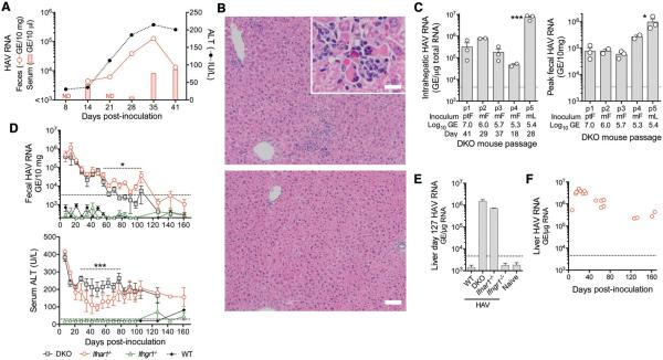
Figure 1.
HAV infection in DKO (Ifnar1−/−Ifngr1−/−), Ifnar1−/−, and Ifngr1−/− mice. (A) Representative course of infection in a DKO mouse inoculated i.v. with 107 GE human HAV (chimpanzee fecal extract). (B) H&E-stained liver from representative (top) infected and (bottom) control DKO mice 41 d.p.i. showing inflammatory infiltrates and (inset) apoptotic hepatocytes (bar = 50 μm, inset bar = 12.5 μm). (C) Summary of serial passage of HAV in DKO mice showing (left) intrahepatic HAV RNA and (right) fecal HAV RNA, source and magnitude of HAV inocula (Pt-F, chimpanzee feces; M-F, DKO mouse feces; M-L, DKO mouse liver), and day of harvest. Data are mean ± SEM or range, n=2–3 animals as shown. *p<0.05, ***p<0.001 p1 vs. p5 by 1-way ANOVA. (D) (top) Fecal HAV RNA and (bottom) serum ALT in DKO, Ifnar1−/−, Ifngr1−/− and wild-type (WT) BL6 mice challenged with 4th passage DKO liver extract (2.6 ×10⁸ GE). Shown are means ± SEM, n=4. *p<0.05, *** p<0.001 for Ifnar1−/− vs. DKO by ANOVA. (E) Intrahepatic HAV RNA in WT, DKO, Ifnar1−/− and Ifngr1−/− mice 127 d.p.i. (mean ± range, n=2). (F) Intrahepatic HAV RNA in Ifnar1−/− mice infected with 4th passage liver extract. Symbols represent individual mice. Dotted horizontal lines in panels indicate level of detection (RNA) or upper limit of normal (ALT).

