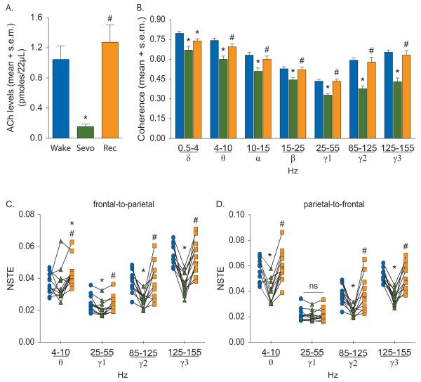
Figure 3.
Effect of sevoflurane on cortical acetylcholine (ACh) (A), corticocortical coherence (B), and frontal-parietal directed connectivity (C-D). The symbol-line plots (C-D) show the individual rat data while the significance symbols represent the outcome of group level statistical testing using repeated measures analysis of variance with Tukey’s multiple comparisons test. Significance symbols denote a statistical difference at an alpha of p<0.05. The actual p values are reported in the text in the results section. *significant as compared to wake, #significant as compared to sevoflurane-induced unconsciousness, blue bars/circles: wake, green bars/triangles: sevoflurane-induced unconsciousness, orange bars/squares: post-sevoflurane recovery wake. ns: not significant, NSTE: normalized symbolic transfer entropy, Rec: recovery wake, s.e.m.: standard error of the mean, Sevo: sevoflurane.

