Abstract
A series of novel quaternary ammonium 4-deoxypyridoxine derivatives was synthesized. Two compounds demonstrated excellent activity against a panel of Gram-positive methicillin-resistant S. aureus strains with MICs in the range of 0.5–2 μg/mL, exceeding the activity of miramistin. At the same time, both compounds were inactive against the Gram-negative E. coli and P. aeruginosa strains. Cytotoxicity studies on human skin fibroblasts and embryonic kidney cells demonstrated that the active compounds possessed similar toxicity with benzalkonium chloride but were slightly more toxic than miramistin. SOS-chromotest in S. typhimurium showed the lack of DNA-damage activity of both compounds; meanwhile, one compound showed some mutagenic potential in the Ames test. The obtained results make the described chemotype a promising starting point for the development of new antibacterial therapies.
1. Introduction
Growing antibiotic resistance has become a major clinical problem in recent decades and has encouraged many researchers to search for novel antibacterial drugs. In particular, methicillin-resistant Staphylococcus aureus (MRSA), vancomycin-resistant enterococci (VRE), and multidrug-resistant Pseudomonas aeruginosa are associated with increased rates of illness and death [1].
Since the 1930s, quaternary ammonium compounds (QACs) are widely used for the control of bacterial growth in clinical and industrial environments. Broad-spectrum antimicrobial and antifungal activity [2–5] and surfactant properties have made QACs such as benzalkonium chloride [6], fluomizin [7], miramistin, and cetylpyridinium chloride [8] the favored hygienic adjuncts in disinfectant cleansing formulations, and they have also been increasingly deployed in the treatment of bacterial infections.
According to literature data [7], QACs generally act by disrupting the cytoplasmic and outer membrane lipid bilayers through association of the positively charged quaternary nitrogen with the anionic head groups of acidic phospholipids and interaction of the lipophilic tail with the hydrophobic membrane core. As a result, QACs form mixed-micelle aggregates with hydrophobic membrane components that solubilize membrane and lyse the cells. Bacterial cell lethality occurs through generalized and progressive leakage of cytoplasmic materials [7]. At the same time, other biomolecular complexes within the bacterial cells are potential targets for action of cationic surfactants which can disrupt critical intermolecular interactions and tertiary structures in such highly specific biochemical systems.
In our group, we have systematically studied chemistry and biology of the biologically active pyridoxine derivatives [9–12]. Recently, we have described a wide series of mono- and bis-ammonium derivatives of pyridoxine and 6-hydroxymethyl pyridoxine [13]. Some of the described compounds possess potent antibacterial activity against several Gram-positive pathogens with minimum inhibitory concentrations (MICs) in the range of 4–64 μg/mL. These promising results encouraged us to synthesize and study their 4-deoxypyridoxine analogs, bearing long alkyl substituents at the quaternary nitrogen. The obtained compounds were tested in vitro for their ability to inhibit growth of a wide number of Gram-positive and Gram-negative bacterial pathogens. The most active compounds were also studied for their cytotoxicity and genotoxicity. Our special interest was focused on activity of the obtained compounds against the methicillin-resistant Staphylococcus aureus pathogens.
2. Materials and Methods
2.1. Synthetic Procedures
2.1.1. General Information
1H and 13C NMR spectra were recorded on a Bruker AVANCE 400 spectrometer at operating frequency of 400 and 101.56 MHz, respectively. Chemical shifts were measured with reference to the residual protons of the solvents (DMSO-d6, 1H, 2.50 ppm, 13C, 39.52 ppm; CDCl3, 1H, 7.26 ppm, 13C, 77.16 ppm; СD3OD, 1H, 4.87 ppm, 13C, 49.00 ppm). Coupling constants (J) are given in Hertz (Hz). The following abbreviations are used to describe coupling: s = singlet; d = doublet; t = triplet; m = multiplet; q = quartet; br s = broad singlet. Melting points were determined using a Stanford Research Systems MPA-100 OptiMelt melting point apparatus and are uncorrected. For TLC analysis silica gel plates from Sorbfil (Krasnodar, Russia) were used with UV light (254 nm/365 nm) or iron (III) chloride as developing agent. Column chromatography was performed on silica gel (60–200 mesh) from Acros.
HRMS mass spectra were obtained on a quadrupole time-of-flight (qTOF) AB Sciex Triple TOF 5600 mass spectrometer using turbo-ion spray source (nebulizer gas nitrogen, a positive ionization polarity, and needle voltage 5500 V). Recording of the spectra was performed in a TOF MS mode with collision energy of 10 eV, declustering potential of 100 eV, and resolution more than 30 000 full-width half-maximum. Samples with the analytes concentration of 5 μmol/L were prepared by dissolving of the test compounds in a mixture of methanol (HPLC-UV Grade, LabScan) and water (LC-MS Grade, Panreac) with a ratio of 1 : 1.
2.1.2. General Procedure for Preparation of the Quaternary Ammonium Salts 4a–c
Tertiary amine (1 equiv.) was added to a solution of compound 3 (1 equiv.) in 30 mL of DMF. The reaction mixture was heated at 50°C for 20 h and then the solvent was evaporated under reduced pressure. The product was recrystallized from acetone : diethyl ether mixture (10 : 1).
2.1.3. N-((5-Acetoxy-4,6-dimethylpyridin-3-yl)methyl)-N,N-dimethyloctan-1-aminium Chloride (4a)
The compound was obtained from 3 (150 mg, 0.70 mmol) and N,N-dimethyloctylamine (0.144 mL, 0.70 mmol) following the general procedure. Yield 54% (140 mg); white solid; mp 169-170°C (dec). 1H NMR (400 MHz, CDCl3, δ, ppm): 0.82 (t, 3 J нн = 6.8 Hz, C7H14, 3H); 1.18–1.26 (m, 5СН2, 10Н); 1.72 (br s, СН2, 2Н); 2.34 (s, СН3, 3Н); 2.36 (s, СН3, 3Н); 2.43 (s, СН3, 3Н); 3.27 (s, 2CH3N+, 6H); 3.57–3.61 (m, CH2N+, 2H); 5.29 (s, CH2N+, 2H); 8.51 (s, СН pyr, 1Н). 13С NMR (100 MHz, CDCl3, δ, ppm): 14.08 (s, CH3); 14.43 (s, CH3); 19.68 (s, CH3); 20.50 (s, CH2); 22.59 (s, CH2); 23.00 (s, CH2); 26.39 (s, CH2); 29.05 (s, CH2); 29.26 (s, CH2); 31.64 (s, CH2); 49.26 (s, CH3N+); 62.97 (s, CH2N+); 63.72 (s, CH2N+); 122.04 (s, С pyr); 143.03 (s, С pyr); 145.55 (s, С pyr); 151.19 (s, С pyr); 153.83 (s, С pyr); 168.20 (s, С=O). HRМS-ESI: found [М–Cl]+ 335.2693, C20H35N2O2. Calculated [М–Cl]+ 335.2693.
2.1.4. N-((5-Acetoxy-4,6-dimethylpyridin-3-yl)methyl)-N,N-dimethyldodecan-1-aminium Chloride (4b)
The compound was obtained from 3 (175 mg, 0.82 mmol) and N,N-dimethyldodecylamine (0.222 mL, 0.82 mmol) following the general procedure. Yield 50% (175 mg); white solid; mp 169-170°C (dec). 1H NMR (400 MHz, CDCl3, δ, ppm): 0.84 (t, 3 J нн = 6.8 Hz, C11H22, 3H); 1.20–1.28 (m, 9СН2, 18Н); 1.73 (br s, СН2, 2Н); 2.35 (s, СН3, 3Н); 2.37 (s, СН3, 3Н); 2.44 (s, СН3, 3Н); 3.28 (s, 2CH3N+, 6H); 3.57–3.61 (m, CH2N+, 2H); 5.29 (s, CH2N+, 2H); 8.51 (s, СН pyr, 1Н). 13С NMR (100 MHz, CDCl3, δ, ppm): 14.19 (s, CH3); 14.46 (s, CH3); 19.72 (s, CH3); 20.52 (s, CH2); 22.74 (s, CH2); 23.04 (s, CH2); 26.42 (s, CH2); 29.35 (s, CH2); 29.38 (s, CH2); 29.43 (s, CH2); 29.50 (s, CH2); 29.64 (s, CH2); 49.31 (s, CH3N+); 63.03 (s, CH2N+); 63.74 (s, CH2N+); 122.01 (s, Cpyr); 143.06 (s, Cpyr); 145.59 (s, Cpyr); 151.20 (s, Cpyr); 153.91 (s, Cpyr); 168.23 (s, С=O). HRМS-ESI: found [М–Cl]+ 391.3319, C25H43N2O2. Calculated [М–Cl]+ 391.3319.
2.1.5. N-((5-Acetoxy-4,6-dimethylpyridin-3-yl)methyl)-N,N-dimethyloctadecan-1-aminium Chloride (4c)
The compound was obtained from 3 (150 mg, 0.70 mmol) and N,N-dimethyloctadecylamine (0.261 mL, 0.70 mmol) following the general procedure. Yield 53% (190 mg); white solid; mp 159-160°C (dec). 1H NMR (400 MHz, CDCl3, δ, ppm): 0.85 (t, 3 J нн = 6.7 Hz, C17H34, 3H); 1.21–1.29 (m, 15СН2, 30Н); 1.74 (br s, СН2, 2Н); 2.36 (s, СН3, 3Н); 2.39 (s, СН3, 3Н); 2.46 (s, СН3, 3Н); 3.29 (s, 2CH3N+, 6H); 3.57–3.61 (m, CH2N+, 2H); 5.30 (s, CH2N+, 2H); 8.50 (s, СН pyr, 1Н). 13С NMR (100 MHz, CDCl3, δ, ppm): 14.22 (s, CH3); 14.51 (s, CH3); 19.75 (s, CH3); 20.55 (s, CH2); 22.79 (s, CH2); 23.07 (s, CH2); 26.44 (s, CH2); 29.39 (s, CH2); 29.46 (s, CH2); 29.55 (s, CH2); 29.69 (s, CH2); 29.76 (s, CH2); 29.80 (s, CH2); 32.02 (s, CH2); 49.36 (s, CH3N+); 63.10 (s, CH2N+); 63.79 (s, CH2N+); 122.01 (s, Cpyr); 143.13 (s, Cpyr); 145.64 (s, Cpyr); 153.18 (s, Cpyr); 153.98 (s, Cpyr); 168.26 (s, С=O). HRМS-ESI: found [М–Cl]+ 475.4258, C30H55N2O2. Calculated [М–Cl]+ 475.4258.
2.1.6. General Procedure for Preparation of 5a–c
A mixture of quaternary ammonium salt 4a–c (1 equiv.) and 1 mL of concentrated HCl in 20 mL of water was stirred at 60°C for 24 h. The solvent was evaporated under reduced pressure to obtain 5a–c in quantitative yield.
2.1.7. 5-((Octyldimethylammonio)methyl)-3-hydroxy-2,4-dimethylpyridin-1-ium Dichloride (5a)
The compound was obtained from 4a (100 mg, 0.27 mmol) following the general procedure. Yield quantitative (0.99 mg); white solid; mp 186–188°C (dec.). 1H NMR (400 MHz, DMSO-d6, δ, ppm): 0.87 (t, 3 J нн = 6.8 Hz, C11H22, 3H); 1.27–1.31 (m, 5СН2, 5Н); 1.77 (br s, СН2, 2Н); 2.50 (s, СН3, 3Н); 2.68 (s, СН3, 3Н); 2.99 (s, 2CH3N+, 6H); 3.42–3.46 (m, CH2N+, 2H); 4.78 (s, CH2N+, 2H); 8.49 (s, СН pyr, 1Н); 10.96 (s, OH, 1H). 13С NMR (100 MHz, DMSO-d6, δ, ppm): 13.94 (s, CH3); 14.87 (s, CH3); 15.43 (s, CH3); 21.83 (s, CH2); 22.03 (s, CH2); 25.84 (s, CH2); 28.41 (s, CH2); 28.48 (s, CH2); 31.14 (s, CH2); 48.66 (s, CH3N+); 60.43 (s, CH2N+); 64.48 (s, CH2N+); 124.81 (s, Cpyr); 136.27 (s, Cpyr); 142.15 (s, Cpyr); 152.87 (s, Cpyr). HRМS-ESI: found [М–Cl]+ 293.2587, C18H34N2O. Calculated [М–Cl]+ 293.2587.
2.1.8. 5-((Dodecyldimethylammonio)methyl)-3-hydroxy-2,4-dimethylpyridin-1-ium Dichloride (5b)
The compound was obtained from 4b (100 mg, 0.23 mmol) following the general procedure. Yield quantitative (0.99 mg); white solid; mp 187-188°C (dec.). 1H NMR (400 MHz, CD3OD, δ, ppm): 0.88 (t, 3 J нн = 6.7 Hz, C11H22, 3H); 1.28–1.42 (m, 18СН2, 9Н); 1.90 (br s, СН2, 2Н); 2.60 (s, СН3, 3Н); 2.70 (s, СН3, 3Н); 3.09 (s, 2CH3N+, 6H); 3.50 (br s, CH2N+, 2H); 4.82 (s, CH2N+, 2H); 8.49 (s, СН pyr, 1Н). 13С NMR (100 MHz, CD3OD, δ, ppm): 14.43 (s, CH3); 15.47 (s, CH3); 15.63 (s, CH3); 23.73 (s, CH2); 23.81 (s, CH2); 27.50 (s, CH2); 30.32 (s, CH2); 30.46 (s, CH2); 30.58 (s, CH2); 30.65 (s, CH2); 30.74 (s, CH2); 33.06 (s, CH2); 50.29 (s, CH3N+); 62.69 (s, CH2N+); 67.40 (s, CH2N+); 126.59 (s, Cpyr); 136.93 (s, Cpyr); 143.83 (s, Cpyr); 148.57 (s, Cpyr); 155.46 (s, Cpyr). HRМS-ESI: found [М–H–2Cl]+ 349.3213, C22H42N2O. Calculated [М–H–2Cl]+ 349.3213.
2.1.9. 5-((Dimethyl(octadecyl)ammonio)methyl)-3-hydroxy-2,4-dimethylpyridin-1-ium Dichloride (5c)
The compound was obtained from 4c (100 mg, 0.20 mmol) following the general procedure. Yield quantitative (0.99 mg); white solid; mp 185–187°C (dec.). 1H NMR (400 MHz, DMSO-d6, δ, ppm): 0.85 (t, 3 J нн = 6.7 Hz, C17H34, 3H); 1.23–1.33 (m, 15СН2, 30Н); 1.77 (br s, СН2, 2Н); 2.50 (s, СН3, 3Н); 2.68 (s, СН3, 3Н); 2.99 (s, 2CH3N+, 6H); 3.42–3.46 (m, CH2N+, 2H); 4.77 (s, CH2N+, 2H); 8.49 (s, CHpyr, 1Н); 10.94 (s, OH, 1H). 13С NMR (100 MHz, DMSO-d6, δ, ppm): 13.94 (s, CH3); 14.86 (s, CH3); 15.46 (s, CH3); 21.85 (s, CH2); 22.08 (s, CH2); 25.85 (s, CH2); 28.56 (s, CH2); 28.68 (s, CH2); 28.79 (s, CH2); 29.03 (s, CH2); 31.27 (s, CH2); 48.66 (s, CH3N+); 60.42 (s, CH2N+); 64.50 (s, CH2N+); 124.81 (s, Cpyr); 136.29 (s, Cpyr); 142.17 (s, Cpyr); 152.87 (s, Cpyr). HRМS-ESI: found [М–H–2Cl]+ 433.4152, C28H54N2O. Calculated [М–H–2Cl]+ 433.4152.
2.2. Biological Experiments
2.2.1. Antibacterial Activity
The antibacterial activity of compounds was evaluated on the number of Gram-positive (Staphylococcus aureus ATCC® 29213™, Staphylococcus epidermidis (clinical isolate), Micrococcus luteus (clinical isolate), and Bacillus subtilis 168) and Gram-negative bacteria (Escherichia coli АТСС 25922™, Pseudomonas aeruginosa АТСС 27853™, and Salmonella typhimurium TA100). Clinical isolates of Staphylococcus epidermidis and Micrococcus luteus were obtained from the Kazan Institute of Epidemiology and Microbiology (Kazan, Russia). The antibacterial activity of compounds 4c and 5c was additionally evaluated on Staphylococcus aureus ATCC 700699™ and a number of clinical isolates: Staphylococcus aureus 100 MRSA, Staphylococcus aureus 5 MRSA, Staphylococcus aureus 6 MRSA, Staphylococcus aureus 3797 MRSA, Staphylococcus aureus 3798 MRSA, Staphylococcus aureus 4603 MRSA, Staphylococcus haemolyticus 161, Staphylococcus haemolyticus 1025, Staphylococcus haemolyticus 602, Staphylococcus haemolyticus 585, Staphylococcus epidermidis 681, Staphylococcus epidermidis 9, Enterococcus faecalis 560, Enterococcus faecium 569, Escherichia coli 396, and Pseudomonas aeruginosa 43.
The MICs of compounds were determined by the broth microdilution method in Mueller-Hinton (MH) broth (pH = 7.3) in 96-well plates. The double-dilution series of compounds with the final concentrations ranging from 1000 to 0.5 μg/mL in the initial screening experiment and 64–0.5 μg/mL in the evaluation of 4c and 5c on clinical isolate strains were used. The bacterial suspension (2–9 × 10⁴ CFU/mL) 200 μL aliquots were seeded into 96-well plates and their incubation was followed. The MIC was determined as the lowest concentration of compound for which no visible bacterial growth could be observed after 24 h of incubation at 37°C.
2.2.2. Cytotoxic Activity
Human skin fibroblasts (HSFs) were isolated from the skin explant according to the conventional protocol [17]. HEK 293 (human embryonic kidney) cells were obtained from the ATCC collection. HSFs cells were cultured in the minimum essential medium Eagle (α-MEM) supplemented with 10% fetal bovine serum, 2 mM L-glutamine, 100 μg/mL streptomycin, and 100 U/mL penicillin under standard conditions (37°C, 5% CO2 atmosphere). HEK 293 cells were grown in the same conditions, but in the Dulbecco's modified Eagle's medium (DMEM). Adhered cells were collected from the culture flask by detaching them with trypsin-EDTA solution. Suspended cells were washed by centrifugation at 200 g in PBS.
Cytotoxic concentrations (IC50) of compounds were determined with the use of MTT assay. Cells were preseeded in 96-well plate at the density of 1000–2000 cells per well and cultured with adding a series of diluted water solutions of compounds for 3 days under standard conditions. Culture medium in the plate was then replaced by the fresh one supplemented with 0.5 mg/mL MTT and additionally kept for 4 h to allow for reduction of MTT into colored product (formazan) by metabolically active cells. Optical absorbance of produced formazan, proportional to viable cell number, was registered on Infinite 200 PRO analyzer at 550 nm.
2.2.3. Genotoxicity
S. typhimurium strain TA100 [18] was grown overnight in 5 mL of LB medium, diluted 4 times by prewarmed LB and incubation was continued for 2 h. Cells were harvested, washed once by 1x salt base solution (g/L: sodium citrate × 3Н2О − 0.5; К2НРО3 × 3Н2О − 14; КН2РО3 − 6; (NH4)2SO4 − 1; MgSO4 × 7Н2О − 0.5) and resuspended in 6 mL of 1x salt base. 100 μL of bacterial suspension was mixed with top agar (0.5% agar, 0.5% NaCl, 50 mM L-histidine, 50 mM biotin, pH 7.4, 42°C) in a final volume of 3 mL and the substance to be tested. Each mixture was then seeded onto the minimal agar plates (1.5% agar in the 1x salt base supplemented with 0.5% glucose and ampicillin 10 μg/mL). Next the plates were incubated at 37°C for 72 hours and colonies were counted. Sodium azide (10 μg/mL) was used as a positive control.
The SOS-chromotest was performed by using the Salmonella typhimurium TA1535/pSK1002 as described in [19]. Briefly, aliquots of 0.5 mL of an overnight culture of the tester strains were diluted in 5 mL of LB medium and then incubated with rigorous agitation in presence of the ficin substances. The Mitomycin C (Sigma) at concentration of 1 μg/mL was used as a positive control. After 4 h of incubation, the cell density (A600) and the β-galactosidase activity were measured by Miller's protocol [20] with modifications. Cells were harvested from 0.5 to 1.5 mL of culture liquid and resuspended in 800 μL of Z-buffer [60 mM Na2HPO4·7H2O, 40 mM NaH2PO4·H2O, 10 mM KCl, and 1 mM MgSO4·7H2O (pH 7.0)] containing additionally 0.005% cetyl trimethylammonium bromide (CTAB) and 50 mM β-mercaptoethanol was added. After preincubation at 30°C for 5 min, the reaction was started by adding of 200 μL of 4 mg/mL o-nitrophenyl-β-D-galactopyranoside in Z-buffer. When the yellow color appeared, the reaction was stopped by 500 μL of 1 M Na2CO3. For the blank solution, the Na2CO3 was added prior to the incubation. The β-galactosidase activity was measured at A420 nm. To calculate the Miller units, we used the following formula: [A420/(A600 of 1 : 10 dilution of cells × time of incubation)] × 1000.
3. Results and Discussion
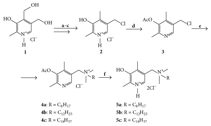
Key chloride intermediate 2 was obtained from pyridoxine hydrochloride 1 according to literature method [15] (Scheme 1). Interaction of 2 with acetyl chloride in the presence of triethylamine led to acetyl derivative 3 [16]. The latter was used for alkylation of the tertiary amines in dimethylformamide, which led to the quaternary ammonium salts 4a–c and, after deacetylation under acidic conditions, to compounds 5a–c. All attempts to obtain compounds 5a–c directly from amines and chloride 2 without protection of hydroxyl group were unsuccessful, probably, due to instability of 2 under the reaction conditions.
Scheme 1.
Reagents and conditions: (a) Zn, AcOH, reflux [15]; (b) HCl, EtOH-H2O, 25°C [15]; (c) SOCl2, DMF, reflux [15]; (d) AcCl, NEt3, CH2Cl2, reflux [16]; (e) DMF, (CH3)2RN, 50°C; (f) H2O, HCl, 25°C.
Compounds 4a–c and 5a–c were evaluated for antibacterial activity against three Gram-positive and three Gram-negative bacterial strains. Table 1 shows MICs of the tested compounds in comparison with miramistin and benzalkonium chloride. Two compounds 4c and 5c with the longest octadecyl substituent demonstrated high antibacterial activity with MICs in the range of 2–32 μg/mL for all pathogens with the exception of P. aeruginosa. In general, their activity in this test was comparable with that of the reference drugs.
Table 1.
In vitro antimicrobial activity of the obtained 4-deoxypyridoxine ammonium salts.
| Compounds | MICs (µg/mL) | |||||
|---|---|---|---|---|---|---|
| Gram-positive bacteria | Gram-negative bacteria | |||||
| S. aureus | S. epidermidis | M. luteus | E. coli K12 | S. typhimurium TA100 | P. aeruginosa | |
| 4a | >64 | >64 | >64 | >64 | >64 | >64 |
| 4b | >64 | 32 | >64 | >64 | >64 | >64 |
| 4c | 32 | 2 | 4 | 4 | 8 | >64 |
| 5a | >64 | >64 | >64 | >64 | >64 | >64 |
| 5b | >64 | >64 | >64 | >64 | >64 | >64 |
| 5c | 8 | 4 | 8 | 2 | 16 | >64 |
| Miramistin | 4 | 2 | 2 | 32 | >64 | 16 |
| Benzalkonium chloride | 2 | 2 | 32 | 4 | 16 | 64 |
Compounds 4c and 5c with promising activities in the primary assay were selected for further evaluation. First, they were studied on an extended panel of clinical Gram-positive and Gram-negative bacterial strains in comparison with miramistin (Table 2). Both compounds exhibited strong activity against all Staphylococcus strains and, moreover, compound 5c was significantly more active than miramistin. At the same time, compounds 4c and 5c were inactive against the studied Gram-negative E. coli and P. аeruginosa strains thus demonstrating a good selectivity for the Gram-positive pathogens. Interestingly, the clinical isolates of S. aureus were more sensitive to both 4c and 5c than S. aureus ATCC 29213 cells, while the clinical isolate E. coli 396 was not sensitive to 4c and 5c in contrast to E. coli K12. We attribute these effects to the different phenotypic resistance of the strains to antimicrobials [21, 22].
Table 2.
In vitro antimicrobial activity of 4c and 5c on an extended panel of clinical bacterial pathogens.
| Strains | MICs (µg/mL) | ||
|---|---|---|---|
| 4c | 5c | Miramistin | |
| S. aureus 700699 ATCC | 1 | 1 | 2 |
| S. aureus 100 (MRSA) | 2 | 0.5 | 2 |
| S. aureus 5 (MRSA) | 1 | 0.5 | 8 |
| S. aureus 6 (MRSA) | 1 | 0.5 | 2 |
| S. aureus 3797 (MRSA) | 2 | 1 | 4 |
| S. aureus 3798 (MRSA) | 1 | 0.5 | 2 |
| S. aureus 4603 (MRSA) | 2 | 0.5 | 4 |
| S. haemolyticus 161 | 2 | 1 | 4 |
| S. haemolyticus 1025 | 1 | 0.5 | 4 |
| S. haemolyticus 602 | 2 | 1 | 4 |
| S. haemolyticus 585 | 4 | 2 | 2 |
| S. epidermidis 681 | 4 | 2 | 1 |
| S. epidermidis 9 | 4 | 4 | 2 |
| E. faecalis 560 | 4 | 2 | 2 |
| E. faecalis 569 | 2 | 4 | 1 |
| E. faecalis 101 | 4 | 8 | 2 |
| E. coli 396 | >64 | >64 | 32 |
| P. aeruginosa 43 | >64 | >64 | 64 |
Compounds 4c and 5c were further examined for their cytotoxicity on human skin fibroblasts (HSF) and human embryonic kidney (HEK 293) cells with the use of MTT assay. According to Table 3, both compounds possessed similar benzalkonium chloride cytotoxicity but were slightly more toxic than miramistin. Despite the satisfactory results obtained in the MTT assay, it should be noted that further studies with the use of lactate dehydrogenase (LDH) release assay or by imaging the uptake of cell-impermeable fluorescent dyes are desirable to more thoroughly assess possible membrane-related toxicity of the described compounds.
Table 3.
Cytotoxicity of the ammonium salts 4c and 5c.
| Compounds | CC50, µg/mL | |
|---|---|---|
| HSF | HEK 293 | |
| 4c | 2.88 ± 0.59 | 2.32 ± 0.75 |
| 5c | 3.25 ± 0.45 | 2.96 ± 0.9 |
| Miramistin | 4.09 ± 0.42 | 4.08 ± 0.77 |
| Benzalkonium chloride | 2.11 ± 0.12 | 2.04 ± 0.9 |
Genotoxicity of compounds 4c and 5c was evaluated by using SOS-chromotest in Salmonella typhimurium TA1535/pSK1002 strain as described by Oda et al. [18]. Mitomycin C was used as a positive control. The optical density at 420 nm (OD420) was measured, and β-galactosidase activity was normalized to the amount of cells estimated from the OD600 values. SOS induction factor was calculated as a ratio of β-galactosidase activity in the presence of compounds and the solvent control (Table 4). No significant dose-dependent increase more than 2-fold was observed thus indicating the lack of DNA-damage activity of compounds 4c and 5c under the tested concentrations.
Table 4.
DNA-damage activity of 4c and 5c in the SOS-chromotest (ratio, fold increase over the solvent control).
| Compounds | Concentration, μg/mL | ||
|---|---|---|---|
| 4 | 20 | 40 | |
| 4c | 0.9 ± 0.32 | 1.3 ± 0.25 | 1.4 ± 0.45 |
| 5c | 1.2 ± 0.35 | 1.5 ± 0.43 | 1.3 ± 0.53 |
| Mitomycin C | 11.7 ± 2.5 | — | — |
The Ames test was then performed using Salmonella typhimurium TA100 [19]. The compounds were tested in concentrations 2, 4, and 8 μg/mL close to their CC50 values for Salmonella cells. Sodium azide was used as a positive mutagenic control. No increase in the number of revertant colonies as well as no dose dependency was observed for 4c, thus suggesting the lack of mutagenicity (Table 5). At the same time, 5c caused some dose-dependent increase in the number of revertant colonies. Probably, this suggests some mutagenic potential of this compound.
Table 5.
Mutagenicity of 4c and 5c in the Ames test (ratio, fold increase over the solvent control).
| Compounds | Concentration, μg/mL | ||
|---|---|---|---|
| 2 | 4 | 8 | |
| 4c | 0.9 ± 0.21 | 0.6 ± 0.15 | 0.2 ± 0.12 |
| 5c | 1.8 ± 0.74 | 2.3 ± 0.53 | 2.4 ± 0.57 |
| Sodium azide | 6.5 ± 2.5 | 8.3 ± 1.4 | 9.8 ± 4.6 |
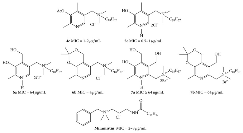
It was further interesting to compare the obtained results with our recently reported data [13] on antibacterial activity of a series of close structural analogs, the quaternary ammonium salts of pyridoxine and 6-hydroxymethyl pyridoxine (Figure 1).
Figure 1.
Structures of antibacterial QACs studied in this work (4c and 5c), their recently reported structural analogs (6a, b and 7a, b) [13], and miramistin. MIC values are shown for S. aureus.
In that study, we observed a strong correlation between the length of the substituent's chain and antimicrobial activity. The most active compounds had the same octadecyl substituent at the quaternary nitrogen (structures 6a, 6b and 7a, 7b) and were bactericidal against several Gram-positive pathogens including S. aureus. Compounds 6a, 6b and 7a, 7b exhibited a moderate activity against a panel of Gram-positive pathogens (MIC > 4 μg/mL, S. aureus), and this activity demonstrated some dependence on compound's lipophilicity. In particular, such a dependency led to activity decrease upon removal of the acetonide protective group.
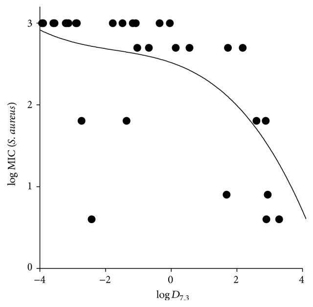
This dependency is more general, though not absolute, for the studied QACs. Figure 2 demonstrates the relationship between the experimental MICs for S. aureus (expressed in a decimal logarithm scale) and calculated logD values at pH 7.3 for the combined set of 32 quaternary ammonium pyridoxine derivatives reported in our recent study [13] and in the current paper. Probably, it reflects the important feature of the active compounds essential for their effective interaction with the hydrophobic membrane core of bacterial cells.
Figure 2.

Log MIC (S. aureus) versus calculated logD 7.3 [14] relationship for the combined set of the quaternary ammonium pyridoxine derivatives reported in our recent study [13] and in the current paper. A polynomial trend line is shown.
In this work, we obtained more active antibacterial agents (MIC in the range of 0.5–2 μg/mL against the studied methicillin-resistant S. aureus strains) than those described in the recent paper [13] but with similar selectivity profile and, as may be supposed, similar mechanism of action.
So far, no specific target has been identified for most QACs; it is assumed that the effect is rather generalized than specific to one target. However, as discussed in literature [7], there should be some target specificities, for example, as shown for the bisquaternary bisnaphthalimide MT02 [23], because the activity of QACs toward different bacterial species varies substantially and cannot be explained simply by the structure of cationic charge and hydrophobic portions. It is possible that the most potent agents exert membrane damage, leading to disruption of the cell envelope and arresting intracellular activity by binding targets in the cytoplasm [24].
It can be suggested that all the studied substances interact with bacterial membranes, given their clear structural similarity to other QACs such as miramistin. However, the role of intracellular targets in their antibacterial action remains unclear. Pyridoxine molecule is a well-established cofactor for many enzymes. Therefore, pyridoxine derivatives can participate in many intracellular interactions thus leading to enhanced or more specific antibacterial action. The observed effect of 5c in the Ames test may suggest some specific interaction with DNA; however, this hypothesis requires further experimental investigation.
Interestingly, Gram-positive bacteria are generally more sensitive to the obtained compounds than Gram-negative bacteria, which is in agreement with literature data on QACs [7]. This has been attributed to the outer membrane of Gram-negative bacteria, which is absent in Gram-positive strains. Importantly, many antibiotic-resistant staphylococci (such as MRSA or methicillin-resistant S. epidermidis) have acquired plasmids such as qacA, qacB, qacC, or qacD, which encode efflux pumps and thus confer resistance determinants to QACs [25]. This suggests that intracellular accumulation of QACs is important for full activity and underscores the importance of the interaction between QACs and intracellular targets for antibacterial activity.
4. Conclusion
In this work we have synthesized a small series of novel quaternary ammonium 4-deoxypyridoxine derivatives and studied their antibacterial activity, cytotoxicity, and genotoxicity. Two compounds demonstrate promising in vitro antibacterial activity and toxicity when compared with the reference antibacterials miramistin and benzalkonium chloride. The obtained results make the described chemotype a promising starting point for the development of new antibacterial therapies with selective effect on Gram-positive pathogens including their drug-resistant forms.
Supplementary Material
The Supplementary Information contains synthetic procedures, analytical characteristics of compounds 4a-c and 5a-c, and biological experimental methods associated with this paper.
Acknowledgments
This work was funded by the subsidy allocated to Kazan Federal University by Federal Targeted Programme for Research and Development in Priority Areas of Development of the Russian Scientific and Technological Complex for 2014–2020 (Project no. 14.575.21.0037 from 27.06.2014, the unique identifier of the agreement RFMEF157514X0037).
Competing Interests
The authors declare that there is no conflict of interests regarding the publication of this manuscript.
References
- 1.Nordmann P., Naas T., Fortineau N., Poirel L. Superbugs in the coming new decade; multidrug resistance and prospects for treatment of Staphylococcus aureus, Enterococcus spp. and Pseudomonas aeruginosa in 2010. Current Opinion in Microbiology. 2007;10(5):436–440. doi: 10.1016/j.mib.2007.07.004. [DOI] [PubMed] [Google Scholar]
- 2.Minbiole K. P. C., Jennings M. C., Ator L. E., et al. From antimicrobial activity to mechanism of resistance: the multifaceted role of simple quaternary ammonium compounds in bacterial eradication. Tetrahedron. 2016;72(25):3559–3566. doi: 10.1016/j.tet.2016.01.014. [DOI] [Google Scholar]
- 3.Obłak E., Piecuch A., Krasowska A., Łuczyński J. Antifungal activity of gemini quaternary ammonium salts. Microbiological Research. 2013;168(10):630–638. doi: 10.1016/j.micres.2013.06.001. [DOI] [PubMed] [Google Scholar]
- 4.Massi L., Guittard F., Levy R., Duccini Y., Géribaldi S. Preparation and antimicrobial behaviour of gemini fluorosurfactants. European Journal of Medicinal Chemistry. 2003;38(5):519–523. doi: 10.1016/S0223-5234(03)00059-X. [DOI] [PubMed] [Google Scholar]
- 5.Ohkura K., Sukeno A., Yamamoto K., Nagamune H., Maeda T., Kourai H. Analysis of structural features of bis-quaternary ammonium antimicrobial agents 4,4′-(α,ω-polymethylenedithio)bis (1-alkylpyridinium iodide)s using computational simulation. Bioorganic and Medicinal Chemistry. 2003;11(23):5035–5043. doi: 10.1016/j.bmc.2003.08.027. [DOI] [PubMed] [Google Scholar]
- 6.Domagk G. Eine neue Klasse von Desinfektionsmitteln. Deutsche Medizinische Wochenschrift. 1935;61(21):829–832. doi: 10.1055/s-0028-1129654. [DOI] [Google Scholar]
- 7.Tischer M., Pradel G., Ohlsen K., Holzgrabe U. Quaternary ammonium salts and their antimicrobial potential: targets or nonspecific interactions? ChemMedChem. 2012;7(1):22–31. doi: 10.1002/cmdc.201100404. [DOI] [PubMed] [Google Scholar]
- 8.Fromm-Dornieden C., Rembe J.-D., Schäfer N., Böhm J., Stuermer E. K. Cetylpyridinium chloride and miramistin as antiseptic substances in chronic wound management—prospects and limitations. Journal of Medical Microbiology. 2015;64(4):407–414. doi: 10.1099/jmm.0.000034. [DOI] [PubMed] [Google Scholar]
- 9.Shtyrlin N. V., Lodochnikova O. A., Pugachev M. V., et al. Theoretical and experimental study on cyclic 6-methyl-2,3,4- tris(hydroxymethyl)pyridin-5-ol acetonides. Russian Journal of Organic Chemistry. 2010;46(4):561–567. doi: 10.1134/s1070428010040202. [DOI] [Google Scholar]
- 10.Pugachev M. V., Shtyrlin N. V., Sapozhnikov S. V., et al. Bis-phosphonium salts of pyridoxine: the relationship between structure and antibacterial activity. Bioorganic and Medicinal Chemistry. 2013;21(23):7330–7342. doi: 10.1016/j.bmc.2013.09.056. [DOI] [PubMed] [Google Scholar]
- 11.Shtyrlin N. V., Pavelyev R. S., Pugachev M. V., Sysoeva L. P., Musin R. Z., Shtyrlin Yu. G. Synthesis of novel 6-substituted sulfur-containing derivatives of pyridoxine. Tetrahedron Letters. 2012;53(31):3967–3970. doi: 10.1016/j.tetlet.2012.05.086. [DOI] [Google Scholar]
- 12.Pugachev M. V., Shtyrlin N. V., Sysoeva L. P., et al. Synthesis and antibacterial activity of novel phosphonium salts on the basis of pyridoxine. Bioorganic and Medicinal Chemistry. 2013;21(14):4388–4395. doi: 10.1016/j.bmc.2013.04.051. [DOI] [PubMed] [Google Scholar]
- 13.Shtyrlin N. V., Sapozhnikov S. V., Koshkin S. A., et al. Synthesis and antibacterial activity of novel quaternary ammonium pyridoxine derivatives. Medicinal Chemistry. 2015;11(7):656–665. doi: 10.2174/1573406411666150504122930. [DOI] [PubMed] [Google Scholar]
- 14.Kah M., Brown C. C. LogD: lipophilicity for ionisable compounds. Chemosphere. 2008;72(10):1401–1408. doi: 10.1016/j.chemosphere.2008.04.074. [DOI] [PubMed] [Google Scholar]
- 15.Serwa R., Nam T.-G., Valgimigli L., et al. Preparation and investigation of vitamin B6-derived aminopyridinol antioxidants. Chemistry—A European Journal. 2010;16(47):14106–14114. doi: 10.1002/chem.201001382. [DOI] [PubMed] [Google Scholar]
- 16.Shtyrlin N. V., Vafina R. M., Pugachev M. V., et al. Synthesis and biological activity of tertiary phosphonium salts based on 3-hydroxypyridine and 4-deoxypyridoxine. Russian Chemical Bulletin. 2016;2:537–545. (Rus). [Google Scholar]
- 17.Rittié L., Fisher G. J. Isolation and culture of skin fibroblasts. Methods in Molecular Medicine. 2005;117:83–98. doi: 10.1385/1-59259-940-0:083. [DOI] [PubMed] [Google Scholar]
- 18.Oda Y., Nakamura S.-I., Oki I., Kato T., Shinagawa H. Evaluation of the new system (umu-test) for the detection of environmental mutagens and carcinogens. Mutation Research. 1985;147(5):219–229. doi: 10.1016/0165-1161(85)90062-7. [DOI] [PubMed] [Google Scholar]
- 19.McCann J., Ames B. N. A simple method for detecting environmental carcinogens as mutagens. Annals of the New York Academy of Sciences. 1976;271:5–13. doi: 10.1111/j.1749-6632.1976.tb23086.x. [DOI] [PubMed] [Google Scholar]
- 20.Miller J. H. Experiments in Molecular Genetics. New York, NY, USA: Cold Spring Harbor Laboratory; 1972. [Google Scholar]
- 21.Cosgrove S. E., Kaye K. S., Eliopoulous G. M., Carmeli Y. Health and economic outcomes of the emergence of third-generation cephalosporin resistance in Enterobacter species. Archives of Internal Medicine. 2002;162(2):185–190. doi: 10.1001/archinte.162.2.185. [DOI] [PubMed] [Google Scholar]
- 22.Blair J. M. A., Webber M. A., Baylay A. J., Ogbolu D. O., Piddock L. J. V. Molecular mechanisms of antibiotic resistance. Nature Reviews Microbiology. 2015;13(1):42–51. doi: 10.1038/nrmicro3380. [DOI] [PubMed] [Google Scholar]
- 23.González-Bulnes L., Gallego J. Indirect effects modulating the interaction between DNA and a cytotoxic bisnaphthalimide reveal a two-step binding process. Journal of the American Chemical Society. 2009;131(22):7781–7791. doi: 10.1021/ja901505p. [DOI] [PubMed] [Google Scholar]
- 24.Locher H. H., Ritz D., Pfaff P., et al. Dimers of nostocarboline with potent antibacterial activity. Chemotherapy. 2010;56(4):318–324. doi: 10.1159/000320033. [DOI] [PubMed] [Google Scholar]
- 25.Paulsen I. T., Brown M. H., Littlejohn T. G., Mitchell B. A., Skurray R. A. Multidrug resistance proteins QacA and QacB from Staphylococcus aureus: membrane topology and identification of residues involved in substrate specificity. Proceedings of the National Academy of Sciences of the United States of America. 1996;93(8):3630–3635. doi: 10.1073/pnas.93.8.3630. [DOI] [PMC free article] [PubMed] [Google Scholar]
Associated Data
This section collects any data citations, data availability statements, or supplementary materials included in this article.
Supplementary Materials
The Supplementary Information contains synthetic procedures, analytical characteristics of compounds 4a-c and 5a-c, and biological experimental methods associated with this paper.


