Abstract
SL3-2 is a polytropic murine leukemia virus with a limited species tropism. We cloned the envelope gene of this virus, inserted it into a bicistronic vector, and found that the envelope protein differs from other, similar envelope proteins that also utilize the polytropic receptor (Xpr1) in that it is severely impaired in mediating infection of human and mink cells. We found that two adjacent amino acid mutations (G212R and I213T), located in a previously functionally uncharacterized segment of the surface subunit, are responsible for the restricted tropism of the SL3-2 wild-type envelope. By selection from a two-codon library, several hydrophobic amino acids at these positions were found to enable the SL3-2 envelope to infect human TE 671 cells. In particular, an M212/V213 mutant had a titer at least 6 orders of magnitude higher than that of the wild-type envelope for human TE 671 cells and infected human, mink, and murine cells with equal efficiencies. Notably, these two amino acids are not found at homologous positions in known murine leukemia virus isolates. Functional analysis and library selection were done on the basis of sequence and tropism analyses of the SL3-2 envelope gene. Similar approaches may be valuable in the design and optimization of retroviral envelopes with altered tropisms for biotechnological purposes.
The murine leukemia virus (MLV) group of gammaretroviruses has been divided into the ecotropic, amphotropic, and polytropic-xenotropic subfamilies according to their host range and interference properties, as determined primarily on the basis of the structures of their respective envelope glycoproteins.
The envelope precursor protein is cleaved into two subunits in the endoplasmic reticulum by a cellular protease. The N-terminal surface subunit (SU) is mainly responsible for receptor recognition and binding, while the C-terminal transmembrane subunit is involved in the fusion of the viral and cellular membranes. Accordingly, the SU subunits of different MLV subfamilies show a considerable degree of residue disparity, especially in the N-terminal 200 to 250 residues, while the transmembrane subunits are very much identical. Interestingly, the N terminus of the SU subunit constitutes a receptor-binding domain (RBD), which can bind to the appropriate receptor independently of the rest of the envelope protein. Thus, the ecotropic RBD, which consists of the first 230 (4) or 245 (22) residues, contains almost all of the unique ecotropic envelope sequences. This domain is followed by a proline-rich region that can tolerate large insertions or deletions (15, 36).
Three segments of the RBD that show large variations in MLV subfamilies are conveniently known as hypervariable regions A, B, and C (VRA, VRB, and VRC, respectively). Several studies have suggested that the determinants of the tropisms of ecotropic and amphotropic envelopes are found in the N terminus of RBD, particularly VRA, while segments further downstream are partially responsible for polytropic envelope tropism (4, 7, 8, 25, 29).
Ecotropic and amphotropic viruses, as defined by their usage of the mCAT-1 (2, 3, 35) and Pit-2 (24) receptors, show different but consistent species tropisms. Xenotropic and polytropic viruses, on the other hand, utilize the same (Xpr1) receptor (10, 34, 38) but differ in species tropism. Most remarkably, the xenotropic viruses are not able to infect laboratory mice, while the polytropic viruses are. A third variation of the polytropic-xenotropic tropism theme is that of SL3-2 virus, isolated from a mouse leukemia cell line (31). Early studies showed that this virus has a host range similar to those of mouse ecotropic viruses in that it replicates in mouse cells but not in cells from other species, such as mink. By RNA oligonucleotide fingerprinting (31) and by receptor interference studies with mouse cells (33), it was found that SL3-2 is related to mink cell focus-forming (MCF) viruses, even though it does not have the polytropic species host range characteristics of MCF viruses.
We have cloned and sequenced the envelope gene of SL3-2, and we show that the SL3-2 envelope protein confers infection through the polytropic receptor but is severely impaired in infecting human and mink cells. Mutating two amino acids at the C terminus of the RBD, upstream of the proline-rich region of this envelope protein, to several different hydrophobic residues is sufficient to enable it to infect nonmurine cells.
Using a mutational library approach, we found that the R212M/T213V mutant is the most efficient in mediating infection of human cells, with a titer at least 106 times higher than that of the wild-type envelope. Methionine and valine at these positions are not found in any known MLV species. Interestingly, the mutated SL3-2 envelope is much more efficient in mediating infection of human cells than the prototype polytropic envelope of MCF virus 247 (MCF 247).
MATERIALS AND METHODS
Cloning of the SL3-2 envelope.
The envelope of SL3-2 was PCR amplified from the genomic DNA of infected NIH 3T3 cells with primers 5′-CTCTCCAAGCT CACTTACAGGCCCTC-3′ and 5′-TGCGGCCGCGTCGACTGGCTAAGCCTTATGAA-3′. The upstream primer was chosen to match a sequence upstream of the splice acceptor site which is conserved in various MLV strains. The downstream primer was designed according to the known sequence of the SL3-2 long terminal repeat (14). The resulting fragment was digested with restriction enzymes NcoI and CelII, inserted into the NcoI and CelII sites of vector Neoenvmo (5) after several steps involving standard cloning procedures, and subsequently sequenced. The complete nucleotide sequence of the env gene as determined in this work can be found in GenBank under accession number AY438266.
Construction of mutants.
All mutants and hybrid constructs were created by overlap extension PCR as described elsewhere (5, 23). Oligonucleotide primer sequences are available upon request. The 212/213 mutants were made by using templates containing stop codons at the mutation sites to minimize the risk of contamination with the wild-type envelope. The PCR product of the overlap extension reaction was transfected directly into packaging cells by using carrier DNA and the calcium phosphate precipitation method.
A library of five positions in VR3 was made by using the following overlapping primers: 5′-GGTAAAAGGGCCAGCTGGGACGSNYCNAAAGYNTGGGGACTAAGACTGTACCGATCCACARGRAYHGACCCGGTGACCCGGTTCTCT-3′ (where S is C or G; N is A, G, C, or T; Y is C or T; R is A or G; and H is A, T, or C) for the downstream fragment and 5′-GTCCCAGCTGGCCCTTTTACC-3′ for the upstream fragment.
A VR3 NNK library (where N is A, G, C, or T and K is T or G) was made by using the following primers: 5′-GTAAAAGGGCCAGCTGGGACGCCTCCAAAGCATGGGGACTAAGACTGTACCGATCCACANNKNNKGACCCGGTGACCCGGTTCTCT-3′ for the downstream fragment and 5′-GTCCCAGCTGGCCCTTTTACC-3′ for the upstream fragment.
Cell cultures.
NIH 3T3, CeB (5), Psi-2 (26), and TE 671 cells were grown in Dulbecco's modified Eagle's medium with Glutamax-1 and 10% (vol/vol) newborn calf serum. HEK 293, HeLa, HeLa mCAT (1), BOSC 23 (30), and Plat-E (28) cells were grown in the same medium containing 10% fetal calf serum. CCL-64 cells were grown in Eagle's minimal essential medium with Glutamax-1, Earle's salts, and 10% fetal calf serum. All growth media contained 100 U of penicillin/ml and 100 μg of streptomycin/ml. All cells were incubated at 37°C in 90% relative humidity and 5.7% CO2.
Titers were measured by 10-fold sequential dilutions, selection of infected cells, and counting of the resulting colonies.
Expression of the MCF 247 envelope in human TE 671 cells.
MCF 247 does not spread in a culture of TE 671 cells. Therefore, the expression of the virus in TE 671 cells was achieved through cotransfection of TE 671 cells with a linearized plasmid expressing wild-type MCF 247 and linearized pPUR (conferring resistance to puromycin). Plasmid p247 was digested with restriction enzyme PstI to excise the MCF 247 fragment, which was subsequently purified from a 1% agarose gel. The MCF 247 fragment lacked the complete long terminal repeat. In order to compensate, the fragments were self-ligated to create concatemers, which were subsequently transfected into TE 671 cells.
The cells were selected with puromycin for 4 weeks, and colonies were isolated. Envelope-expressing cells were sorted by fluorescence-activated cell sorting (FACS) with antienvelope antibody 83A25 (17).
RESULTS
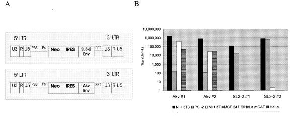
In this study, we used a conditionally replication-competent vector system, the minivirus system, which has been described elsewhere (5). Briefly, this system consists of (i) a bicistronic vector expressing the neomycin phosphotransferase II gene and env and (ii) an NIH 3T3-derived complementing cell line, designated a semipackaging cell line, expressing the Gag and Pol proteins. The expression of Env is achieved by using an internal ribosome entry site element from encephalomyocarditis virus between the marker and the envelope genes.
In semipackaging cells, the minivirus vector behaves as a replication-competent virus, which at the same time offers the convenience of resistance marker-based selection of infected cells. Figure 1A shows the design of the minivirus vector.
FIG. 1.
Bicistronic vector designs and titers. (A) The vector backbone is based on the Akv MLV, in which the gag and pol genes are replaced by the neomycin phosphotransferase II (Neo) selection marker and an internal ribosome entry site (IRES). The envelope genes are derived from either Akv or SL3-2 MLV. LTR, long terminal repeat. PBS, primer binding site; PPT, polypurine tract. (B) Titers of two SL3-2 and two Akv envelope-expressing vectors were measured on murine NIH 3T3 and human TE 671 cells. Interference properties were assessed by measuring titers on MCF 247-expressing murine NIH 3T3 cells (which do not permit infection through Xpr1) and Psi-2 cells (which do not permit infection through mCAT-1). Note the logarithmic scale of the titers.
Determination of the host range of SL3-2.
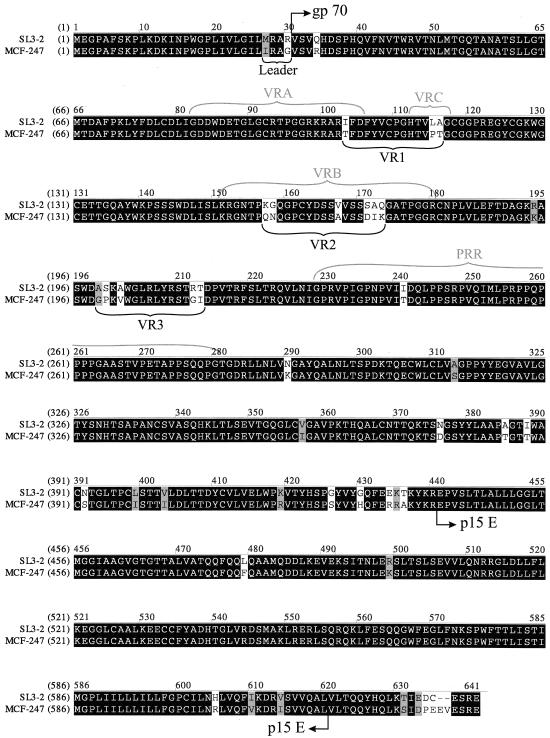
The SL3-2 envelope shows 93% identity at the amino acid level with the envelope protein of polytropic MCF 247. The latter has a wide host range and is able to infect cells from several species (12, 21), in contrast to the original report, which suggested that SL3-2 has a host range similar to that of ecotropic viruses (31). To determine the host range of the cloned vector, BOSC 23 packaging cells (30) were transfected with two clones of the SL3-2 minivirus vector (as well as two clones of the Akv minivirus as controls). After 24 h, the supernatant from these cells was used to transduce semipackaging cells (CeB cells), which express Moloney MLV gag-pol from a single construct (13). CeB cells were selected for G418 resistance until a confluent and stable culture of infected semipackaging cells was established. The supernatant from these cells was used to measure the titers of vectors expressing the SL3-2 envelope on a number of different cells.
As shown in Fig. 1B, vectors containing the Akv and SL3-2 envelopes have similar titers on NIH 3T3 cells. Psi-2 cells express the ecotropic envelope, which blocks infection through the ecotropic receptor (26). Accordingly, the vector expressing the ecotropic Akv envelope has a titer that is approximately 10,000-fold lower on these cells, whereas the SL3-2 vector is unaffected. Therefore, the SL3-2 envelope does not use the ecotropic receptor for infection. The reason for the lower interference levels seen in Psi-2 cells than in MCF 247-infected NIH 3T3 cells most likely is that Psi-2 cells express the ecotropic envelope less consistently because these cells do not contain a replication-competent virus (26). MCF 247-infected NIH 3T3 cells show interference with the SL3-2 envelope but not with that of Akv, confirming that the SL3-2 envelope uses the polytropic receptor for entry.
Thus, SL3-2 uses the polytropic receptor and not the ecotropic receptor (mCAT-1). Neither vector in this study was able to mediate infection of human HeLa cells. HeLa cells expressing the ecotropic receptor were efficiently infected by the Akv vector but not by the SL3-2 vector. Since the Akv and SL3-2 vectors are identical except in the envelope gene, the inability of the SL3-2 vector to infect human cells must be related to the SL3-2 envelope and not to a postentry defect. Similar results were obtained in repeated experiments and confirm previous reports that SL3-2 has a polytropic envelope with a limited host range (31, 33).
Determinants of the restricted tropism of SL3-2.
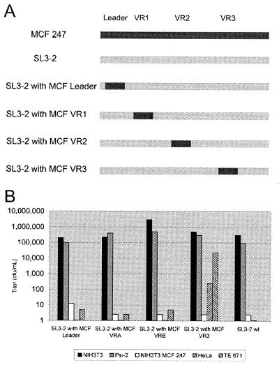
Alignment of the amino acid sequences of the SL3-2 and MCF 247 envelope proteins showed three regions containing clusters of nonidentical residues in the RBD (Fig. 2). Two of these regions (VR1 and VR2) overlap parts of VRA or VRC and VRB that show great variations within subfamilies of MLV, whereas the third (VR3) is a 15-amino-acid-long segment upstream of the proline-rich region. To further investigate the determinants of the tropism differences in these two viruses, we used chimeras in which these segments in the MCF 247 envelope protein replaced the corresponding segments in an SL3-2 backbone.
FIG. 2.
Alignment of the amino acid sequences of the MCF 247 and SL3-2 envelope proteins. The segments exchanged for the creation of SL3-2-MCF 247 chimeras (Fig. 3) are indicated. VRA, VRB, and VRC are defined according to sequence comparisons of the SL3-2 and ecotropic and amphotropic MLVs (37). PRR, the proline-rich region.
As a control, a fourth mutant was created by replacing a segment of the putative signal peptide of the SL3-2 envelope with the corresponding segment of the MCF 247 envelope. There are two amino acid differences in the signal peptide, but since the signal peptide is not a part of the mature envelope, no change in tropism was expected.
These constructs were transfected into Plat-E packaging cells (28). The virions produced were used to infect CeB semipackaging cells. After selection for G418 resistance, the titers of the minivirus constructs on murine NIH 3T3 cells, human TE 671 and HeLa cells, NIH 3T3 cells infected with MCF 247, and Psi-2 cells were measured.
As shown in Fig. 3, all of the constructs had comparable titers on NIH 3T3, MCF 247-infected NIH 3T3, and Psi-2 cells, indicating that all four chimeras utilize the same receptor with the same efficiency in murine cells. Interestingly, the SL3-2 construct with MCF VR3 had a titer that was 5,000- to 10,000-fold higher on human TE 671 cells and was the only construct capable of infecting human HeLa cells, indicating that a major determinant for utilizing the human receptor is located in this region.
FIG. 3.
Tropism and interference properties of SL3-2-MCF 247 chimeras on murine and human cells. (A) Schematic representations of chimeras. VR1, VR2, and VR3 are defined in Fig. 2. (B) Titers of mutants on murine NIH 3T3 cells, human HeLa and TE 671 cells, murine Psi-2 cells in which the ecotropic mCAT-1 receptor is blocked, and MCF 247-expressing NIH 3T3 cells in which the polytropic Xpr1 receptor is blocked. wt, wild type. Note the logarithmic scale of the titers.
Library analysis of the five nonidentical amino acids in VR3.
Five amino acids in VR3 differ between the MCF 247 and the SL3-2 envelopes. Any combination of these amino acids can be responsible for the different tropisms of these viruses. To clarify this issue, we used a randomized library in which the amino acids found at these five positions alternated between those found either in SL3-2 or in MCF 247.
Minivirus-based libraries are described elsewhere (5). Briefly, a pool of vectors in which random codons are found at positions of interest is generated. The DNA library is made by using two successive PCRs. The first PCR creates two fragments, one of which contains the randomized sequences. Each fragment is made by a single PCR regardless of the diversity of the library, since the random codons of the library originate from the degenerate primers, which can be made (purchased) as a pool of the desired sequences. These fragments are assembled in the second PCR.
The PCR-generated DNA library pool is directly transfected into Plat-E packaging cells (28), which express all of the proteins necessary for virion formation, including the envelope protein of the Moloney ecotropic virus. The vectors are packaged into virions and used to transduce semipackaging cells. This infection step occurs regardless of the envelope genes of the vectors, since the virions are produced by packaging cells containing a wild-type envelope gene. The semipackaging cells are selected for resistance to G418 and hence the presence of the minivirus vector. A stable cellular library pool is thus established. The cells in the cellular library contain all of the necessary trans-elements for producing virions, including a single copy of the vector that encodes a mutant envelope protein. The same copy is also packaged into the newly formed virions. Thus, the virions contain the gene for the envelope protein carried on the surface. Hence, the genotype of the envelope follows its phenotype. The supernatant of the selected semipackaging cells contains the virion library.
When the virion library is used to transduce a cell culture (in this study, human TE 671 cells), only virions containing functional envelopes are able to infect target cells. Selection for G418 resistance yields colonies each containing an integrated minivirus vector that encodes a functional envelope protein, the primary structure of which can be determined by sequencing of the integrated vector. The result is the selection of functional envelopes in a pool of random mutants.
To distinguish any functional requirements of the nucleotide sequence from those at the amino acid level, the library was designed so that each alternating amino acid could be translated from at least two different codons (see Materials and Methods). The library was selected for the ability to infect human TE 671 cells. Thirty-three colonies were isolated and sequenced (Table 1).
TABLE 1.
Frequency of emergence of SL3-2 or MCF virus residues at randomized positions among 33 envelope sequences isolated through selection of a five-amino-acid library for infection of human TE 671 cellsa
| Position | Residue allowed by the library design (virus in which it is found) | No. (%) of isolates containing the residue |
|---|---|---|
| 199b | Ala (SL3-2) | 18 (55) |
| Gly (MCF) | 11 (33) | |
| 200 | Ser (SL3-2) | 20 (60) |
| Pro (MCF) | 13 (40) | |
| 202 | Ala (SL3-2) | 17 (51) |
| Val (MCF) | 16 (49) | |
| 212c | Arg (SL3-2) | 2 (6) |
| Gly (MCF) | 30 (91) | |
| 213 | Thr (SL3-2) | 5 (15) |
| Ile (MCF) | 28 (85) |
The library was designed to encode two possible amino acids at each of the indicated positions in random combinations, totaling 32 different possible amino acid combinations. The two possibilities at each randomized position are the residues found at the corresponding positions in either the SL3-2 or the MCF 247 envelope protein.
Two serine and two threonine residues, encoded by codons not predicted by the library design, were also found.
A single serine residue, encoded by a codon not predicted by the library design, was also found.
While the first three positions showed a more or less random occurrence of amino acids, there was a strong bias toward glycine and isoleucine at positions 212 and 213, suggesting that these two amino acids are the determinants for the different tropisms of SL3-2 and MCF 247. The random occurrence of codons confirmed that the mutations affect tropism at the amino acid level and not at the nucleotide level (data not shown).
We confirmed the effect of the mutations at positions 212 and 213 on the tropism of SL3-2 by making four constructs and measuring titers on human or mouse cells (Table 2). These constructs represented the four possible combinations of residues found at positions 212 and 213 of either the MCF 247 or the SL3-2 envelope protein. The construct containing R212/T213 was the wild-type SL3-2 envelope, while the G212/I213 construct contained the MCF 247 primary structure at these positions. The other two constructs were mutants containing combinations of the SL3-2 and MCF 247 primary structures. The codons used at positions 212 and 213 of these constructs were the same as those found in wild-type viruses.
TABLE 2.
Titers of SL3-2 mutants with various residues at positions 212 and 213 on human TE 671 and murine NIH 3T3 cells
| Construct | Titer (CFU/m) on:
|
|
|---|---|---|
| TE 671 cells | NIH 3T3 cells | |
| R212/T213 (wild type) | 4.8 | 5.0 × 10⁶ |
| R212/I213 | 2.3 × 101 | 3.3 × 10⁶ |
| G212/T213 | 1.5 × 10³ | 2.0 × 10⁶ |
| G212/I213 | 4.5 × 10⁵ | 2.0 × 10⁶ |
All of the mutants had similar titers on mouse cells. As expected, the G212/I213 mutant had a titer 10,000-fold higher than that of the wild type on human cells. Other mutants showed intermediate titers.
A few isolated colonies contained amino acids other than those found in either SL3-2 or MCF 247. Four of these contained threonine or the structurally similar serine at position 199. Since we found that mutation of codon 199 to encode threonine (in the context of either SL3-2 or MCF 247 codons in the rest of VR3) caused no increase in the titer on TE 671 cells (data not shown), the emergence of Thr-199 and Ser-199 codons must have resulted from a bias in the DNA library, probably through erroneous incorporation of an A at position 1 of codon 199 during primer synthesis.
Improving the efficiency of the SL3-2 envelope in infecting human cells.
The discovery of the effect of glycine at position 212 and isoleucine at position 213 on the tropism of the SL3-2 envelope was based on the assumption that polytropic MCF 247 containing these two residues is able to infect human cells. We hypothesized that since MCF viruses have not evolved to infect humans, infection of human cells by polytropic envelopes might be improved through replacement of residues 212 and 213 with other amino acid residues. To address this issue, a randomized library of residues 212 and 213 in which all 20 amino acid residues can be encoded at either position was constructed. The randomized amino acids were encoded by NNK codons (N is A, T, G, or C and K is T or G) to minimize the occurrence of stop codons from three to one while allowing at least one codon for every amino acid. The library was synthesized and selected as described above. We also constructed a minivirus vector containing the envelope protein of MCF 247 for direct titer comparisons.
Incorporation of residues other than those found in known viral isolates at positions 212 and 213. Selection of the library on human TE 671 cells yielded several amino acid residues not found in wild-type isolates (Table 3). Interestingly, 46% of the isolated colonies contained methionine at position 212, a residue not found in wild-type MLVs. Another 23% of the colonies contained glycine at this position, in agreement with our previous findings.
TABLE 3.
Codons derived from colonies isolated by Neo selection of human TE 671 cells transduced by a randomized library introduced at positions 212 and 213 of the SL3-2 envelope protein
| Codons | Amino acids |
|---|---|
| ATG GTG | Met Val |
| ATG GTG | Met Val |
| ATG GTG | Met Val |
| ATG GTT | Met Val |
| ATG GTT | Met Val |
| ATG GTT | Met Val |
| ATG GCT | Met Ala |
| ATG GCT | Met Ala |
| ATG TTT | Met Phe |
| ATG TTT | Met Phe |
| ATG CAT | Met His |
| ATG TTG | Met Leu |
| GGT GTG | Gly Val |
| GGT GTT | Gly Val |
| GGG GTG | Gly Val |
| GGG AAG | Gly Lys |
| GGG TGG | Gly Trp |
| GGT TTG | Gly Leu |
| CTG ACT | Leu Thr |
| CTT GTT | Leu Val |
| CTT GTT | Leu Val |
| CTG GCT | Leu Ala |
| TGT CGT | Cys Arg |
| CAG GGT | Gln Gly |
| CGG GCG | Arg Ala |
| TCT GTT | Ser Val |
| CGG ACC | Wild type |
| TAG TAG | Stopa |
| GTT T | Stopb |
Two consecutive stop codons.
Frameshift.
None of the isolated colonies contained isoleucine at position 213. The most abundant residues at this position were valine (46%) and alanine (12%). In experiments involving the NNK randomization principle, different codons for the same amino acids have been found at nearly equal frequencies, confirming that the selection of the library occurred at the amino acid level and not at the nucleotide level.
Interestingly, the frequencies of the different mutants corresponded well with what would be statistically expected if the effects of mutations at the two positions were independent. For example, 46% of the isolated colonies contained methionine at the first randomized position, and another 46% contained valine at the second randomized position. If the occurrences of these amino acids were independent of each other, then 46% × 46% of the isolated colonies (corresponding to five or six colonies) would be expected to contain both methionine and valine. Indeed, six colonies with M212/V213 were isolated. The same correspondence was observed for all of the mutants found, suggesting that amino acid residues 212 and 213 in the SL3-2 envelope protein have independent effects on the tropism of this envelope protein.
Two of the colonies contained stop codons or frameshift mutations. These were probably the result of reinfection of the semipackaging cells that contained these mutants by other vectors that encode a functional envelope in the cellular library. Such reinfected cells can produce infectious virions that contain defective vectors, in which case the selection procedure can be bypassed.
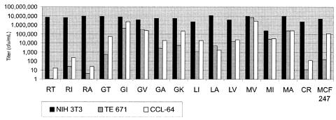
To verify the results obtained from this library selection, we made 15 mutants with different amino acid residues at positions 212 and 213. The different residues were chosen from among the three most frequently found in the library selection and the residues in wild-type SL3-2 and MCF viruses. We also included a mutant with cysteine at position 212 and arginine at position 213 and another with glycine at position 212 and lysine at position 213, which were found only once in the library screening, to examine the significance of such rare incidents. Titers on murine, human, and mink cells were measured (Fig. 4).
FIG. 4.
Effects of different substitutions at positions 212 and 213 on the tropism of the SL3-2 envelope. Titers of 15 mutants on murine NIH 3T3 cells, human TE 671 cells, and mink CCL-64 cells were measured. The single-letter code for amino acids at positions 212 and 213 is used to designate the mutants.
The mutants had comparable titers on murine cells after 4 weeks of selection of the semipackaging cells but widely different titers on human and mink cells. The titers on mink and human cells corresponded to each other for most of the mutants, suggesting that the tropism determinants for human and mink cell infections are similar. As expected, the mutant found most abundantly in the library screening (M212/V213) had the highest titer on human cells, approximately 1 order of magnitude higher than that of the G212/I213 mutant. This mutant infected human TE 671 and murine cells with equal efficiencies. The MCF virus envelope vector infected murine cells with an efficiency similar to that of the SL3-2 virus envelope vector but, surprisingly, had a limited ability to infect human cells compared to its SL3-2 VR3 equivalent (G212/I213 mutant). These data indicate that there are other determinants for the tropism of polytropic viruses.
Receptor usage of the SL3-2 VR3 mutants.
In order to determine whether the SL3-2 mutants also use the polytropic Xpr1 receptor for infection of nonmurine cells, interference studies were conducted with human TE 671 and mink CCL-64 cells expressing the MCF virus envelope protein.
Wild-type MCF 247 was used to chronically infect mink CCL-64 cells. The same virus was unable to infect human TE 671 cells. MCF virus envelope expression in these cells was achieved by cotransfection of the cells with proviral DNA and a selection marker. Following selection, the envelope-expressing cells were sorted by FACS with antienvelope antibody 83A25 (17). The sorted cells were expanded and sorted twice more to achieve a significant level of envelope expression.
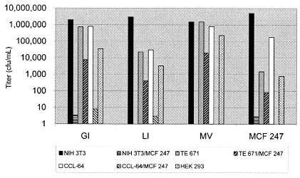
The titers of three SL3-2 mutants as well as the MCF minivirus on murine, human, and mink cells that either did or did not express the MCF 247 envelope protein as well as HEK 293 cells were measured (Fig. 5). HEK 293 cells are of human origin and were included in the study to ensure that the expanded tropism of the SL3-2 mutants was a general feature for human cells.
FIG. 5.
Tropism and interference properties of SL3-2 VR3 mutants. Titers of three SL3-2 mutants and an MCF 247 envelope-expressing vector on NIH 3T3, CCL-64, and TE 671 cells that did or did not express MCF 247 (to assess the interference properties of the mutants in different cell types) as well as human HEK 293 cells were measured. The single-letter code for amino acids at positions 212 and 213 is used to designate the mutants. Note the logarithmic scale of the titers.
Mink and murine cells showed nearly complete interference by both the SL3-2 mutants and the MCF minivirus, as expected, while the titers of all three mutants examined were decreased by a factor of approximately 100 as a result of the expression of MCF 247 in TE 671 cells. These data confirmed that the SL3-2 mutants use the polytropic receptor for infecting these cells (Fig. 5).
The less efficient level of interference on human cells probably reflects the fact that the murine and mink cells were chronically infected with wild-type virus (reaching a maximum level of interference as a result of virus spreading), while the human cells were sorted according to the envelope expression level. FACS enrichment cannot be expected to be 100% efficient, and if even as few as 1% of the sorted cells do not express high levels of the envelope protein, interference levels of more than 100 times reduction of titer will be impossible to observe. Alternatively, the inefficiency of the MCF virus envelope in mediating infection of human cells (observed both as measured low titers of the MCF minivirus and as the inability of wild-type MCF 247 to spread in TE 671 cell cultures) may also contribute to the low interference levels observed in human cells.
DISCUSSION
The tropism of murine leukemia viruses is diverse. Known mouse ecotropic viruses can infect only rodent cells, and the amphotropic viruses show a consistent wide tropism, being capable of infecting cells from several species, including humans, cats, and dogs (12).
MLVs that utilize the Xpr1 receptor show great variations in tropism. These viruses occur in three variants: xenotropic, MCF, and SL3-2. Xenotropic viruses cannot use the murine receptor but are able to use receptors of other species, such as mink. The polytropic viruses can infect both murine and nonmurine (mink) cells. SL3-2 is able to infect only murine cells (12, 31).
We have cloned and sequenced the SL3-2 envelope and constructed a bicistronic vector that expresses this envelope as well as the neo gene. Using this vector system, we have confirmed that the previously reported restricted tropism of SL3-2 (31) is indeed dependent on the envelope protein. We have also mapped the determinant for this restricted tropism to two amino acids upstream of the proline-rich region.
Using a mutational library approach, we found that mutation of arginine at position 212 and threonine at position 213 to several hydrophobic amino acids enables the SL3-2 envelope to use the human version of the polytropic receptor (Xpr1). Most interestingly, methionine and valine, which are not found in any wild-type isolates, are the most effective residues with regard to infection of human TE 671 cells. All of the SL3-2 mutants that we examined had the same efficiencies in infecting human and mink cells; in contrast, MCF 247 was more efficient in infecting mink cells. The SL3-2 G212/I213 mutant was more efficient than MCF 247 in infecting human cells, even though the latter contains the same residues at positions 212 and 213. This result suggests that there are other determinants for tropism of polytropic envelopes. Sequences downstream of VR3 are not likely to be involved in receptor binding and hence tropism determination, although this possibility cannot be ruled out. However, it is more likely that VRA and VRB play role in this scenario, since these regions have been implicated in receptor binding in other MLV subfamilies (4, 7, 8, 25). Experiments similar to those conducted in this study on the background of the MCF 247 envelope should facilitate the description of other factors involved in the tropism of polytropic envelopes.
Alignment of MLV envelope sequences shows that almost all of the nonidentical regions are found in the N-terminal region of the SU subunit, while there is a high degree of identity in VR3. In accordance with these observations, the receptor determinants of the ecotropic and amphotropic viruses are mapped to the N-terminal region of the RBD (29, 32). More specifically, a 14-amino-acid fragment from residues 50 to 64 of amphotropic SU (7) and several residues from 81 to 95 of ecotropic SU (4, 25) are important for receptor binding and usage.
A more complex picture emerges with respect to the determinant for the receptor specificity of the polytropic envelope. Previous studies indicated that, although the main determinant for the receptor usage of the polytropic envelope is also found within the first 120 residues of SU (9), more C-terminally located segments in the RBD or even in the proline-rich region (8) are involved in envelope-receptor interactions. Ott and Rein suggested that hybrids made of an amphotropic N terminus (up to an EcoRI site which corresponds to residue 187 in the SL3-2 envelope) and a polytropic C terminus, although amphotropic, can still bind to the polytropic receptor (29). Our finding that residues 212 and 213 are important determinants of envelope function is in agreement with the idea that some polytropic determinants are found near the proline-rich region of the SU subunit. It should be noted that the residues homologous to SL3-2 residues 212 and 213 in the amphotropic 4070A envelope are glycine and threonine, and it is therefore unlikely that these two amino acids are directly responsible for the peculiar receptor specificity of the amphotropic-polytropic hybrid envelope.
The residues corresponding to SL3-2 R212 and T213 in Friend MLV, glycine at position 249 and arginine at position 250 (G215 and R216, as numbered in the coordinates of the pdb file obtained from The Protein Data Bank at http://www.rcsb.org/pdb/), and in feline leukemia virus subtype B, glycine at position 224 and tyrosine at position 225 (G190 and Y191, as numbered in the coordinates of the pdb file obtained from The Protein Data Bank), are found in small loops on the surface of the SU subunit, according to the crystal structures of the RBDs (6, 18). The high degree of sequence identity at this region between Friend and SL3-2 viruses makes it likely that residues 212 and 213 in SL3-2 are also found on the surface of the protein, in which case these residues might be involved in a direct interaction with the receptor.
Glycine at position 212 seems to be well conserved in all subclasses of MLVs. Indeed, among the 30 envelope sequences that we found in the National Center for Biotechnology Information database (http://www.ncbi.nlm.nih.gov/), all had glycine in the corresponding position. However, in our studies, it does not seem to play a crucial role for polytropic envelope function in mouse cells. In fact, several mutants with amino acid substitutions at this position yielded similar titers on NIH 3T3 cells. On the other hand, an envelope with a methionine at position 212 was more infectious for human TE 671 cells. The significance of glycine at position 212, if any, remains to be elucidated.
Attempts to change the tropism of a virus by engineering its RBD, either by insertion of ligands to known cell surface proteins (16, 19, 20, 27, 37) or by selection of randomized libraries (11), have had various degrees of success. Common to all of the previously engineered viral envelopes is that none of them was as efficient as the wild-type envelope. We used mutational libraries to successfully retarget an envelope protein toward human cells and achieved wild-type levels of infection of the new target. A similar method was used by Bupp and Roth (11) to redirect the feline leukemia virus subtype A envelope toward an unknown receptor, although the titers achieved by the mutant envelope (ca. 103 CFU/ml) were low and were only 2 orders of magnitude higher than those of the wild-type feline leukemia virus subtype A envelope (ca. 101 CFU/ml). The SL3-2 mutant M212/V213 envelope is much more efficient in infecting human TE 671 cells than the two wild-type polytropic envelopes tested. This mutant envelope infects TE 671 cells with the same efficiency as the parent envelope infects murine cells and has a titer 6 to 7 orders of magnitude higher than that of the parent envelope on these cells. These results could be due to the fact that the SL3-2 mutants continue to use Xpr1 instead of a novel cellular receptor.
A problem with this approach may be representing the diversity of the possible combinations in the cellular library. Although we cannot eliminate the possibility that mutants with a greater capacity for infection of human cells exist, it is unlikely that we would not find them as a result of library bias, since the diversity of the library that we used included only 400 different combinations at the amino acid level.
Acknowledgments
We thank Lene S. Jøhnke for technical assistance and Alexander Schmitz for helping with FACS. The MCF 247 envelope plasmid was kindly provided by N. DiFronzo.
This work was supported by the Karen Elise Jensen Foundation, the John and Birte Meyer's Foundation, the Lundbeck Foundation, and the Danish Research Agency.
REFERENCES
- 1.Aagaard, L., J. G. Mikkelsen, S. Warming, M. Duch, and F. S. Pedersen. 2002. Fv1-like restriction of N-tropic replication-competent murine leukaemia viruses in mCAT-1-expressing human cells. J. Gen. Virol. 83:439-442. [DOI] [PubMed] [Google Scholar]
- 2.Albritton, L. M., J. W. Kim, L. Tseng, and J. M. Cunningham. 1993. Envelope-binding domain in the cationic amino acid transporter determines the host range of ecotropic murine retroviruses. J. Virol. 67:2091-2096. [DOI] [PMC free article] [PubMed] [Google Scholar]
- 3.Albritton, L. M., L. Tseng, D. Scadden, and J. M. Cunningham. 1989. A putative murine ecotropic retrovirus receptor gene encodes a multiple membrane-spanning protein and confers susceptibility to virus infection. Cell 57:659-666. [DOI] [PubMed] [Google Scholar]
- 4.Bae, Y., S. M. Kingsman, and A. J. Kingsman. 1997. Functional dissection of the Moloney murine leukemia virus envelope protein gp70. J. Virol. 71:2092-2099. [DOI] [PMC free article] [PubMed] [Google Scholar]
- 5.Bahrami, S., T. Jespersen, F. S. Pedersen, and M. Duch. 2003. Mutational library analysis of selected amino acids in the receptor binding domain of envelope of Akv murine leukemia virus by conditionally replication competent bicistronic vectors. Gene 315:51-61. [DOI] [PubMed] [Google Scholar]
- 6.Barnett, A. L., D. L. Wensel, W. Li, D. Fass, and J. M. Cunningham. 2003. Structure and mechanism of a coreceptor for infection by a pathogenic feline retrovirus. J. Virol. 77:2717-2729. [DOI] [PMC free article] [PubMed] [Google Scholar]
- 7.Battini, J. L., O. Danos, and J. M. Heard. 1998. Definition of a 14-amino-acid peptide essential for the interaction between the murine leukemia virus amphotropic envelope glycoprotein and its receptor. J. Virol. 72:428-435. [DOI] [PMC free article] [PubMed] [Google Scholar]
- 8.Battini, J. L., O. Danos, and J. M. Heard. 1995. Receptor-binding domain of murine leukemia virus envelope glycoproteins. J. Virol. 69:713-719. [DOI] [PMC free article] [PubMed] [Google Scholar]
- 9.Battini, J. L., J. M. Heard, and O. Danos. 1992. Receptor choice determinants in the envelope glycoproteins of amphotropic, xenotropic, and polytropic murine leukemia viruses. J. Virol. 66:1468-1475. [DOI] [PMC free article] [PubMed] [Google Scholar]
- 10.Battini, J. L., J. E. Rasko, and A. D. Miller. 1999. A human cell-surface receptor for xenotropic and polytropic murine leukemia viruses: possible role in G protein-coupled signal transduction. Proc. Natl. Acad. Sci. USA 96:1385-1390. [DOI] [PMC free article] [PubMed] [Google Scholar]
- 11.Bupp, K., and M. J. Roth. 2003. Targeting a retroviral vector in the absence of a known cell-targeting ligand. Hum. Gene Ther. 14:1557-1564. [DOI] [PubMed] [Google Scholar]
- 12.Cloyd, M. W., M. M. Thompson, and J. W. Hartley. 1985. Host range of mink cell focus-inducing viruses. Virology 140:239-248. [DOI] [PubMed] [Google Scholar]
- 13.Cosset, F. L., Y. Takeuchi, J. L. Battini, R. A. Weiss, and M. K. Collins. 1995. High-titer packaging cells producing recombinant retroviruses resistant to human serum. J. Virol. 69:7430-7436. [DOI] [PMC free article] [PubMed] [Google Scholar]
- 14.Dai, H. Y., M. Etzerodt, A. J. Baekgaard, S. Lovmand, P. Jorgensen, N. O. Kjeldgaard, and F. S. Pedersen. 1990. Multiple sequence elements in the U3 region of the leukemogenic murine retrovirus SL3-2 contribute to cell-dependent gene expression. Virology 175:581-585. [DOI] [PubMed] [Google Scholar]
- 15.Erlwein, O., C. J. Buchholz, and B. S. Schnierle. 2003. The proline-rich region of the ecotropic Moloney murine leukaemia virus envelope protein tolerates the insertion of the green fluorescent protein and allows the generation of replication-competent virus. J. Gen. Virol. 84:369-373. [DOI] [PubMed] [Google Scholar]
- 16.Erlwein, O., W. Wels, and B. S. Schnierle. 2002. Chimeric ecotropic MLV envelope proteins that carry EGF receptor-specific ligands and the Pseudomonas exotoxin A translocation domain to target gene transfer to human cancer cells. Virology 302:333-341. [DOI] [PubMed] [Google Scholar]
- 17.Evans, L. H., R. P. Morrison, F. G. Malik, J. Portis, and W. J. Britt. 1990. A neutralizable epitope common to the envelope glycoproteins of ecotropic, polytropic, xenotropic, and amphotropic murine leukemia viruses. J. Virol. 64:6176-6183. [DOI] [PMC free article] [PubMed] [Google Scholar]
- 18.Fass, D., R. A. Davey, C. A. Hamson, P. S. Kim, J. M. Cunningham, and J. M. Berger. 1997. Structure of a murine leukemia virus receptor-binding glycoprotein at 2.0 angstrom resolution. Science 277:1662-1666. [DOI] [PubMed] [Google Scholar]
- 19.Gollan, T. J., and M. R. Green. 2002. Redirecting retroviral tropism by insertion of short, nondisruptive peptide ligands into envelope. J. Virol. 76:3558-3563. [DOI] [PMC free article] [PubMed] [Google Scholar]
- 20.Gollan, T. J., and M. R. Green. 2002. Selective targeting and inducible destruction of human cancer cells by retroviruses with envelope proteins bearing short peptide ligands. J. Virol. 76:3564-3569. [DOI] [PMC free article] [PubMed] [Google Scholar]
- 21.Hartley, J. W., N. K. Wolford, L. J. Old, and W. P. Rowe. 1977. A new class of murine leukemia virus associated with development of spontaneous lymphomas. Proc. Natl. Acad. Sci. USA 74:789-792. [DOI] [PMC free article] [PubMed] [Google Scholar]
- 22.Heard, J. M., and O. Danos. 1991. An amino-terminal fragment of the Friend murine leukemia virus envelope glycoprotein binds the ecotropic receptor. J. Virol. 65:4026-4032. [DOI] [PMC free article] [PubMed] [Google Scholar]
- 23.Jespersen, T., M. Duch, and F. S. Pedersen. 1997. Efficient non-PCR-mediated overlap extension of PCR fragments by exonuclease “end polishing.” BioTechniques 23:48, 50, 52. [DOI] [PubMed] [Google Scholar]
- 24.Kavanaugh, M. P., D. G. Miller, W. Zhang, W. Law, S. L. Kozak, D. Kabat, and A. D. Miller. 1994. Cell-surface receptors for gibbon ape leukemia virus and amphotropic murine retrovirus are inducible sodium-dependent phosphate symporters. Proc. Natl. Acad. Sci. USA 91:7071-7075. [DOI] [PMC free article] [PubMed] [Google Scholar]
- 25.MacKrell, A. J., N. W. Soong, C. M. Curtis, and W. F. Anderson. 1996. Identification of a subdomain in the Moloney murine leukemia virus envelope protein involved in receptor binding. J. Virol. 70:1768-1774. [DOI] [PMC free article] [PubMed] [Google Scholar]
- 26.Mann, R., R. C. Mulligan, and D. Baltimore. 1983. Construction of a retrovirus packaging mutant and its use to produce helper-free defective retrovirus. Cell 33:153-159. [DOI] [PubMed] [Google Scholar]
- 27.Marin, M., D. Noel, S. Valsesia-Wittman, F. Brockly, M. Etienne-Julan, S. Russell, F. L. Cosset, and M. Piechaczyk. 1996. Targeted infection of human cells via major histocompatibility complex class I molecules by Moloney murine leukemia virus-derived viruses displaying single-chain antibody fragment-envelope fusion proteins. J. Virol. 70:2957-2962. [DOI] [PMC free article] [PubMed] [Google Scholar]
- 28.Morita, S., T. Kojima, and T. Kitamura. 2000. Plat-E: an efficient and stable system for transient packaging of retroviruses. Gene Ther. 7:1063-1066. [DOI] [PubMed] [Google Scholar]
- 29.Ott, D., and A. Rein. 1992. Basis for receptor specificity of nonecotropic murine leukemia virus surface glycoprotein gp70SU. J. Virol. 66:4632-4638. [DOI] [PMC free article] [PubMed] [Google Scholar]
- 30.Pear, W. S., G. P. Nolan, M. L. Scott, and D. Baltimore. 1993. Production of high-titer helper-free retroviruses by transient transfection. Proc. Natl. Acad. Sci. USA 90:8392-8396. [DOI] [PMC free article] [PubMed] [Google Scholar]
- 31.Pedersen, F. S., R. L. Crowther, D. Y. Tenney, A. M. Reimold, and W. A. Haseltine. 1981. Novel leukaemogenic retroviruses isolated from cell line derived from spontaneous AKR tumour. Nature 292:167-170. [DOI] [PubMed] [Google Scholar]
- 32.Peredo, C., L. O'Reilly, K. Gray, and M. J. Roth. 1996. Characterization of chimeras between the ecotropic Moloney murine leukemia virus and the amphotropic 4070A envelope proteins. J. Virol. 70:3142-3152. [DOI] [PMC free article] [PubMed] [Google Scholar]
- 33.Rein, A., and A. Schultz. 1984. Different recombinant murine leukemia viruses use different cell surface receptors. Virology 136:144-152. [DOI] [PubMed] [Google Scholar]
- 34.Tailor, C. S., A. Nouri, C. G. Lee, C. Kozak, and D. Kabat. 1999. Cloning and characterization of a cell surface receptor for xenotropic and polytropic murine leukemia viruses. Proc. Natl. Acad. Sci. USA 96:927-932. [DOI] [PMC free article] [PubMed] [Google Scholar]
- 35.Wang, H., M. P. Kavanaugh, R. A. North, and D. Kabat. 1991. Cell-surface receptor for ecotropic murine retroviruses is a basic amino-acid transporter. Nature 352:729-731. [DOI] [PubMed] [Google Scholar]
- 36.Weimin Wu, B., P. M. Cannon, E. M. Gordon, F. L. Hall, and W. F. Anderson. 1998. Characterization of the proline-rich region of murine leukemia virus envelope protein. J. Virol. 72:5383-5391. [DOI] [PMC free article] [PubMed] [Google Scholar]
- 37.Wu, B. W., J. Lu, T. K. Gallaher, W. F. Anderson, and P. M. Cannon. 2000. Identification of regions in the Moloney murine leukemia virus SU protein that tolerate the insertion of an integrin-binding peptide. Virology 269:7-17. [DOI] [PubMed] [Google Scholar]
- 38.Yang, Y. L., L. Guo, S. Xu, C. A. Holland, T. Kitamura, K. Hunter, and J. M. Cunningham. 1999. Receptors for polytropic and xenotropic mouse leukaemia viruses encoded by a single gene at Rmc1. Nat. Genet. 21:216-219. [DOI] [PubMed] [Google Scholar]


