Abstract
The germ line genome of ciliates is extensively rearranged during development of the somatic macronucleus. Numerous sequences are eliminated, while others are amplified to a high ploidy level. In the Paramecium aurelia group of species, transformation of the maternal macronucleus with transgenes at high copy numbers can induce the deletion of homologous genes in sexual progeny, when a new macronucleus develops from the wild-type germ line. We show that this trans-nuclear effect correlates with homology-dependent silencing of maternal genes before autogamy and with the accumulation of ∼22- to 23-nucleotide (nt) RNA molecules. The same effects are induced by feeding cells before meiosis with bacteria containing double-stranded RNA, suggesting that small interfering RNA-like molecules can target deletions. Furthermore, experimentally induced macronuclear deletions are spontaneously reproduced in subsequent sexual generations, and reintroduction of the missing gene into the variant macronucleus restores developmental amplification in sexual progeny. We discuss the possible roles of the ∼22- to 23-nt RNAs in the targeting of deletions and the implications for the RNA-mediated genome-scanning process that is thought to determine developmentally regulated rearrangements in ciliates.
Paramecium tetraurelia, like all ciliates, is a unicellular eukaryote possessing two different kinds of nuclei, both of which develop after fertilization from mitotic copies of the zygotic nucleus. The diploid micronuclei divide mitotically and are transcriptionally silent during vegetative growth. They serve only germ line functions, undergoing meiosis during sexual events. The highly polyploid macronucleus, which divides amitotically, is a somatic nucleus: it is responsible for all vegetative transcription but is lost shortly after sexual events, to be replaced by a new one. During development of the new macronucleus, the germ line genome is amplified from 2n to ∼800n and is extensively rearranged by two distinct types of DNA elimination events. Approximately 60,000 short, single-copy elements called internal eliminated sequences (IESs) are precisely removed from coding and non coding sequences (14). In addition, longer germ line-specific sequences, typically containing repeated sequences such as transposons or minisatellites, are removed by an imprecise mechanism that generates heterogeneity among the ∼800 macronuclear copies. Imprecise deletions can lead either to the rejoining of flanking sequences or to their capping by de novo telomere addition (23). The latter results in the fragmentation of germ line chromosomes into shorter, acentric macronuclear chromosomes with heterogeneous telomere positions distributed over telomere addition regions (TARs) of 0.2 to 2 kb. Furthermore, some macronuclear chromosomes can end at one of several alternative TARs spaced by 2 to 10 kb, implying the elimination of variable lengths of germ line sequences.
Both types of developmentally regulated deletions can be determined by epigenetic mechanisms, and a number of individual rearrangements have been shown to be controlled by homology-dependent maternal effects (28, 29). This was first revealed by genetic analyses of strain d48, a non-Mendelian mutant that lacks the A surface antigen gene in its macronuclear genome (9). In the wild-type macronucleus, the micronuclear chromosome carrying the A gene is reproducibly fragmented at three alternative TARs downstream of the gene. In d48, fragmentation is associated with elimination of an extended DNA region that includes the A gene, so that all macronuclear copies end at a single TAR located at the 5′ end of the gene (10). Although the d48 germ line genome is strictly identical to that of the wild type, the alternative rearrangement pattern is maternally inherited during sexual reproduction (9, 20). Thus, the A gene appears to be maintained in the developing macronucleus only if it is already present in the maternal macronucleus, which is still present in the cytoplasm at that time. Indeed, introduction of the missing A gene into the d48 macronucleus can rescue the developmental amplification of the germ line A gene in sexual progeny (18, 21, 42). Epigenetic inheritance of macronuclear deletions has also been observed with other genes (26, 36). By using a cell line carrying macronuclear deletions for two different genes, the rescue effect was shown to be gene specific (36). Since maternal and zygotic nuclei do not fuse, a gene-specific signal must be transmitted through the cytoplasm.
Surprisingly, the opposite effect was observed in another set of experiments using the sibling species Paramecium primaurelia. Transformation of the wild-type macronucleus with a cloned sequence at high copy numbers, followed by induction of meiosis, was shown to cause imprecise deletions of the homologous germ line sequence in the developing zygotic macronucleus (26). Like the A gene deletion in d48, the induced macronuclear deletion is maternally transmitted to sexual progeny, although genetic analyses confirmed that the germ line genome of the micronuclei remains wild type. The maternal deletion effect was observed with all sequences tested, including noncoding sequences; targeting a subtelomeric gene resulted in terminal deletions, while targeting an internal sequence resulted in heterogeneous internal deletions of the homologous zygotic sequence (23, 27). The sequence specificity of this trans-nuclear effect is therefore likely to be achieved by the pairing of homologous nucleic acids.
To reconcile these conflicting results, we sought to determine the conditions of maternal transformation that lead to the deletion or to the rescue effects in P. tetraurelia. We found that high copy numbers of maternal transgenes induced the deletion of sufficiently homologous genes in the new macronucleus, but only when transformation first induced homology-dependent silencing of the endogenous maternal gene before meiosis. We further show that prezygotic silencing and postzygotic deletions correlate with the accumulation of homologous short RNA molecules of 22 to 23 nucleotides (nt). Feeding cells with bacteria containing long double-stranded RNA (dsRNA) induces the same effects, suggesting that transgene-induced deletions are mediated through the cytoplasm by dsRNA. In contrast, low copy numbers of maternal transgenes are sufficient for the rescue effect, which is not associated with homology-dependent silencing but does not require production of full-length mRNAs, either. We discuss possible models for RNA-mediated programming of rearrangements.
MATERIALS AND METHODS
Paramecium strains, cultivation, autogamy, and serotype tests.
The P. tetraurelia strains 29A and 51A used in this study are entirely homozygous strains carrying the A29 and A51 alleles of the A surface antigen gene, respectively. 29A is strain d4.2, a standard laboratory strain carrying a few genes from strain 29 (including A29) in the genetic background of strain 51; 51A is largely isogenic with d4.2 and was obtained by introducing the A51 allele into d4.2 by two rounds of backcrossing with strain 51. Cells were grown in a wheat grass powder (Pines International Co., Lawrence, Kans.) infusion medium bacterized the day before use with Klebsiella pneumoniae, unless otherwise stated, and supplemented with 0.8 mg of beta-sitosterol (Merck, Darmstadt, Germany)/liter. Cultivation and autogamy were carried out at 27°C as previously described (26). The expressed surface antigen was determined by adding serotype-specific antisera (1:200 to 1:1,000) to small volumes of medium in depression slides. Cells expressing the corresponding antigen were immobilized within 1 h at 27°C.
Constructs and probes.
Wild-type A29 and A51 transgenes were constructed by cloning a 10.2-kb SalI-XhoI macronuclear fragment containing the gene and regulatory sequences (22, 25) into the pCRScriptAmpSK+ vector (Stratagene). The plasmids were injected either as circular molecules or after linearization in the vector sequence. Minichromosomes were obtained by inserting 165 bp of natural Paramecium telomeric repeats, ending with a synthetic BamHI site, into both the SalI and XhoI sites. After BamHI digestion, the minichromosomes were purified from the vector by agarose gel electrophoresis. The frameshifted version of A51 was obtained by Klenow fill-in of the BglII site and religation. The 3′-truncated A51 transgene was constructed by subcloning the SalI-BbsI fragments, yielding a construct lacking the 3′ untranslated region (3′ UTR) and the last 201 bp of the coding sequence. The 5′-truncated A51 transgene was constructed by subcloning the XmnI-XhoI fragments, yielding a construct lacking the promoter and the first 13 bp of the coding sequence. The A51 internal fragment is a 2.2-kb HindIII fragment from the central part of the coding sequence. Probes were either PCR products or gel-purified restriction fragments. PCR probes are as follows: a29 (A29 specific), covering nt 5035 to 5227 of accession number AY585741; a51 (A51 specific), covering nt 3470 to 4292 of M65163; b (B specific), covering nt 2901 to 3815 of L04795; and e (used for quantification of both A alleles), covering nt 1765 to 2644 of M65163. The sequences of the primers used for these (and ND7) PCRs, as well as that of the tRNA oligonucleotide probe, are available upon request. Probe c (C specific) is a 523-bp AvaII-HindIII fragment from plasmid pSC3.35H, provided by J. R. Preer, Jr. Probe d (hybridizing to both alleles) is a ∼3-kb EcoRI fragment located ∼5 kb upstream of the A gene and was isolated from plasmid p29B, provided by L. Amar. The telomeric probe is a 30-bp degenerate oligonucleotide, 5′-[(C/A)AACCC]5-3′.
Microinjections.
Young cells (less than 15 divisions after autogamy), from a single caryonidal clone in each experiment, were microinjected into Volvic mineral water (Volvic, France) containing 0.2% bovine serum albumin, under an oil film (Nujol), while being visualized with a phase-contrast inverted microscope (Axiovert 35 M; Zeiss). Column-purified (QIAGEN) plasmids were used for injection. Linearized plasmids were cut in the vector sequence at the BamHI restriction site. Minichromosomes were cut from the plasmids, gel purified, and extracted (with a Qiaquick gel extraction kit). All DNA solutions were filtered with a 0.22-μm-pore-size Ultrafree-MC filter (Millipore), precipitated with ethanol, and dissolved in filtered water. Approximately 5 pl of a 5-mg/ml solution was delivered into the macronucleus.
Dot blot copy number quantification.
For rapid screening of transformed clones and quantification of dsRNA-induced deletions, about 500 cells were pipetted from depression slide cultures and lysed in 500 μl of 0.4 N NaOH-50 mM EDTA. Lysates were incubated for 30 min at 68°C and loaded on a Hybond N+ membrane (Amersham, Little Chalfont, Buckinghamshire, United Kingdom) using a dot blot apparatus. For precise quantification, total genomic DNA samples (0.1 to 2 μg) were treated in the same way. Three different amounts were loaded for each sample to check for the linearity of the hybridization signal. The membrane was kept wet with 0.4 N NaOH for 15 min, washed in 2× SSC (1× SSC is 0.15 M NaCl plus 0.015 M sodium citrate), and further treated as a Southern blot. Hybridization signals were quantified with a Fuji Bas 1000 imager. Measured signals were normalized by rehybridizing the membrane with a telomeric oligonucleotide probe, as previously described (7). The reference value of 1 copy per haploid genome (1 cphg) was arbitrarily defined as the average of normalized signals from uninjected control clones of the same experiment, based on the assumption that all genes are present in similar copy numbers (∼800 copies) in the macronucleus. Pre- and postautogamous samples were treated separately because postautogamous clones were younger at the time of DNA extraction, and the A gene/telomere ratio was found to decrease with clonal age. Due to the high relative error in the quantification of very weak signals, values of <0.1 cphg are not significantly different from background.
DNA and RNA extraction.
Four hundred-milliliter cultures of exponentially growing cells at ∼1,000 cells/ml or of autogamous cells at ∼4,000 cells/ml were centrifuged. For DNA extraction, cell pellets were washed in 10 mM Tris-HCl (pH 7.0), resuspended in 1 volume of the same buffer, and quickly added to 4 volumes of lysis solution (0.44 M EDTA [pH 9.0], 1% sodium dodecyl sulfate [SDS], 0.5% N-laurylsarcosine [Sigma], and 1 mg of proteinase K [Merck]/ml). The lysates were incubated at 55°C for at least 5 h, gently extracted with phenol, and dialyzed twice against TE (10 mM Tris-HCl, 1 mM EDTA [pH 8.0]) containing 20% ethanol and once against TE. RNA was extracted from unwashed cell pellets by the method described in reference 5, modified by the addition of glass beads.
Feeding vectors and Escherichia coli strains.
For the ND7 experiment, a 397-bp PCR fragment was introduced into the plasmid and strain described in reference 11. dsRNA synthesis was induced by adding 0.4 mM isopropyl-β-d-thiogalactopyranoside (IPTG) at an optical density at 595 nm of 0.4 for 3 h, which results in transcription of the Plac-driven T7 polymerase gene in the chromosome. Bacteria were then washed and resuspended in Paramecium medium. The 0.8-kb HindIII fragment from the A gene was cloned into a pCRScriptAmpSK+ vector modified by the insertion of a second T7 promoter and then introduced into the same E. coli strain. We found that IPTG induction was not required for the effects of this plasmid in paramecia, presumably because titration of the LacI repressor by the LacO operator present in the high-copy-number plasmid made dsRNA production constitutive. As a control, the same plasmid was introduced into the SURE2 strain (Stratagene).
Southern and Northern blotting.
DNA restriction and electrophoresis (1 to 2 μg per sample) were carried out according to standard procedures (34). DNA was transferred from agarose gels to Hybond N+ membranes (Amersham) in 0.4 N NaOH after depurination in 0.25 N HCl. RNA samples (∼15 μg) were denatured and run on polyacrylamide-urea gels (Long Ranger; BioWhittaker Molecular Applications, Rockland, Maine), transferred to Hybond N+ membranes under a vacuum in 20× SSC, and UV cross-linked. Hybridization was carried out in a solution containing 7% SDS, 0.5 M sodium phosphate, 1% bovine serum albumin, and 1 mM EDTA (pH 7.2) at 60°C (decreasing to 30°C for oligonucleotide probes). Probes were labeled by random priming with [α-32P]dATP (3,000 Ci/mmol; Amersham). The telomeric oligonucleotide was labeled with [γ-32P]ATP (5,000 Ci/mmol; Amersham) and T4 polynucleotide kinase. Membranes were then washed for at least 30 min in 0.2× SSC-0.1% SDS at 60°C (or 2× SSC-0.1% SDS at 32°C for Northern blots) prior to autoradiography or image plate exposure.
RESULTS
High copy numbers of a maternal transgene induce homology-dependent deletions.
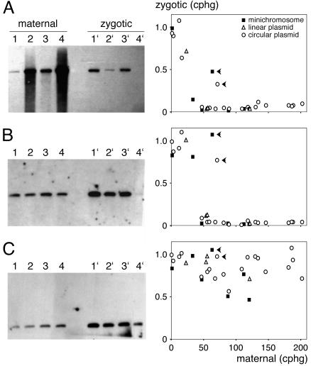
To characterize the deletion effect in P. tetraurelia, a transgene containing the A29 gene and regulatory sequences was microinjected into the macronuclei of cells of strain A29, which carries the same allele of the A gene. The transgene was injected either as a circular or linearized plasmid or as a minichromosome capped with telomeres (see Materials and Methods). DNA was extracted from transformed clones maintaining various transgene copy numbers, and aliquots of the cultures were starved to induce autogamy, a self-fertilization process. To check for deletions in new macronuclei, 20 postautogamous cells from each clone were grown together. Pre- and postautogamous DNA samples were analyzed on Southern blots, and relative A gene copy numbers were determined by a dot blot procedure (see Materials and Methods). Figure 1A shows that A gene copy numbers in zygotic macronuclei were reduced to background levels when maternal macronuclei contained ≥40 copies of the transgene per haploid genome (1 cphg = ∼800 copies per macronucleus) but were not significantly affected for maternal transgene copy numbers of <15 cphg. No significant difference was observed among the three forms of the injected molecule.
FIG. 1.
Induction of macronuclear deletions by maternal A29 transgenes. (A) Deletions of the zygotic A29 gene. A Southern blot of MfeI-restricted total DNA from representative clones transformed with the circular plasmid (maternal) and their postautogamous progeny (zygotic) was hybridized with the A29-specific probe a29 (see Materials and Methods), revealing a 5.6-kb fragment. Because the micronuclear/macronuclear ploidy ratio is ∼1:200 in vegetative cells, only macronuclear DNA is detectable. Lane 1, uninjected control. Lanes 2 to 4, clones transformed with various copy numbers of the circular A29 transgene; the smear arises from molecules linearized within the MfeI fragment after microinjection. Twice as much DNA was loaded onto the gel for postautogamous samples (lanes 1′ to 4′). In the graph on the right, A29 gene copy numbers in the zygotic macronucleus (zygotic) are plotted as a function of A29 gene copy numbers in the maternal macronucleus (maternal). Uninjected controls contained 1 cphg on average (maximum variation, 19%). Symbols used for the different forms of injected A29 transgenes are explained in the key. Solid arrowheads indicate the two clones that were expressing serotype A before autogamy. (B and C) Effects of maternal A29 transgenes on zygotic B and C genes, respectively. The same blot was rehybridized with the B-specific probe b or the C-specific probe c, revealing a 2.2- or 1.3-kb MfeI fragment, respectively. Graphs on the right show copy numbers of the B and C genes in the zygotic macronucleus.
In this experiment, two highly injected clones showed only a moderate reduction in A gene copy numbers (Fig. 1A). Interestingly, these were the only clones to express the A gene at the time of autogamy, as determined by immunological tests of live cells, suggesting that transgene expression is antagonistic to the deletion effect (see below). Note that expression is here defined by the production of protein, which implies the synthesis of stable mRNAs, and not simply by transcription, which can result in the production of other types of RNAs (see Discussion).
P. tetraurelia surface antigens are encoded by a family of paralogous genes with various levels of sequence identity. To examine the homology requirement of the deletion effect, we determined the macronuclear copy numbers of the G, B, and C genes in the same postautogamous DNA samples. The coding sequences of these genes (6.7 to 8.2 kb) are globally 78, 74, and 56% identical to that of A, respectively. Figure 1B shows that the B gene, like the A gene, was completely deleted in the progeny of clones transformed with ≥40 cphg of the A transgene, except for the two A-expressing clones mentioned above. With moderate transgene copy numbers (between 15 and 40 cphg), zygotic B gene copy numbers were not significantly reduced, whereas a slight effect was observed for the A gene. Similar results were obtained for the G gene (data not shown). Although it is quite variable, the amplification of the more divergent C gene was not significantly affected by the A transgene, even at high copy numbers (Fig. 1C). Thus, the efficiency of the maternal deletion effect depends on the level of sequence identity between the inducing transgene and the target zygotic gene.
Transgene-induced deletions correlate with prezygotic silencing.
Members of the surface antigen gene are expressed in a mutually exclusive manner: only one protein is produced at a time. The expressed protein defines the serotype of the cell, which is clonally inherited during vegetative growth (1). These genes are regulated at multiple levels (13), including transcription (22). In the course of the experiment described above, we noted that injection of A29 transgenes at high copy numbers into A-expressing recipient cells usually led to silencing of the endogenous A29 gene, causing cells to switch to another serotype. The lack of a strong deletion effect in the two exceptional A-expressing clones could thus be linked to the lack of silencing.
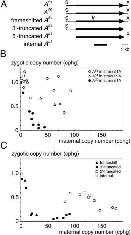
We then tested a different allele of the A gene. A51 is 99.5% identical to A29 over most of the coding sequence, except for a central 850-bp portion where homology drops sharply to 77%. This allele is much more readily expressed than A29, which may be due to differences in the promoter region. Indeed, A is a rare serotype in strain A29 but the most frequent serotype in strain A51, which carries the A51 allele but is otherwise largely isogenic with A29. Unlike A29 transgenes, A51 transgenes did not silence A expression when injected at high copy numbers into the macronuclei of cells of strain A51. Transformed A51 clones expressing serotype A were allowed to undergo autogamy, and macronuclear A gene copy numbers were quantified in postautogamous progeny as described above. Figure 2B shows that high copy numbers of A51 transgenes did not induce efficient deletions of the A51 gene in zygotic macronuclei. Neither did they induce efficient deletions of the zygotic A29 gene when A29 cells were used as recipients (Fig. 2B). Conversely, high copy numbers of A29 transgenes resulted in the same deletion effect in strain A51 as in strain A29 (Fig. 2B). Thus, it is the transgene allele that makes the difference, not the allele of the recipient strain.
FIG. 2.
Effects of different maternal transgenes on A gene amplification in the zygotic macronucleus. (A) Map of the sequences present in the different constructs. S, SalI; X, XhoI; fs, frameshift. (B) Effects of A51 and A29 transgenes (as minichromosomes or circular plasmids) in strain 51A or 29A. Symbols used for each injection experiment are explained in the key. (C) Effects of A51 nonexpressible transgenes (as minichromosomes or circular plasmids) in strain 51A or 29A.
To confirm that the incapacity of the A51 transgene to induce efficient deletions is linked to its stable expression in transformed clones, we constructed nonexpressible versions (Fig. 2A). We first introduced a frameshift resulting in a premature stop codon about 3 kb into the coding sequence. Although only a few high-copy-number transformants were obtained for this construct, Fig. 2C shows that it induced deletion of the zygotic A gene as efficiently as A29 transgenes. Three other nonexpressible constructs were tested: a 3′-truncated version in which the 3′ UTR was deleted, a 5′-truncated construct carrying a deletion of the promoter, and a 2.2-kb fragment from the central part of the coding sequence. The 3′-truncated version also induced an efficient maternal deletion effect (Fig. 2C). The other two constructs, which did not contain the transcription promoter, showed a weaker effect: very high copy numbers resulted only in a moderate decrease of A gene copy numbers in the zygotic macronucleus (Fig. 2C). Like A29 transgenes, all nonexpressible A51 constructs caused silencing of endogenous A expression when injected at high copy numbers, as shown by immunological detection of the proteins at the cell surface. The 5′-truncated construct, however, only transiently silenced expression of the endogenous A gene. This is consistent with the previous observation that the presence of the 3′ UTR inhibits the homology-dependent silencing induced by high copy numbers of promoterless constructs containing different genes (12).
Silencing and deletions correlate with accumulation of short RNAs.
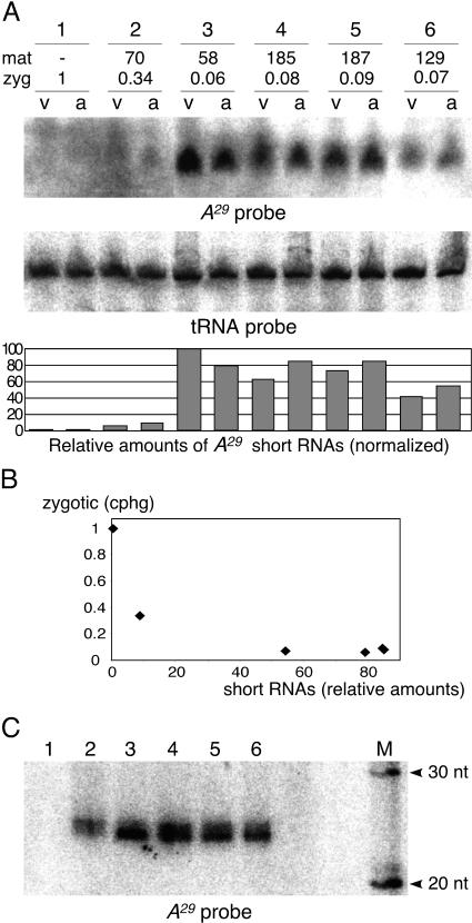
In one of the above transformation experiments (Fig. 1A, circular A29 transgene), total RNA was extracted from transformed clones, both during vegetative growth and after meiosis, during the development of new macronuclei. RNA samples were analyzed on acrylamide-urea denaturing gels (Fig. 3). By use of an A gene probe, specific short RNAs could be detected at both stages in high-copy-number transformants that showed the silencing and deletion effects but not in uninjected controls (Fig. 3A), indicating that these short RNAs originated from transgene transcripts. Strikingly, these RNAs were much less abundant in one of the two high-copy-number transformants in Fig. 1A that did not show the silencing effect and showed only a moderate deletion effect (clone 2 in Fig. 3A; RNA was not extracted from the other clone). A gene copy numbers in zygotic macronuclei thus appear to be better predicted by the amount of short RNA (after normalization with a tRNA probe [see the plot in Fig. 3B]) than by transgene copy numbers. A higher-resolution analysis revealed two closely migrating species in both vegetative (data not shown) and autogamous (Fig. 3C) samples. These species were estimated to be ∼22 to 23 nt long by comparison with RNA oligonucleotides, after taking into account the effect of purine content on electrophoretic mobility (M. Nowacki and E. Meyer, unpublished data). Thus, these short RNAs, which are within the size range of small interfering RNA molecules (siRNAs) in other organisms, are likely to mediate silencing through the RNA interference (RNAi) pathway and may also be responsible for the deletion effect.
FIG. 3.
Northern blot detection of A29 short RNAs. (A) Analysis of total RNA samples from clones transformed with the circular A29 transgene. RNAs were extracted during vegetative growth (v) and autogamy (a) and were run on a 6% acrylamide sequencing gel. The blot was successively hybridized with the A29-specific probe a29 (top panel) and, as a loading control, with a 75-nt tRNA probe (bottom panel). Clone 1, uninjected control. For clones 2 to 6, copy numbers of the maternal A29 transgene (mat) and copy numbers of the endogenous A29 gene in the macronuclei of postautogamous progeny (zyg) (copies per haploid genome) are given. The histogram below shows the relative amount of A29 short RNAs in each sample, after normalization with the tRNA signal. (B) A gene copy numbers in the sexual progeny of transformed clones (zygotic) are plotted as a function of the relative amounts of A29 short RNAs in autogamous samples, as determined in panel A. (C) High-resolution analysis of autogamous samples. The same autogamous samples used for panel A were run on a 15% acrylamide gel and hybridized with the same probe. RNA molecular weight markers (M) were 32P labeled before electrophoresis.
Silencing and deletions can be induced by dsRNA feeding.
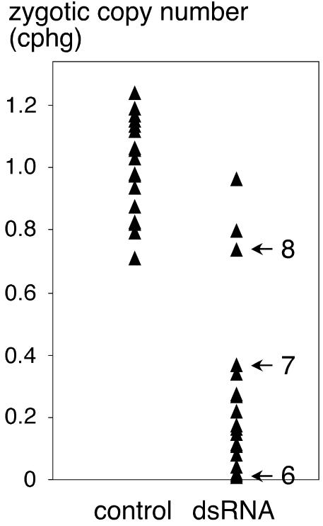
Feeding bacteria containing long dsRNAs to Paramecium cells has been shown to trigger homology-dependent silencing during vegetative growth (11). To test whether direct delivery of dsRNA would also induce deletions in the developing macronucleus, a 0.8-kb fragment of the A51 coding sequence was cloned between convergent T7 promoters in an E. coli strain, allowing the production of dsRNA (see Materials and Methods). A-expressing cells of strain 51A were first fed dsRNA-containing bacteria during vegetative growth (which, as expected, led to the silencing of endogenous A51) and then starved to induce autogamy. Postautogamous cells were grown individually on the normal food bacterium (Klebsiella), and A gene copy numbers in new macronuclei were determined by the dot blot procedure. To control for a possible effect of the A gene DNA sequence present in the bacterial plasmid, another batch of cells was fed a different E. coli strain that contained the same plasmid but could not produce dsRNA because it lacked the T7 polymerase gene. Figure 4 shows that A gene copy numbers in sexual progeny were dramatically lower when parental cells were fed the dsRNA-producing E. coli strain than when they received the control strain. In other experiments we found that the bacterial dsRNA, which must escape the phagosomes to target the degradation of mRNAs, is also processed into ∼22- to 23-nt pieces that persist after autogamy is induced, during macronuclear development (data not shown) (Nowacki and Meyer, unpublished). Thus, these short RNAs are associated with both transgene-induced and dsRNA-induced deletions.
FIG. 4.
A gene deletions induced by dsRNA feeding. Copy numbers of the A51 gene were determined in individual postautogamous clones. Clones for which results are shown on the left (control) were derived from parental 51A cells that were fed the E. coli control strain prior to meiosis; clones for which results are shown on the right (dsRNA) were derived from parental 51A cells that were fed the dsRNA-producing E. coli strain. The reference value of 1 cphg was defined as the average copy number in control clones. A gene deletions in clones 6, 7, and 8 (arrows) are molecularly analyzed in Fig. 5C.
Physical extent and maternal inheritance of macronuclear A gene deletions.
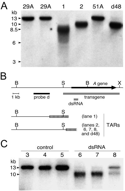
The extents of transgene-induced A gene deletions were mapped by Southern blot analysis of postautogamous DNA samples. Digestion with BglII, which cuts within the gene and 13 kb upstream, yields a single fragment in wild-type clones (Fig. 5A). The shorter, smeared band observed for first-generation macronuclear mutants is typical of microheterogeneous telomeric fragments. This indicates that A gene deletions are terminal truncations of the macronuclear chromosome, as previously observed in the targeting of subtelomeric genes in P. primaurelia. The new TAR extends over ≥2.5 kb upstream of the gene and partially overlaps the sequence present in the transgene.
FIG. 5.
Mapping of induced and inherited A gene deletions. (A) Southern blot of BglII-restricted genomic DNA samples. The blot was hybridized with probe d (see map in panel B). The same 13-kb BglII-BglII fragment is obtained with wild-type strains 29A and 51A. The original d48 cell line shows a diffuse band migrating at ∼10 kb, which represents a BglII telomere fragment. Lane 1, first-generation macronuclear deletion obtained by transformation with A29; lane 2, stable 29AΔmac cell line derived from the clone for which results are shown in lane 1 by a few rounds of autogamy. (B) Map showing the deduced positions of the different TARs (striped boxes). The positions of probe d (solid rectangle) and the sequences covered by the injected transgene and by the E. coli dsRNA (shaded rectangles) are indicated. B, BglII; S, SalI; X, XhoI. (C) Mapping of dsRNA-induced A gene deletions. A Southern blot of BglII-restricted DNA was hybridized with probe d. Samples are individual clones from the postautogamous progeny of cells fed the control E. coli strain (lanes 3 to 5) or the dsRNA-producing strain (lanes 6 to 8) (see Fig. 4).
As previously described, macronuclear deletions of the A gene were spontaneously reproduced after further rounds of autogamy. Although occasional reversions were observed, selection for complete deletions led to the establishment of stable cell lines, designated 29AΔmac, which behaved like the original d48 cell line. The TAR used in these cell lines was slightly different from that of the first-generation mutants but was undistinguishable from that used in the d48 cell line (Fig. 5A).
A gene deletions induced by dsRNA feeding were analyzed in the same way (Fig. 5C). In this case the TAR observed in first-generation mutants was identical to that of the stable 29AΔmac and d48 cell lines. This may be related to the fact that, unlike the A29 transgene, the inducing dsRNA did not extend upstream of the gene. Hybridization with different probes, however, confirmed that targeting a short portion of the coding sequence resulted in the deletion of the entire gene (data not shown), as observed in P. primaurelia transgene-induced deletions. Analysis of one of the few postautogamous clones that did not show a marked deletion effect (clone 8 in Fig. 4 and 5C) showed that a fraction of macronuclear copies were still truncated at the same TAR, although most copies appeared to be wild type.
dsRNA-induced internal deletions of the ND7 gene.
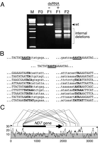
To test the generality of dsRNA-induced deletions, we then targeted ND7, a nonessential gene located at an internal position in a macronuclear chromosome, which is involved in the regulated discharge of trichocysts (37). Feeding cells with an E. coli strain producing dsRNA homologous to a 0.4-kb portion of the coding sequence resulted in a mutant phenotype (no discharge of trichocysts after picric acid stimulation), indicating that the gene was silenced. After induction of autogamy, individual cells were grown on a Klebsiella-containing medium. The phenotypes of the resulting clones ranged from wild type to intermediate (discharge of a limited number of trichocysts) or mutant. To test for macronuclear deletions, a 3.2-kb fragment containing the entire gene was amplified by PCR. The wild-type fragment was clearly absent from phenotypically mutant clones; instead, a set of shorter fragments revealed the occurrence of heterogeneous internal deletions of 1.5 to 2.2 kb (Fig. 6A). The same set of deletions was present in phenotypically wild-type or intermediate clones, but these also contained a variable fraction of wild-type copies (data not shown).
FIG. 6.
dsRNA-induced deletions of the ND7 gene. (A) PCR amplification of the ND7 gene in wild-type and mutant clones. The primers used (see panel C) produce a 3.2-kb fragment from the wild-type parental clone (F0). F1 lanes show products obtained from individual postautogamous clones derived from the F0 clone after feeding with Klebsiella (−) or with the dsRNA-producing E. coli strain (+). Lane F2 shows products obtained from an individual clone after a second autogamy of clone F1(+). (B) Sequences of the junctions of internal deletions in clone F1(+). Deletions occurred between short direct repeats (boldfaced), one of which is maintained in the rearranged sequence. Deleted sequences are lowercased. (C) Map of internal deletions. Thin bent lines above the map show the positions of deletion boundaries for each of the 10 sequenced examples. The tips of the solid arrowheads on both sides indicate the 3′ ends of PCR primers. The open rectangle within the ND7 coding sequence (solid arrow) indicates the sequence used for dsRNA production in E. coli. The plot below the map shows the local density of 5′-TA-3′ dinucleotides, computed in a 40-bp window sliding along the sequence (expressed as percent dinucleotides).
The PCR products were then cloned and sequenced. Figure 6B shows that deletions always occurred between two short (2- to 6-bp) direct repeats containing at least one TA dinucleotide. The deletions mostly encompassed the entire ND7 gene, and the heterogeneous breakpoints on either side were preferentially located in short regions particularly rich in 5′-TA-3′ dinucleotides (Fig. 6C). These molecular features are strikingly similar to those of transgene-induced internal deletions in P. primaurelia (27).
Phenotypically mutant clones, which were maintained on a Klebsiella-containing medium, were then allowed to undergo a second autogamy. Again, the progeny showed variable phenotypes ranging from mutant to wild type, indicating that the ND7 gene was still present in the micronuclei of mutant clones. The pattern of heterogeneous deletions in a second-generation mutant clone was found to be very similar to that of the first-generation mutant (Fig. 6A). The reversion frequency decreased with repeated rounds of autogamy and selection of mutant phenotypes, suggesting that stabilization of the new rearrangement pattern is achieved when macronuclei are completely devoid of ND7 sequences. One important conclusion is that imprecise internal deletions, like terminal deletions, can be epigenetically inherited in sexual progeny.
Transgene-induced rescue of maternally inherited A gene deletions.
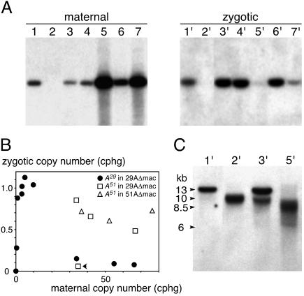
To determine the conditions of transformation leading to the rescue effect, the circular A29 transgene was injected into the macronucleus of the stable 29AΔmac cell line, which reproducibly deletes the germ line A29 gene during macronuclear development. The transformed clones, which did not express the A gene, were allowed to undergo autogamy. The Southern blots in Fig. 7A and the plot in Fig. 7B show that developmental amplification of the gene was fully restored in the progeny of clones with low transgene copy numbers (1 to 10 cphg); as little as 0.1 cphg of the transgene induced the amplification of ∼0.3 cphg of the gene in the new macronucleus. This further shows that the rescue effect does not require transgene expression. However, A gene amplification was not restored in the progeny of clones with high (≥40 cphg) copy numbers of the A29 transgene, i.e., under conditions that lead to silencing and deletion in wild-type cells.
FIG. 7.
Trangene-induced reversion of inherited A gene macronuclear deletions. (A) Southern blot of HindIII-digested genomic DNA from representative transformed clones and controls (lanes 1 to 7) and their postautogamous progeny (lanes 1′ to 7′), hybridized with probe a29. Lane 1, 29A control clone; lane 2, 29AΔmac uninjected control clone; lanes 3 to 7, A29-transformed 29AΔmac clones. (B) Copy number of the A29 or A51 gene in the zygotic macronucleus as a function of the A29 or A51 transgene copy number in the maternal macronucleus. The only A51-transformed clone that did not express the A gene is indicated by a solid arrowhead. (C) Mapping of TARs in rescued and nonrescued clones. Shown is a Southern blot of BglII-digested genomic DNA, hybridized with probe d (see Fig. 5). Samples are the same as in panel A.
The positions of TARs in rescued and nonrescued postautogamous clones were analyzed by Southern blotting (Fig. 7C). The stabilized mutant TAR of the parental 29AΔmac cell line was faithfully reproduced in sexual progeny of uninjected controls. In addition to the wild-type fragment, some of the rescued clones still contained a minor fraction of macronuclear copies ending in this TAR. In the nonrescued progeny of high-copy-number transformants, however, TARs were displaced upstream and were more extended, just as in first-generation deletion mutants (compare Fig. 7C and 5A). The B and G genes were also deleted in these clones, although they were present in the transformed maternal macronucleus (data not shown). High copy numbers of the maternal A29 transgene therefore induce the same deletions in the developing macronucleus, whether the maternal macronucleus contains endogenous copies of the gene or not. Importantly, this experiment shows that injection of the same transgene into the same cell line can promote either the amplification or the deletion of the homologous germ line gene, depending on the transgene copy number.
To determine whether rescue can be observed with high transgene copy numbers in the absence of silencing, the readily expressed A51 transgene was injected as a minichromosome into the macronuclei of the 29AΔmac and 51AΔmac cell lines (Fig. 7B). All but one of the transformed clones were of serotype A, indicating expression of the A51 transgene. Analysis of the macronuclear genome of postautogamous progeny showed that, in contrast to the A29 transgene, high copy numbers (>35 cphg) of the A51 transgene did restore amplification of the A29 and A51 zygotic genes, except for the one clone in which the transgene was not expressed. This exception suggests that even the wild-type A51 transgene can occasionally induce silencing and deletions at high copy numbers. Taken together, these experiments show that rescue can be observed at all transgene copy numbers. However, under conditions that lead to homology-dependent silencing, the deletion effect is dominant over the rescue effect.
DISCUSSION
It has been known for some time that the DNA rearrangements that occur during development of a new macronucleus are sensitive to the presence of homologous sequences in the maternal macronucleus, but contradictory results have been reported. This study has shown that the same maternal transgene can induce either the deletion or the amplification of the homologous germ line gene in the developing macronucleus, indicating that these opposite effects are not due to different types of DNA sequences. Instead, our results suggest that they are determined by differential processing of transcripts from the maternal transgene.
Involvement of short RNAs in the deletion effect.
One of the main conclusions from this work is that the deletion effect is observed only in the progeny of clones in which transformation establishes homology-dependent silencing; it is not observed when the maternal transgene is expressed. Expression was monitored in our experiments by immunological tests and is therefore defined by the production of protein, which requires not only transcription but also the processing of primary transcripts into mRNAs and their incorporation into stable mRNPs. Initially revealed by the different effects of the A29 and A51 alleles, the link with expression was confirmed by introducing mutations that prevent expression of the A51 transgene: these also made it capable of inducing deletions at high copy numbers.
High copy numbers of nonexpressible transgenes have previously been shown to trigger homology-dependent silencing of endogenous genes during vegetative growth (33). Under these conditions, even promoterless constructs give rise to sense and antisense transcripts (12). It was therefore proposed that, above a certain copy number threshold, such promiscuous transcription results in the formation of dsRNA, leading to silencing through the production of siRNAs. The RNAi pathway indeed appears to be conserved in paramecia, since dsRNA delivery through bacterial feeding results in the silencing of homologous genes (11).
The lack of silencing or deletion effects of expressed transgenes, even at high copy numbers, may mean that promiscuous bidirectional transcription does not occur when transgenes are engaged in the production of stable mRNAs. Among nonexpressible constructs, those that retained the A gene promoter (3′ truncation and frameshift) were the most efficient at inducing deletions, suggesting a role for promoter-initiated transcripts that are not processed into mRNAs. Increasing the concentration of sense transcripts may drive the more efficient formation of dsRNA from rare promiscuous transcripts. The sharp transition observed, as transgene copy numbers increase, between no effect and a full deletion effect could reflect the concentration-dependent formation of dsRNA. Alternatively, aberrant (untranslatable) transcripts could be detected by mRNA surveillance systems and fed into processing pathways that ultimately result in the production of siRNAs by other mechanisms, for instance, through the action of RNA-dependent RNA polymerases. Interestingly, incorrect 3′ ends appear to facilitate RNAi, since the presence of the 3′ UTR has been reported to inhibit the silencing caused by promoterless transgenes at high copy numbers (12). In the case of the frameshift construct, promoter-initiated transcripts may be degraded by non-sense-mediated RNA decay, a conserved pathway that is genetically linked to RNAi in Caenorhabditis elegans (6).
Wild-type transgenes, when unexpressed, also induce strong silencing and deletion effects. This was observed in most cases of transformation with A29 transgenes and in one case of transformation with the wild-type A51 transgene (in the rescue experiment). It may be more difficult to explain the production of siRNAs in such cases, since the mutually exclusive expression of surface antigen genes appears to be regulated primarily at the level of transcription (22). However, other levels of regulation have been described (13), and one expressible transgene was found to be posttranscriptionally regulated under some conditions (L. Martin and E. Meyer, unpublished data). Whatever the mechanism may be, we have shown that the silencing and deletion effects of wild-type A29 transgenes indeed correlate with the accumulation of short RNAs of ∼22 to 23 nt. Such sizes are typically produced by the dsRNA-specific endonuclease Dicer, which further supports the involvement of dsRNA in these effects. Although RNA was extracted only from a limited number of clones, the deletion effect appears to correlate better with the amounts of short RNA than with transgene copy numbers: indeed, one of the high-copy-number transformants that showed only a moderate deletion effect had a correspondingly low level of short RNAs (Fig. 1A and 3B). Most transgene copies may have been producing mRNA in this clone, which was expressing serotype A.
We have further shown that dsRNA feeding also results in the deletion of homologous genes during macronuclear development, in the sexual progeny of treated clones. Molecular analysis of dsRNA-induced deletions cannot distinguish them from transgene-induced deletions: in both cases, internal deletions have heterogeneous boundaries on either side of the targeted gene and occur between short direct repeats containing TA dinucleotides (27), like the spontaneous deletions of germ line transposons and other repeated sequences (23). The only common consequence of the two methods for inducing deletions appears to be the processing of dsRNA into ∼22- to 23-nt molecules that are still present during macronuclear development. It is therefore very likely that these short RNAs, in addition to their role in the degradation of homologous mRNAs, are involved in programming the deletions. This would be consistent with the observed effects of the A29 transgene on paralogous genes. Indeed, the coding sequence of A29 shares with the G gene 15 segments of perfect identity that are at least 23 bp long, totaling 456 bp, and it shares 10 such segments with B, for a total of 310 bp, whereas the longest segment of perfect identity with C is only 18 bp.
Short RNAs may target deletions through chromatin modification.
The ∼22- to 23-nt RNAs could induce deletions in different ways. One possibility, which would imply that they can enter the developing macronucleus, is that they recognize the homologous gene by pairing directly with homologous DNA or with nascent transcripts. This would target an epigenetic modification of chromatin which would spread in both directions until it reached TA-rich boundaries outside the gene. The modified chromatin would then be recognized by the deletion machinery. Such a model is supported by recent evidence from other systems implicating the RNAi pathway in the regulation of chromatin structure (for reviews, see references 2, 17, and 35). In Schizosaccharomyces pombe (15, 39, 40) and Drosophila melanogaster (32), RNAi genes are required for the establishment of heterochromatin, and short RNAs have been proposed to target the methylation of histone H3 on K9 to specific loci.
In the ciliate Tetrahymena thermophila, H3-K9 methylation was shown to target DNA elimination during macronuclear development (24, 38), and endogenous short RNAs (26 to 31 nt) that accumulate at that stage may be involved in the process (30). Furthermore, injection of long dsRNA was recently shown to target deletions in the developing macronucleus of T. thermophila (41), suggesting that similar mechanisms are operating in both ciliates. The larger size of the Tetrahymena short RNAs might reflect a difference between Dicer enzymes in the two species. However, siRNAs associated with transgene-induced silencing in Arabidopsis thaliana were recently shown to fall into two distinct size classes that appear to have different roles: 21- to 22-nt RNAs would be responsible for the degradation of homologous mRNA, while 24- to 26-nt RNAs would mediate the methylation of homologous DNA in the nucleus and the systemic spreading of silencing (16). Thus, it is also possible that two size classes with different functions exist in both ciliate species.
A genome-wide comparison of maternal and zygotic sequences?
Like terminal deletions of the A gene and other subtelomeric genes (26, 36), we found that dsRNA-induced internal deletions of the ND7 gene are spontaneously reproduced in subsequent sexual generations. Epigenetic inheritance of experimentally induced macronuclear deletions may thus be quite general. The recurrent elimination of these genes cannot be determined by short RNAs produced from the maternal macronucleus, since the maternal macronucleus is devoid of these sequences. This is also the case for the spontaneously eliminated transposons and IESs, and there is some evidence that a similar epigenetic mechanism is involved in the elimination of these elements. Indeed, introduction of some IESs at low copy numbers into the maternal macronucleus was shown to cause the retention of homologous IESs in the developing macronucleus in both P. tetraurelia (7, 8) and T. thermophila (3), a homology-dependent effect which is analogous to the rescue of inherited macronuclear deletions. Thus, different types of sequences appear to be maintained in the developing macronucleus only if they are already present in the maternal macronucleus, suggesting that elimination could be a default fate.
These observations suggest that epigenetic programming of rearrangements may involve a global comparison of the germ line genome to be rearranged with the previously rearranged genome of the maternal macronucleus (29). Based on the evidence that micronucleus-specific sequences are transcribed on both strands during early stages of conjugation in Tetrahymena (4), it has been proposed that dsRNA originating from the micronucleus during conjugation could be exported to the parental macronucleus and processed by a Dicer-like activity into the observed ∼26- to 31-nt RNAs. These were called “scan RNAs” (scnRNAs) because their proposed function is to scan the rearranged genome of the parental macronucleus by pairing interactions: scnRNAs that can pair with homologous DNA would be destroyed, while scnRNAs that cannot would be sent to the developing macronucleus to target the deletion of micronucleus-specific sequences (30; reviewed in reference 31). It is not known whether the entire micronuclear genome produces scnRNAs; should this be so, then elimination would be a default state: only germ line sequences that have homologous counterparts in the parental macronucleus could be maintained in the developing macronucleus.
Such a mechanism may well explain the epigenetic inheritance of macronuclear deletions and the rescue effect in paramecia, but it is more difficult to reconcile with the transgene-induced deletion effect. Indeed, the large number of transgene copies that are present in the maternal macronucleus would be expected to destroy homologous short RNAs and prevent them from inducing deletions in the developing macronucleus. It remains possible that transgene-induced short RNAs are too abundant to be titrated by the high-copy-number maternal transgenes, or that silenced transgenes are in an altered chromatin state that prevents their recognition by the short RNAs. However, the ∼22- to 23-nt RNAs identified in this study differ from the Tetrahymena scnRNAs not only in size but, more importantly, by the fact that they are not developmentally regulated: they are produced from macronuclear transgenes during the vegetative growth of silenced clones, and they remain unaltered in abundance and mobility during autogamy. The ∼22- to 23-nt RNAs are likely to target the degradation of homologous mRNAs before meiosis, a siRNA function involving RNA-RNA pairing interactions, and it is not clear whether the same molecules could also be involved in RNA-DNA pairing during autogamy.
The ∼22- to 23-nt RNAs may target deletions indirectly.
If the function of the ∼22- to 23-nt RNAs is limited to the degradation of other RNA molecules, their involvement in both transgene-induced and dsRNA-induced deletions may be explained by an alternative scanning model. Developmentally regulated scnRNAs from the germ line genome (which in paramecia would be distinct from the ∼22- to 23-nt RNAs) could scan the maternal genome through pairing interactions with maternal transcripts rather than with maternal DNA itself. By acting as a sink for homologous scnRNAs, the maternal transcripts would prevent them from reaching the developing macronucleus, thereby protecting homologous genomic sequences from elimination. In such an RNA-RNA scanning model, the gene-specific ∼22- to 23-nt RNAs could target deletions indirectly. They could degrade the protecting maternal transcripts during autogamy just as they degrade homologous mRNAs before meiosis, thereby allowing homologous scnRNAs to target deletions in the developing macronucleus. This would explain how an excess of maternal A gene copies can have the same effect as a complete absence of A gene copies, but only when this excess results in the production of ∼22- to 23-nt RNAs: in both cases the pool of maternal transcripts would lack A gene sequences, leading to the deletion of the zygotic A gene.
The protecting maternal transcripts may be distinct from normal mRNAs, since the rescue effect does not require expression of the maternal transgene and does not even require the entire coding sequence (19, 43). In support of this model, cytoplasm taken from wild-type cells during macronuclear development has been shown to restore A gene amplification when injected into d48 cells at early developmental stages (21). Such a developmentally regulated cytoplasmic factor with a positive action on A gene amplification, which cannot be accounted for by the RNA-DNA scanning model, may represent the protecting maternal transcripts. Future studies will determine whether several classes of short RNAs with different functions indeed exist in both tetrahymenae and paramecia and whether the scanning process requires a specialized transcription of the maternal genome in addition to the production of scnRNAs from the germ line genome.
Acknowledgments
We thank J. Forney, J. R. Preer, Jr., L. Amar, and J. Cohen for the gifts of phages and plasmids, and all members of the lab for helpful comments on the manuscript.
O. Garnier and V. Serrano were recipients of fellowships from the Fondation pour la Recherche Médicale and the Association pour la Recherche sur le Cancer, respectively. Work in our lab was supported by the Centre National de la Recherche Scientifique (Programme Génome and Soutien aux Jeunes Equipes), the Ministère de l'Education Nationale, de la Recherche et de la Technologie (Programme de Recherche fondamentale en Microbiologie et Maladies infectieuses et parasitaires), the Association pour la Recherche sur le Cancer (grant 5733), and the Comité de Paris de la Ligue Nationale contre le Cancer (grant 75/01-RS/73).
REFERENCES
- 1.Caron, F., and E. Meyer. 1989. Molecular basis of surface antigen variation in paramecia. Annu. Rev. Microbiol. 43:23-42. [DOI] [PubMed] [Google Scholar]
- 2.Cerutti, H. 2003. RNA interference: traveling in the cell and gaining functions? Trends Genet. 19:39-46. [DOI] [PubMed] [Google Scholar]
- 3.Chalker, D. L., and M. C. Yao. 1996. Non-Mendelian, heritable blocks to DNA rearrangement are induced by loading the somatic nucleus of Tetrahymena thermophila with germ line-limited DNA. Mol. Cell. Biol. 16:3658-3667. [DOI] [PMC free article] [PubMed] [Google Scholar]
- 4.Chalker, D. L., and M. C. Yao. 2001. Nongenic, bidirectional transcription precedes and may promote developmental DNA deletion in Tetrahymena thermophila. Genes Dev. 15:1287-1298. [DOI] [PMC free article] [PubMed] [Google Scholar]
- 5.Chomczynski, P., and N. Sacchi. 1987. Single-step method of RNA isolation by acid guanidinium thiocyanate-phenol-chloroform extraction. Anal. Biochem. 162:156-159. [DOI] [PubMed] [Google Scholar]
- 6.Domeier, M. E., D. P. Morse, S. W. Knight, M. Portereiko, B. L. Bass, and S. E. Mango. 2000. A link between RNA interference and nonsense-mediated decay in Caenorhabditis elegans. Science 289:1928-1931. [DOI] [PubMed] [Google Scholar]
- 7.Duharcourt, S., A. Butler, and E. Meyer. 1995. Epigenetic self-regulation of developmental excision of an internal eliminated sequence on Paramecium tetraurelia. Genes Dev. 9:2065-2077. [DOI] [PubMed] [Google Scholar]
- 8.Duharcourt, S., A. M. Keller, and E. Meyer. 1998. Homology-dependent maternal inhibition of developmental excision of internal eliminated sequences in Paramecium tetraurelia. Mol. Cell. Biol. 18:7075-7085. [DOI] [PMC free article] [PubMed] [Google Scholar]
- 9.Epstein, L. M., and J. D. Forney. 1984. Mendelian and non-Mendelian mutations affecting surface antigen expression in Paramecium tetraurelia. Mol. Cell. Biol. 4:1583-1590. [DOI] [PMC free article] [PubMed] [Google Scholar]
- 10.Forney, J. D., and E. H. Blackburn. 1988. Developmentally controlled telomere addition in wild-type and mutant paramecia. Mol. Cell. Biol. 8:251-258. [DOI] [PMC free article] [PubMed] [Google Scholar]
- 11.Galvani, A., and L. Sperling. 2002. RNA interference by feeding in Paramecium. Trends Genet. 18:11-12. [DOI] [PubMed] [Google Scholar]
- 12.Galvani, A., and L. Sperling. 2001. Transgene-mediated post-transcriptional gene silencing is inhibited by 3′ non-coding sequences in Paramecium. Nucleic Acids Res. 29:4387-4394. [DOI] [PMC free article] [PubMed] [Google Scholar]
- 13.Gilley, D., B. M. Rudman, J. R. Preer, Jr., and B. Polisky. 1990. Multilevel regulation of surface antigen gene expression in Paramecium tetraurelia. Mol. Cell. Biol. 10:1538-1544. [DOI] [PMC free article] [PubMed] [Google Scholar]
- 14.Gratias, A., and M. Bétermier. 2001. Developmentally programmed excision of internal DNA sequences in Paramecium aurelia. Biochimie 83:1009-1022. [DOI] [PubMed] [Google Scholar]
- 15.Hall, I. M., G. D. Shankaranarayana, K. Noma, N. Ayoub, A. Cohen, and S. I. Grewal. 2002. Establishment and maintenance of a heterochromatin domain. Science 297:2232-2237. [DOI] [PubMed] [Google Scholar]
- 16.Hamilton, A., O. Voinnet, L. Chappell, and D. Baulcombe. 2002. Two classes of short interfering RNA in RNA silencing. EMBO J. 21:4671-4679. [DOI] [PMC free article] [PubMed] [Google Scholar]
- 17.Jenuwein, T. 2002. Molecular biology. An RNA-guided pathway for the epigenome. Science 297:2215-2218. [DOI] [PubMed] [Google Scholar]
- 18.Jessop-Murray, H., L. D. Martin, D. Gilley, J. R. Preer, Jr., and B. Polisky. 1991. Permanent rescue of a non-Mendelian mutation of Paramecium by microinjection of specific DNA sequences. Genetics 129:727-734. [DOI] [PMC free article] [PubMed] [Google Scholar]
- 19.Kim, C. S., J. R. Preer, Jr., and B. Polisky. 1994. Identification of DNA segments capable of rescuing a non-Mendelian mutant in Paramecium. Genetics 136:1325-1328. [DOI] [PMC free article] [PubMed] [Google Scholar]
- 20.Kobayashi, S., and S. Koizumi. 1990. Characterization of Mendelian and non-Mendelian mutant strains by micronuclear transplantation in Paramecium tetraurelia. J. Protozool. 37:489-492. [Google Scholar]
- 21.Koizumi, S., and S. Kobayashi. 1989. Microinjection of plasmid DNA encoding the A surface antigen of Paramecium tetraurelia restores the ability to regenerate a wild-type macronucleus. Mol. Cell. Biol. 9:4398-4401. [DOI] [PMC free article] [PubMed] [Google Scholar]
- 22.Leeck, C. L., and J. D. Forney. 1994. The upstream region is required but not sufficient to control mutually exclusive expression of Paramecium surface antigen genes. J. Biol. Chem. 269:31283-31288. [PubMed] [Google Scholar]
- 23.Le Mouël, A., A. Butler, F. Caron, and E. Meyer. 2003. Developmentally regulated chromosome fragmentation linked to imprecise elimination of repeated sequences in paramecia. Eukaryot. Cell 2:1076-1090. [DOI] [PMC free article] [PubMed] [Google Scholar]
- 24.Liu, Y., K. Mochizuki, and M. A. Gorovsky. 2004. Histone H3 lysine 9 methylation is required for DNA elimination in developing macronuclei in Tetrahymena. Proc. Natl. Acad. Sci. USA 101:1679-1684. [DOI] [PMC free article] [PubMed] [Google Scholar]
- 25.Martin, L. D., S. Pollack, J. R. Preer, Jr., and B. Polisky. 1994. DNA sequence requirements for the regulation of immobilization antigen A expression in Paramecium tetraurelia. Dev. Genet. 15:443-451. [DOI] [PubMed] [Google Scholar]
- 26.Meyer, E. 1992. Induction of specific macronuclear developmental mutations by microinjection of a cloned telomeric gene in Paramecium primaurelia. Genes Dev. 6:211-222. [DOI] [PubMed] [Google Scholar]
- 27.Meyer, E., A. Butler, K. Dubrana, S. Duharcourt, and F. Caron. 1997. Sequence-specific epigenetic effects of the maternal somatic genome on developmental rearrangements of the zygotic genome in Paramecium primaurelia. Mol. Cell. Biol. 17:3589-3599. [DOI] [PMC free article] [PubMed] [Google Scholar]
- 28.Meyer, E., and S. Duharcourt. 1996. Epigenetic programming of developmental genome rearrangements in ciliates. Cell 87:9-12. [DOI] [PubMed] [Google Scholar]
- 29.Meyer, E., and O. Garnier. 2002. Non-Mendelian inheritance and homology-dependent effects in ciliates. Adv. Genet. 46:305-337. [DOI] [PubMed] [Google Scholar]
- 30.Mochizuki, K., N. A. Fine, T. Fujisawa, and M. A. Gorovsky. 2002. Analysis of a piwi-related gene implicates small RNAs in genome rearrangement in Tetrahymena. Cell 110:689-699. [DOI] [PubMed] [Google Scholar]
- 31.Mochizuki, K., and M. A. Gorovsky. 2004. Small RNAs in genome rearrangement in Tetrahymena. Curr. Opin. Genet. Dev. 14:181-187. [DOI] [PubMed] [Google Scholar]
- 32.Pal-Bhadra, M., B. A. Leibovitch, S. G. Gandhi, M. Rao, U. Bhadra, J. A. Birchler, and S. C. Elgin. 2004. Heterochromatic silencing and HP1 localization in Drosophila are dependent on the RNAi machinery. Science 303:669-672. [DOI] [PubMed] [Google Scholar]
- 33.Ruiz, F., L. Vayssie, C. Klotz, L. Sperling, and L. Madeddu. 1998. Homology-dependent gene silencing in Paramecium. Mol. Biol. Cell 9:931-943. [DOI] [PMC free article] [PubMed] [Google Scholar]
- 34.Sambrook, J., and D. W. Russell. 2001. Molecular cloning: a laboratory manual, 3rd ed. Cold Spring Harbor Laboratory Press, Cold Spring Harbor, N.Y.
- 35.Schramke, V., and R. Allshire. 2004. Those interfering little RNAs! Silencing and eliminating chromatin. Curr. Opin. Genet. Dev. 14:174-180. [DOI] [PubMed] [Google Scholar]
- 36.Scott, J. M., K. Mikami, C. L. Leeck, and J. D. Forney. 1994. Non-Mendelian inheritance of macronuclear mutations is gene specific in Paramecium tetraurelia. Mol. Cell. Biol. 14:2479-2484. [DOI] [PMC free article] [PubMed] [Google Scholar]
- 37.Skouri, F., and J. Cohen. 1997. Genetic approach to regulated exocytosis using functional complementation in Paramecium: identification of the ND7 gene required for membrane fusion. Mol. Biol. Cell 8:1063-1071. [DOI] [PMC free article] [PubMed] [Google Scholar]
- 38.Taverna, S. D., R. S. Coyne, and C. D. Allis. 2002. Methylation of histone H3 at lysine 9 targets programmed DNA elimination in Tetrahymena. Cell 110:701-711. [DOI] [PubMed] [Google Scholar]
- 39.Verdel, A., S. Jia, S. Gerber, T. Sugiyama, S. Gygi, S. I. Grewal, and D. Moazed. 2004. RNAi-mediated targeting of heterochromatin by the RITS complex. Science 303:672-676. [DOI] [PMC free article] [PubMed] [Google Scholar]
- 40.Volpe, T. A., C. Kidner, I. M. Hall, G. Teng, S. I. Grewal, and R. A. Martienssen. 2002. Regulation of heterochromatic silencing and histone H3 lysine-9 methylation by RNAi. Science 297:1833-1837. [DOI] [PubMed] [Google Scholar]
- 41.Yao, M. C., P. Fuller, and X. Xi. 2003. Programmed DNA deletion as an RNA-guided system of genome defense. Science 300:1581-1584. [DOI] [PubMed] [Google Scholar]
- 42.You, Y., K. Aufderheide, J. Morand, K. Rodkey, and J. Forney. 1991. Macronuclear transformation with specific DNA fragments controls the content of the new macronuclear genome in Paramecium tetraurelia. Mol. Cell. Biol. 11:1133-1137. [DOI] [PMC free article] [PubMed] [Google Scholar]
- 43.You, Y., J. Scott, and J. Forney. 1994. The role of macronuclear DNA sequences in the permanent rescue of a non-Mendelian mutation in Paramecium tetraurelia. Genetics 136:1319-1324. [DOI] [PMC free article] [PubMed] [Google Scholar]


