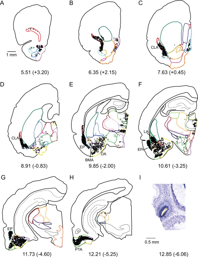
Figure 6.

A–H. Computer generated plots of coronal sections showing the distribution of labeled cells arising from a retrograde tracer injection in the LEA (shown in panel H). Eight of 48 rostrocaudal levels are shown for experiment 124FB. Boundaries of subcortical structures are depicted by colored contours. See Figure 2 for color code. Note the high density of cells in CLA, EP, LA, BMA, OA, and PTA. I. Injection site on a Nissl stained section.
