Abstract
The integrin family comprises 24 transmembrane receptors, each a heterodimeric combination of one of 18α and one of 8β subunits. Their main function is to integrate the cell adhesion and interaction with the extracellular microenvironment with the intracellular signaling and cytoskeletal rearrangement through transmitting signals across the cell membrane upon ligand binding. Integrin αvβ3 is a receptor for the extracellular matrix proteins containing arginine–glycine–aspartic (RGD) tripeptide sequence. The αvβ3 is generally expressed in low levels on the epithelial cells and mature endothelial cells, but it is highly expressed in many solid tumors. The αvβ3 levels correlate well with the potential for tumor metastasis and aggressiveness, which make it an important biological target for development of antiangiogenic drugs, and molecular imaging probes for early tumor diagnosis. Over the last decade, many radiolabeled cyclic RGD peptides have been evaluated as radiotracers for imaging tumors by SPECT or PET. Even though they are called “αvβ3-targeted” radiotracers, the radiolabeled cyclic RGD peptides are also able to bind αvβ5, α5β1, α6β4, α4β1, and αvβ6 integrins, which may help enhance their tumor uptake due to the “increased receptor population.” This article will use the multimeric cyclic RGD peptides as examples to illustrate basic principles for development of integrin-targeted radiotracers and focus on different approaches to maximize their tumor uptake and T/B ratios. It will also discuss important assays for pre-clinical evaluations of the integrin-targeted radiotracers, and their potential applications as molecular imaging tools for noninvasive monitoring of tumor metastasis and early detection of the tumor response to antiangiogenic therapy.
Keywords: Integrin αvβ3, PET and SPECT radiotracers, Tumor imaging
INTRODUCTION
Cancer is the second leading cause of death worldwide (Siegel et al. 2015). Most patients will survive if the cancer can be detected at the early stage. Accurate and rapid detection of rapidly growing and metastatic tumors is of great importance before they become widely spread. There are several imaging modalities available for the diagnosis of cancer, including X-ray computed tomography (CT), ultrasound (US), nuclear magnetic resonance imaging (MRI), and nuclear medicine procedures. While CT, US and MRI are better suited for anatomic analysis of solid tumors, molecular imaging with positron emission tomography (PET) and single-photon emission computed tomography (SPECT) offers significant advantages with respect to sensitivity and specificity because they are able to provide the detailed information related to biochemical changes in tumor tissues at the cellular and molecular levels (Mankoff et al. 2008; Shokeen and Anderson 2009; Tweedle 2009; Correia et al. 2011; Fani and Maecke 2012; Fani et al. 2012; Gaertner et al. 2012; Laverman et al. 2012b; Jamous et al. 2013). The most sensitive molecular imaging modalities are SPECT (~10−10 mol/L) and PET (10−10–10−12 mol/L) using radiotracers (Fani and Maecke 2012; Fani et al. 2012; Gaertner et al. 2012). According to their biodistribution properties, radiotracers are classified as those whose biodistribution is determined by their chemical and physical properties, and those whose ultimate distribution is determined by their receptor or enzyme binding. The latter class is called target-specific radiotracers. Peptides are often used as targeting biomolecules (BM) for receptor binding in order to achieve high tumor specificity. Many radiotracers have been developed to target the receptors overexpressed on tumor cells and/or tumor vasculature (Mankoff et al. 2008; Shokeen and Anderson 2009; Tweedle 2009; Correia et al. 2011; Fani and Maecke 2012; Fani et al. 2012; Gaertner et al. 2012; Laverman et al. 2012b; Jamous et al. 2013).
A large number of radiolabeled cyclic RGD (arginine–glycine–aspartic) peptides have been evaluated as SPECT or PET radiotracers for tumor imaging (Liu et al. 2005; Wu et al. 2005; Jia et al. 2006; Liu et al. 2006; Zhang et al. 2006; Alves et al. 2007; Dijkgraaf et al. 2007a, b; Liu et al. 2007; Wu et al. 2007; Jia et al. 2008; Li et al. 2008b; Liu et al. 2008a; Shi et al. 2008; Wang et al. 2008a, b; Liu et al. 2009a, b; Shi et al. 2009a, b, c; Chakraborty et al. 2010; Kubas et al. 2010; Dumont et al. 2011; Jia et al. 2011; Shi et al. 2011a, b; Zhou et al. 2011b; Nwe et al. 2012; Pohle et al. 2012; Zhou et al. 2012; Ji et al. 2013a, b; Li et al. 2013; Simecek et al. 2013; Tsiapa et al. 2013; Maschauer et al. 2014; Yang et al. 2014; Zheng et al. 2015). Many excellent review articles have appeared to cover their nuclear medicine applications (D’Andrea et al. 2006; Liu 2006; Meyer et al. 2006; Beer and Schwaiger 2008; Cai and Chen 2008; Liu et al. 2008b; Liu 2009; Stollman et al. 2009; Beer and Chen 2010; Chakraborty and Liu 2010; Dijkgraaf and Boerman 2010; Haubner et al. 2010; Beer et al. 2011; Michalski and Chen 2011; Zhou et al. 2011a; Danhier et al. 2012; Tateishi et al. 2012. This article is not intended to be an exhaustive review of current literature on radiolabeled cyclic RGD peptides. Instead, it will use the multimeric cyclic RGD peptides to illustrate some basic principles for new radiotracer development and to address some important issues associated with integrin-targeted radiotracers. It will focus on different approaches to maximize the tumor uptake and T/B ratios. Authors would apologize to those whose work has not been cited in this article.
RADIOTRACER DESIGN
Integrin-targeted radiotracer
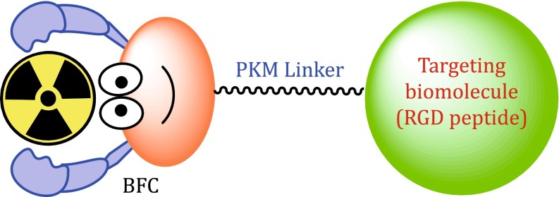
Figure 1 shows the schematic construction of an integrin-targeted radiotracer (Liu 2006, 2009). The cyclic RGD peptide serves as a “vehicle” to carry the isotope to integrins expressed on both tumor cells and activated endothelial cells of tumor neovasculature. BFC is a bifunctional coupling agent to attach the appropriate radionuclide to a cyclic RGD peptide (Liu and Edwards 2001; Liu 2004, 2008; Liu and Chakraborty 2011). The pharmacokinetic modifying (PKM) linker is often used to improve excretion kinetics of radiotracers (Liu and Edwards 2001; Liu 2004, 2008). For a new radiotracer to be successful in clinics, it must show clinical indications for several of high-incidence tumor types (namely breast, lung, and prostate cancers). Renal excretion is necessary in order to maximize the tumor-to-background (T/B) ratios. The main objective of tumor imaging is to achieve the following goals: (1) to detect the presence of tumor at early stage, (2) to distinguish between benign and malignant tumors, (3) to follow the tumor growth and tumor response to a specific therapy (chemotherapy, radiation therapy, or combination thereof), (4) to predict success or failure of a specific therapeutic regimen, and (5) to access the prognosis of a particular tumor.
Fig. 1.
Schematic presentation of the integrin αvβ3-targeted radiotracers. The cyclic RGD peptide is used as the targeting biomolecule (BM) to carry the isotope into the tumor tissue. BFC is used to attach the isotope to the targeting biomolecule. PKM linker is utilized to modify its pharmacokinetics
Radionuclide
The choice of radionuclide depends largely on the modality for tumor imaging. More than 80% of radiotracers for SPECT in nuclear medicine departments are 99mTc compounds due to optimal nuclear properties of 99mTc and its easy availability at low cost (Liu and Edwards 2001; Liu 2004, 2008; Liu and Chakraborty 2011). The 6-h half-life is long enough to allow radiopharmacists to carry out radiosynthesis and for physicians to collect clinically useful images. At the same time, it is short enough to permit administration of 20–30 mCi of 99mTc without imposing a significant radiation dose to the patients. 18F is a cyclotron-produced isotope suitable for PET. It has a half-life of 110 min. Despite its short half-life, the availability of preparative modules makes 18F radiotracers more accessible to clinicians (Anderson et al. 2003). 64Cu is another PET isotope to develop target-specific radiotracers. It has a half-life of 12.7 h and a β+ emission (18%, E max = 0.655 MeV). Despite poor nuclear properties, 64Cu is a viable alternative to 18F for research programs that wish to incorporate high sensitivity and spatial resolution of PET, but cannot afford to maintain the expensive isotope production infrastructure (Anderson et al. 2003). 68Ga is generator-produced PET isotope with the half-life of 68 min. The 68Ge–68Ga generator can be used for more than a year. 68Ga could become as useful for PET as 99mTc for SPECT (Maecke et al. 2005). The 68Ga-labeled somatostatin analogs have been studied for PET imaging of somatostatin-positive tumors in both pre-clinical animal models and cancer patients (Henze et al. 2005; Koukouraki et al. 2006a, b). Gallium chemistry and related nuclear medicine applications have been reviewed recently (Maecke et al. 2005).
Bifunctional coupling agent (BFC)
The choice of BFC depends on the radionuclide (Liu 2004, 2008; Liu and Chakraborty 2011). Among various BFCs for 99mTc-labeling, 6-hydazinonicotinic acid (Fig. 2: HYNIC) is of great interest due to its high efficiency (rapid radiolabeling and high radiolabeling yield), the high solution stability of its 99mTc complexes, and the easy use of co-ligands for modification of biodistribution properties of 99mTc radiotracers (Liu 2004, 2005, 2008; Liu and Chakraborty 2011). In contrast, DOTA (1,4,7,10-tetraazacyclododecane-1,4,7,10-tetraacetic acid), NOTA (1,4,7-triazacyclononane-1,4,7-triacetic acid), and their derivatives (Fig. 2) have been widely used for 68Ga/64Cu-labeling of biomolecules due to the high hydrophilicity and in vivo stability of its 68Ga/64Cu chelates (Anderson et al. 2003; Maecke et al. 2005; Shokeen and Anderson 2009). Organic prosthetic groups (Fig. 2: 4-FB, 4-FBz, 2-FP, and 2-FDG) are often needed for 18F-labeling (Dolle 2005; Li et al. 2007, 2008a; Glaser et al. 2008; Hausner et al. 2008; Hohne et al. 2008; Mu et al. 2008; Becaud et al. 2009; Namavari et al. 2009; Vaidyanathan et al. 2009; Jacobson and Chen 2010; Liu et al. 2010; Wangler et al. 2010; Jacobson et al. 2011; Schirrmacher et al. 2013). However, recent results indicate that the Al(NOTA) chelates is more efficient for routine radiosynthesis of 18F radiotracers using the kit formulation (McBride et al. 2009, 2010, 2012; D’Souza et al. 2011; Lang et al. 2011; Liu et al. 2011; Laverman et al. 2010, 2012a).
Fig. 2.
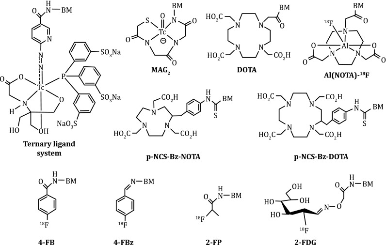
Examples of BFCs useful for radiolabeling of cyclic RGD peptides. HYNIC and MAG2 are useful for 99mTc-labeling while DOTA, NOTA, and their derivatives are better suited for chelation of 64Cu and 68Ga. For 18F-labeling, 4-FB, 4-FBz, 2-FP, and 2-FDG are often used as prosthetic groups. The Al(NOTA) chelate is highly efficient for radiosynthesis of 18F radiotracers using a kit formulation
Integrins as molecular targets for tumor imaging
Angiogenesis is a requirement for tumor growth and metastasis (Hwang and Varner 2004; Weigelt et al. 2005). The angiogenic process depends on the vascular endothelial cell migration and invasion, and is regulated by cell adhesion receptors. Integrins are such a family of receptors that facilitate the cellular adhesion to and the migration on extracellular matrix proteins, and regulate the cellular entry and withdraw from the cell cycle (Albelda et al. 1990; Falcioni et al. 1994; Carreiras et al. 1996; Bello et al. 2001; Sengupta et al. 2001; Cooper et al. 2002; Zitzmann et al. 2002; Hwang and Varner 2004; Jin and Varner 2004; Weigelt et al. 2005; Sloan et al. 2006; Zhao et al. 2007; Hodivala-Dilke 2008; Barczyk et al. 2010; Taherian et al. 2011; Gupta et al. 2012; Sheldrake and Patterson 2014). The integrin family comprises 24 transmembrane receptors (Table 1) (Sheldrake and Patterson 2014). Their main function is to integrate the cell adhesion and interaction with the extracellular microenvironment with the intracellular signaling and cytoskeletal rearrangement through transmitting signals across the cell membrane on ligand binding. Many integrins are crucial to the tumor initiation, progression, and metastasis. Among the 24 members, the αvβ3 is studied most extensively for its role in tumor angiogenesis and metastasis (Albelda et al. 1990; Falcioni et al. 1994; Carreiras et al. 1996; Bello et al. 2001; Sengupta et al. 2001; Cooper et al. 2002; Zitzmann et al. 2002; Hwang and Varner 2004; Jin and Varner 2004; Weigelt et al. 2005; Sloan et al. 2006; Zhao et al. 2007; Hodivala-Dilke 2008; Barczyk et al. 2010; Taherian et al. 2011; Gupta et al. 2012). It is not surprising that radiolabeled cyclic RGD peptides are often called “αvβ3–targeted” radiotracers in majority of the literature (D’Andrea et al. 2006; Liu 2006; Meyer et al. 2006; Beer and Schwaiger 2008; Cai and Chen 2008; Liu et al. 2008b; Liu 2009; Stollman et al. 2009; Beer and Chen 2010; Chakraborty and Liu 2010; Dijkgraaf and Boerman 2010; Haubner et al. 2010; Beer et al. 2011; Michalski and Chen 2011; Zhou et al. 2011a; Danhier et al. 2012; Tateishi et al. 2012).
Table 1.
Natural integrin ligands and their corresponding recognition peptide sequences
| Integrins | Recognition sequence | Natural ligands |
|---|---|---|
| αvβ1, αvβ3, αvβ5, αvβ6, αvβ8, α5β1, α8β1, αIIbβ3 | RGD | Vitronectin, fibronectin, osteopontin, fibrinogen |
| α4β1, α9β1, α4β7, αEβ7, αLβ2, αMβ2, αXβ2, αDβ2 | LDV and related sequences | Fibronectin, vascular cell adhesion molecule 1, mucosal addressin cell adhesion molecule 1, intercellular cell adhesion molecule 1 |
| α1β1, α2β1, α10β1, α11β1 | GFOGER | Collagen, laminin |
| α3β1, α6β1, α7β1, α6β4 | Other | Laminin |
Table were adapted from Sheldrake and Patterson (2014)
The changes in the αvβ3 expression levels and activation state have been well documented during tumor growth and metastasis (Hwang and Varner 2004; Weigelt et al. 2005; Sloan et al. 2006; Zhao et al. 2007; Hodivala-Dilke 2008; Barczyk et al. 2010; Gupta et al. 2012). The αvβ3 is expressed in low levels on epithelial cells and mature endothelial cells, but it is highly expressed in many solid tumors, which include osteosarcomas, glioblastoma, melanomas, and carcinomas of lung and breast (Albelda et al. 1990; Falcioni et al. 1994; Carreiras et al. 1996; Bello et al. 2001; Sengupta et al. 2001; Cooper et al. 2002; Zitzmann et al. 2002; Hwang and Varner 2004; Jin and Varner 2004; Weigelt et al. 2005; Sloan et al. 2006; Zhao et al. 2007; Hodivala-Dilke 2008; Barczyk et al. 2010; Taherian et al. 2011; Gupta et al. 2012). Studies show that αvβ3 is overexpressed on tumor cells and activated endothelial cells of tumor neovasculature (Pilch et al. 2002; Taherian et al. 2011). It is believed that the αvβ3 expressed on endothelial cells modulate cell adhesion and migration during angiogenesis, while the αvβ3 overexpressed on carcinoma cells potentiate metastasis by facilitating invasion and movement of tumor cells across blood vessels (Sloan and Anderson 2002; Minn et al. 2005; Dittmar et al. 2008; Lorger et al. 2009; Omar et al. 2010). It has been shown that the αvβ3 expression levels correlate with the potential for metastasis and aggressiveness of tumors, including glioma, melanoma, and carcinomas of the breast and lungs (Zhao et al. 2007; Hodivala-Dilke 2008). The αvβ3 is considered as an important biological target to develop antiangiogenic drugs (Gottschalk and Kessler 2002; Kumar 2003; Jin and Varner 2004; D’Andrea et al. 2006) and molecular imaging probes for diagnosis of tumors (D’Andrea et al. 2006; Meyer et al. 2006; Liu 2006, 2009; Beer and Schwaiger 2008; Cai and Chen 2008; Liu et al. 2008b; Stollman et al. 2009; Beer and Chen 2010; Chakraborty and Liu 2010; Dijkgraaf and Boerman 2010; Haubner et al. 2010; Beer et al. 2011; Michalski and Chen 2011; Zhou et al. 2011a; Danhier et al. 2012; Tateishi et al. 2012).
Cyclic RGD peptides as targeting biomolecules
The αvβ3 is a receptor for the extracellular matrix proteins with the exposed RGD tripeptide sequence. Theoretically, both linear and cyclic RGD peptides can be used as targeting biomolecules. A major drawback of linear RGD peptides are their low binding affinity (IC50 > 100 nmol/L), lack of specificity (αvβ3 vs. αIIBβ3), and rapid degradation by proteases in serum. Cyclization of RGD peptides via the linkers, such as S-S disulfide, thioether, and rigid aromatic rings, leads to the increased receptor binding affinity and selectivity (Aumailley et al. 1991; Gurrath et al. 1992; Müller et al. 1992; Haubner et al. 1996). It seems that the αIIBβ3 is less sensitive to variations in the RGD peptide backbone and can accommodate a larger distance or spacer than αIIBβ3 and αvβ5 (Pfaff et al. 1994). Incorporation of the RGD sequence into a cyclic pentapeptide framework (Fig. 3: c(RGDfV) and EMD121974) could significantly increase the binding affinity and selectivity of αvβ3/αvβ5 over αIIbβ3 (Aumailley et al. 1991; Gurrath et al. 1992; Müller et al. 1992; Pfaff et al. 1994; Haubner et al. 1996). The addition of a rigid aromatic ring into the cyclic hexapeptide structure (Fig. 3: DMP728 and DMP757) enhances the binding affinity of αIIBβ3 (Liu et al. 2010; Jacobson et al. 2011; Danhier et al. 2012). The structure–activity studies indicated that the amino acid residue in position 5 has little impact on αvβ3/αvβ5 binding affinity (Aumailley et al. 1991; Gurrath et al. 1992; Müller et al. 1992; Haubner et al. 1996). The valine (V) residue in c(RGDfV) can be replaced by lysine (K) or glutamic acid (E) to afford c(RGDfK) and c(RGDfE), respectively, without changing their αvβ3/αvβ5 binding affinity.
Fig. 3.
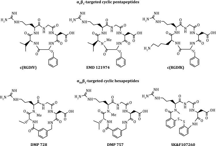
Examples of monomeric cyclic RGD peptides as targeting biomolecules for the development of αvβ3-targeted radiotracers. EMD121974 has been under clinical investigations as an “orphan drug” for treatment of glioblastoma either stand-alone or in combination with radiation therapy. DMP728 and DMP757 were originally developed as anti-thrombotic agents
Figure 4 shows several examples of monomeric cyclic RGD peptides that have high affinity for αvβ3 and αvβ5. Among the radiotracers evaluated in pre-clinical tumor-bearing models, [18F]Galacto-RGD (Fig. 4: 2-[18F]fluoropropanamide c(RGDfK(SAA); SAA = 7-amino-l-glyero-l-galacto-2,6-anhydro-7-deoxyheptanamide) was the first one under clinical investigation for visualization of αvβ3 expression in cancer patients (Beer et al. 2005; 2007, 2008; Haubner et al. 2005). The results from imaging studies in cancer patients showed that there was sufficient αvβ3 for PET imaging. The tumor uptake of [18F]Galacto-RGD correlates with the αvβ3 levels in cancer patients (Haubner et al. 2005; Beer et al. 2007, 2008). However, the radiotracers derived from monomeric cyclic RGD peptides all had low tumor uptake with T/B ratios because of their relatively low αvβ3 binding affinity.
Fig. 4.
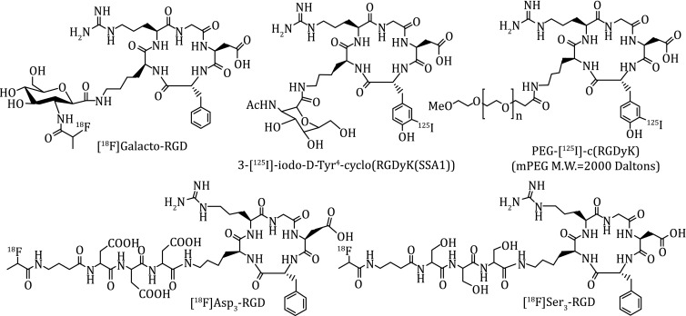
Examples of the radiolabeled monomeric cyclic RGD peptides as radiotracers
It must be noted that cyclic RGD peptides bind not only αvβ3 but also other integrins. While the αvβ3 plays pivotal role in the tumor growth and progression, αIIBβ3 is critical for the platelet aggregation during thrombosis formation. The interaction between αvβ3 and αIIbβ3 facilitates the adhesion of tumor cells to the vasculature and often leads to metastasis (Felding-Habermann et al. 1996; Bakewell et al. 2003). The αvβ5 is very similar to αvβ3 in the ligand binding site region and has a similar expression pattern and function to those of αvβ3. Both αvβ5 and αvβ3 are highly expressed on the activated endothelial cells and have similar roles in angiogenesis, promoting angiogenic response to different growth factors (Bakewell et al. 2003; Goodman et al. 2012). The αvβ5 has been shown to overexpress on a wide range of tumor types (Goodman et al. 2012; Boger et al. 2014). A number of tumors co-express αvβ3 and αvβ5 (Sung et al. 1998; Erdreich-Epstein et al. 2000; Graf et al. 2003; Humphries et al. 2006; Monferran et al. 2008; Bianchi-Smiraglia et al. 2013; Roth et al. 2013; Vogetseder et al. 2013; Boger et al. 2014; Navarro-Gonzalez et al. 2015), because both engage the same ECM ligands and activate complementary cell signaling pathways in order to promote tumor progression (Sung et al. 1998; Bianchi-Smiraglia et al. 2013). It was also reported that the tumor cell expression of αvβ3, αvβ5, α5β1, α6β4, α4β1, and αvβ6 is correlated with the progression of various tumors (Vogetseder et al. 2013; Boger et al. 2014). The structures of other RGD-binding integrins (αvβ6, αvβ8, αvβ1 and α8β1) have not yet been studied in details (Sheldrake and Patterson 2014).
MAXIMIZING BINDING AFFINITY VIA MULTIMERIZATION
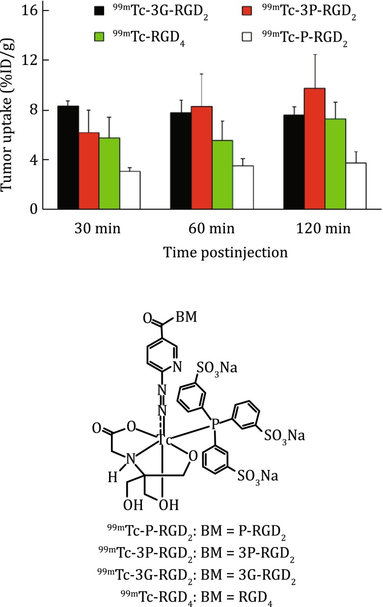
The multivalent concept has been used to develop radiotracers with the increased tumor-targeting capability. For example, E[c(RGDfK)]2 (RGD2) was the first cyclic RGD dimer for development of diagnostic (99mTc) and therapeutic (90Y and 64Cu) radiotracers (Liu et al. 2001a; b; 2005, 2006, 2007, 2008a, 2015; Jia et al. 2006, 2008). RGD tetramers RGD4 was also used to develop SPECT and PET radiotracers (Wu et al. 2005; Liu et al. 2007, 2008a). Both the in vitro assays and biodistribution data showed that the radiolabeled (99mTc, 18F, and 64Cu) multimeric cyclic RGD peptides have higher αvβ3 binding affinity and better tumor uptake than their monomeric analogs (Liu et al. 2008b; Liu 2009). It is important to note that multimeric RGD peptides are not necessarily multivalent (Liu et al. 2008b; Chakraborty et al. 2010). Two factors (Fig. 5: bivalency and enhanced local RGD concentration) contribute to the high αvβ3 binding affinity of cyclic RGD peptides (Liu et al. 2008b; Chakraborty et al. 2010). The concentration factor exists in all multimeric RGD peptides regardless of the linker length. Given the short distance (6 bonds excluding side-arms of K-residues) between two RGD motifs in E[c(RGDfK)]2 and E[c(RGDyK)]2, it is unlikely that they would bind to two adjacent αvβ3 sites simultaneously. However, the binding of one RGD motif to αvβ3 will increase the “local concentration” of second RGD motif in the vicinity of αvβ3 sites (Fig. 5B). The concentration factor may explain the higher tumor uptake of radiolabeled (99mTc, 111In, 90Y, 18F, and 64Cu) E[c(RGDfK)]2 and E[c(RGDyK)]2 than their monomeric derivatives (Beer and Chen 2010; Chakraborty and Liu 2010; Dijkgraaf and Boerman 2010; Beer et al. 2011; Michalski and Chen 2011; Zhou et al. 2011a). The key for bivalency is the distance between two cyclic RGD motifs. For example, this distance is 38 bonds in PEG4-E[c(RGDfK(PEG4))]2 (3P-RGD2: PEG4 = 15-amino-4,7,10,13-tetraoxapentadecanoic acid), and 26 bonds G3-E[c(RGDfK(G3))]2 (3G-RGD2: G3 = Gly-Gly-Gly), which are long enough for them to achieve the bivalency. As a result, HYNIC-3P-RGD2 (IC50 = 60 ± 3 nmol/L) and HYNIC-3G-RGD2 (IC50 = 59 ± 3 nmol/L) have much higher αvβ3 binding affinity than HYNIC-P-RGD2 (P-RGD2 = PEG4-E[c(RGDfK)]2: (IC50 = 89 ± 7 nmol/L)) (Shi et al. 2008; Wang et al. 2008b). 99mTc-3P-RGD2 and 99mTc-3G-RGD2 had higher breast tumor uptake than 99mTc-P-RGD2 (Fig. 6) (Shi et al. 2008; Wang et al. 2008b). Since the tumor uptake of 99mTc-3P-RGD2 and 99mTc-3P-RGD2 is comparable to that of 99mTc-RGD4 suggests that the contribution from “concentration factor” may not be as significant as that from the “bivalency.”
Fig. 5.
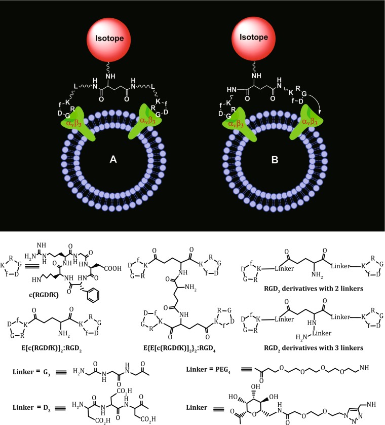
Top: Schematic illustration of the interactions between cyclic RGD peptide dimers and αvβ3. A The distance between two RGD motifs is not long enough for simultaneous integrin αvβ3 binding. However, the RGD concentration is “locally enriched” in the vicinity of neighboring integrin αvβ3 once the first RGD motif is bound. B The distance between two RGD motifs is long due to the presence of two linkers (L). As a result, the cyclic RGD dimer is able to bind integrin αvβ3 in a “bivalent” fashion. In both cases, the end-result would be higher integrin αvβ3 binding affinity for the multimeric cyclic RGD peptides. Bottom: Selected cyclic RGD peptide dimers and tetramers useful for development of αvβ3-targeted radiotracers. The D3, G3, PEG4, and sugar linkers are used to increase the distance between two RGD motifs and to improve radiotracer excretion kinetics from non-cancerous organs
Fig. 6.
Direct comparison of tumor uptake for 99mTc-P-RGD2, 99mTc-3G-RGD2, 99mTc-3P-RGD2, and 99mTc-RGD4 in athymic nude mice bearing MDA-MB-435 breast cancer xenografts. The biodistribution data were adapted from Shi et al. (2008) and Wang et al. (2008b)
MAXIMIZING RADIOTRACER UPTAKE BY TARGETING MULTIPLE RECEPTORS
Two most important factors affecting the radiotracer tumor uptake are receptor binding affinity and receptor population. The receptor binding affinity is critically important for selective tumor localization and tumor uptake of radiolabeled cyclic RGD peptides (Liu et al. 2008b). The receptor population is equally important for the receptor-based molecular imaging. It will not be possible to image the tumor if that it has very limited or no receptor expression even if the receptor ligand has high receptor binding affinity. There are two approaches to maximize the target population. The first approach (Fig. 7A) involves the use of the same cyclic RGD peptide to target two or more integrins (such as αvβ3, αvβ5, α5β1, α6β4, α4β1, and αvβ6). Another approach (Fig. 7B) involves the use of a bifunctional peptide that is able to target two different receptors, such as αvβ3 and bombesin (BBN) receptor. By targeting two different receptors, the radiotracer will have more opportunities to localize in the tumor due to the larger populations of two receptors than that of a single receptor. The so-called “bivalent heterodimers” (Fig. 7) has been used to target the αvβ3 and BBN receptors (Li et al. 2008c; Liu et al. 2009c, d). The xenografted PC-3 and MDA-MB-435 tumor-bearing models were used to evaluate their tumor-targeting capability and biodistribution properties. It is well-established that the xenografted PC-3 tumors have low αvβ3 expression (Zhou et al. 2011b; Ji et al. 2013c). It was also shown that the xenografted MDA-MB-435 tumor has little BBN receptor expression (Liu et al. 2009c, d). Therefore, both PC-3 and MDA-MB-435 tumor-bearing models are not appropriate to prove the concept of “bivalent heterodimers.” For the bifunctional radiotracers to achieve the bivalency, the αvβ3 and BBN receptors must be co-localized and the distance between them must be short. Otherwise, it would not be advantageous even if they might be able to target both individual receptors. Unfortunately, there is lack of concrete experimental data to demonstrate if the c(RGDfK)-BBN(7–14) and c(RGDyK)-BBN(7–14) conjugates are “bivalent” for tumor targeting, and whether there is indeed a “synergetic effect” between the cyclic RGD and BBN(7–14) peptides. Another challenge associated with the “bifunctional heterodimer concept” is which binding unit actually contributes to the radiotracer tumor uptake.
Fig. 7.
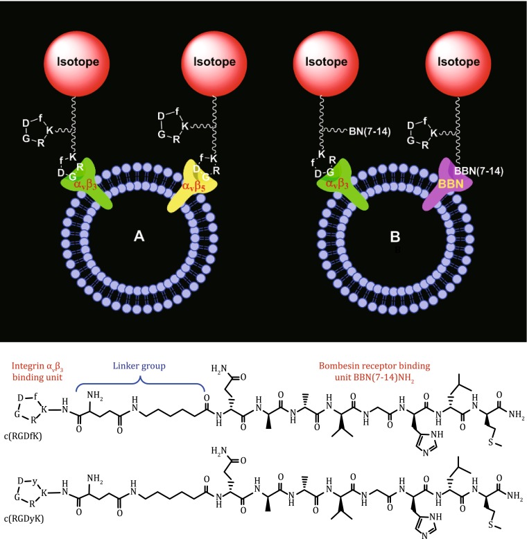
Top: Schematic presentation of the interactions between the dimeric cyclic RGD peptide to target two or more integrins (such as αvβ3, αvβ5, α5β1, α6β4, α4β1, and αvβ6). B Schematic illustration of the interactions between the bifunctional peptide and two different receptors (αvβ3 and BBN receptor). By targeting two different receptors, the radiotracer will have more opportunities to localize in the tumor because of the increased receptor population. The two targeted receptors (e.g., αvβ3/αvβ5 or αvβ3/BBN) must be co-localized and the distance between them must be short for the bifunctional radiotracer to achieve “simultaneous receptor binding.” Bottom: Selected examples of bifunctional peptides containing c(RGDfK)/c(RGDyK) and Aca-BBN(7–14)NH2 (ε-aminocaproic acid-Gln-Trp-Ala-Val-Gly-His-Leu-Met-NH2)
INTEGRIN AND RGD SPECIFICITY
Integrin specificity
Blocking experiment (Fig. 8A) has been used to demonstrate the αvβ3 specificity of radiolabeled RGD peptides with a known αvβ3 antagonist (e.g., c(RGDfK) or RGD2) as the blocking agent. This experiment is often performed by biodistribution or imaging (PET or SPECT). The blocking agent is pre- or co-injected with the radiotracer. Co-injection or pre-injection of excess blocking agents (such as RGD2) will result in partial or complete blockage of the radiotracer tumor uptake (Fig. 8B). It is important to note that there is also a significant reduction in radiotracer uptake in the αvβ3-positive organs (e.g., eyes, intestine, kidneys, lungs, liver, muscle, and spleen). The normal organ uptake is consistent with the β3 and CD31 staining data for the liver, kidneys, and lungs from the tumor-bearing athymic nude mice.
Fig. 8.

A Comparison of organ uptake (%ID/g) for 99mTc-2P-RGD2 in athymic nude mice bearing U87MG glioma xenografts in the absence or presence of excess RGD2 at 60 min p.i. Co-injection of excess RGD2 resulted in significant reduction in the uptake of 99mTc-2P-RGD2 in the tumor and normal organs. B Comparison of the 60-min biodistribution data of 111In-3P-RGD2 and 111In-3P-RGK2 in athymic nude mice bearing U87MG glioma xenografts. The low tumor uptake for 111In-3P-RGK2 indicates that the radiolabeled cyclic RGD dimers are RGD-specific. The experimental data were adapted from Shi et al. (2011a)
RGD specificity
There are several ways to determine the RGD specificity of radiolabeled cyclic RGD peptides, including: (1) the in vitro binding assay using 125I-echistatin as the integrin-specific radioligand (Zhang et al. 2006; Wu et al. 2007; Wang et al. 2008b; Shi et al. 2009c), (2) the in vitro tissue or cellular immunohistochemical (IHC) staining assay using fluorescent probes (Zheng et al. 2014), (3) the in vivo imaging experiment (PET or SPECT) (Zhang et al. 2006; Wu et al. 2007; Wang et al. 2008b; Shi et al. 2009c), and (4) the biodistribution study (Shi et al. 2009a, 2011a, b; Chakraborty et al. 2010). In all cases, a nonsense peptide with the “scrambled sequence” will be used to prepare the corresponding radiotracer or fluorescent probe. For example, 3P-RGK2 is the nonsense peptide with the composition identical to that of 3P-RGD2. The αvβ3 binding affinity of DOTA-3P-RGK2 (IC50 = 596 ± 48 nmol/L) was >20× lower than that of DOTA-3P-RGD2 (IC50 = 29 ± 4 nmol/L). Similar results were also seen with FITC-3P-RGK2 (IC50 = 589 ± 73 nmol/L) and FITC-3P-RGD2 (IC50 = 32 ± 7 nmol/L). Because of the low αvβ3 affinity of DOTA-3P-RGK2 (Chakraborty et al. 2010; Shi et al. 2011a, b), 111In(DOTA-3P-RGK2) had significantly lower (p < 0.01) uptake than 111In(DOTA-3P-RGD2) in the xenografted breast tumors and the αvβ3-positive normal organs, such as eyes, intestine, liver, lungs, and spleen (Fig. 8B) (Shi et al. 2011a). These results clearly show that the uptake of radiolabeled cyclic RGD peptides in tumors and some normal organs is indeed αvβ3-specific.
LINEAR RELATIONSHIP BETWEEN RADIOTRACER TUMOR UPTAKE AND ΑVΒ3 EXPRESSION
It has been shown that the radiolabeled cyclic RGD peptides are useful for non-invasive imaging of tumors in cancer patients (Beer et al. 2005, 2007, 2008; Haubner et al. 2005). It is the total αvβ3 level that will contribute the tumor uptake of a αvβ3-targeted radiotracer. The capability to visualize the αvβ3 expression provides new opportunities to characterize the tumor angiogenesis noninvasively, to select appropriate patients for antiangiogenic treatment, and to monitor the tumor response to antiangiogenic drugs. However, there were only a few reports on the correlation between the αvβ3 expression levels and radiotracer tumor uptake (Beer et al. 2005, 2007, 2008; Haubner et al. 2005; Zhang et al. 2006).
99mTc-3P-RGD2 was studied for its capability to monitor the αvβ3 expression in five different tumor-bearing animal models (U87MG, MDA-MB-435, A549, HT29, and PC-3). IHC staining was performed to determine the αvβ3 and CD31 (a biomarker for tumor vasculature) expression levels in xenografted U87MG, MDA-MB-435, A549, HT29, and PC-3 tumor tissues (Zhou et al. 2011b). It was found that the total αvβ3 expression levels on the tumor cells and tumor neovasculature follow the general ranking trend: U87MG > MDA-MB-435 = A549 = HT29 > PC-3. In contrast, the CD31 expression levels follow the general ranking order of U87MG = HT29 > MDA-MB-435 = A549 > PC-3 (Fig. 9). More importantly, there is an excellent relationship between the tumor uptake and the αvβ3 expression levels (Zhou et al. 2011b). The linear relationship between the tumor uptake (%ID/g) and αvβ3 density suggests that 99mTc-3P-RGD2 is useful for non-invasive monitoring of the αvβ3 expression levels in cancer patients.
Fig. 9.

Relationship between the tumor uptake (%ID/g: radioactivity density) and relative β3 or CD31 levels in five xenografted tumors (U87MG, MDA-MB-435, A549, HT29, and PC-3). The total β3 expression was represented by the percentage of red area over total area in each slice of tumor tissue. Each data point was derived from at least 15 different areas of same tissue (×100 magnification). Experiments were repeated three times independently with similar results. The experimental data were adapted from Zhou et al. (2011b)
MONITORING TUMOR RESPONSE TO ANTIANGIOGENIC THERAPY
99mTc-3P-RGD2 has been used to monitor the tumor response to antiangiogenesis treatment with linifanib (ABT-869) (Ji et al. 2013b, d), a multi-targeted receptor tyrosine kinase inhibitor targeting vascular endothelial growth factor (VEGF) and platelet-derived growth factor (PDGF) receptors (Albert et al. 2006; Shankar et al. 2007; Wong et al. 2009; Zhou et al. 2009; Hernandez-Davies et al. 2011; Jiang et al. 2011; Tannir et al. 2011; Luo et al. 2012). We found that there was a significant decrease in tumor uptake (%ID/cm3) and T/M ratios of 99mTc-3P-RGD2 in the xenografted U87MG model, while no significant changes in tumor uptake of 99mTc-3P-RGD2 were seen in the PC-3 model after linifanib treatment (Ji et al. 2013d). The uptake changes in MDA-MB-435 tumors were between those observed in the U87MG and PC-3 models (Ji et al. 2013b). This is consistent with the tumor αvβ3 expression levels (Zhou et al. 2011b). Highly vascularized tumors (e.g., U87MG) with higher level of αvβ3 and CD31 have better tumor response to linifanib therapy than poorly vascularized tumors (e.g., PC-3) with low levels of αvβ3 and CD31 (Fig. 10). Thus, 99mTc-3P-RGD2 might be a screening tool to select appropriate patients who will benefits most antiangiogenic treatment. If the tumor has a high αvβ3 expression, as indicated by high tumor uptake of 99mTc-3P-RGD2 at the time of diagnosis, antiangiogenic therapy would more likely be effective. If the tumor has little αvβ3 expression (low uptake of 99mTc-3P-RGD2), antiangiogenic therapy would not be effective regardless the amount of antiangiogenic drug administered into the patient.
Fig. 10.
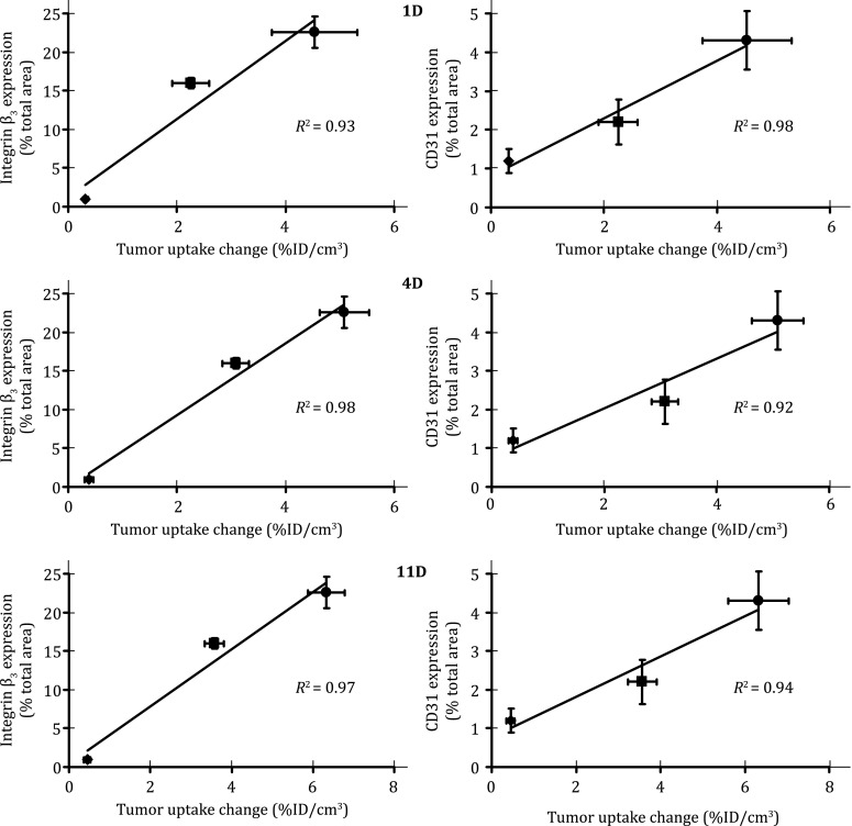
Linear relationship between the %ID/cm3 tumor uptake change at days 1 (top), 4 (middle) and 11 (bottom) after linifanib therapy and the expression levels of the αvβ3 (left) and CD31 (right) in three tumor-bearing animal models. The %ID/cm3 tumor uptake values of 99mTc-3P-RGD2 were calculated from SPECT/CT quantification and reported as the mean plus/minus standard error of the mean based on results from five animals (n = 5). The %ID/cm3 tumor uptake change was calculated by deducting the %ID/cm3 tumor uptake of 99mTc-3P-RGD2 on days 1, 4, and 11 from its original value on −1 day (before linifanib therapy) in the same animal. The average %ID/cm3 tumor uptake change is used as the indicator of tumor response to linifanib treatment. The experimental data were adapted from Zheng et al. (2014)
MONITORING TUMOR METASTASIS
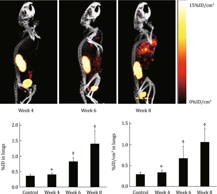
99mTc-3P-RGD2 SPECT/CT has been used as a noninvasive imaging tool to monitor the tumor growth and progression of breast cancer lung metastasis (Albert et al. 2006; Ji et al. 2013d). Figure 11 shows the SPECT/CT images of athymic nude mice (n = 8) with breast cancer lung metastasis. As expected, the SPECT/CT images showed no detectable metastatic breast tumor lesions in the lungs at week 4 (Fig. 11: top). By week 6, small breast cancer lesions started to appear in the mediastinum and lungs. At week 8, SPECT/CT images revealed many metastatic cancer lesions in both lungs (Albert et al. 2006). Figure 11 (bottom) compares the %ID (left) and %ID/cm3 (right) uptake values of 99mTc-3P-RGD2 in the lungs. Even though the lung uptake of 99mTc-3P-RGD2 (0.41 ± 0.05 %ID) at week 4 seemed to be higher than that in the control animals (0.36 ± 0.06 %ID), this difference was not significant (p > 0.05) within the experimental errors. At week 6, the tumor burden in the lungs became significant. The lung uptake of 99mTc-3P-RGD2 was much higher (0.89 ± 0.12 %ID, p < 0.01) than that in the control group. By week 8, the uptake of 99mTc-3P-RGD2 in the lungs was increase to 1.40 ± 0.42 %ID. In all cases, the lung size remained relatively unchanged (1.21–1.32 cm3) during the 8-week study period.
Fig. 11.
Top: The 3D views of SPECT/CT images of an athymic nude mouse at week 4, 6, and 8 after tail-vein injection of 1.0 × 10⁶ MDA-MB-231 cells. Bottom: The %ID (left) and %ID/cm3 (right) uptake values of 99mTc-3P-RGD2 in the lungs obtained from SPECT/CT quantification in the athymic nude mice (n = 8) at week 4, 6, and 8 after tail-vein injection of 1.0 × 10⁶ MDA-MB-231 cells. Normal animals (n = 4) were used in the control group. † p < 0.05, significantly different from the control group; *p > 0.05, no significant difference from the control group. The imaging and SPECT quantification data were from Ji et al. (2013b)
CLINICAL EXPERIENCES WITH 99mTc-3P-RGD2
The excellent in vivo tumor-targeting efficacy of 99mTc-3P-RGD2 in animal models guaranteed its further clinical application. In a first-in-human study, 99mTc-3P-RGD2 was investigated for its capability to noninvasively differentiate solitary pulmonary nodules (SPNs) (Ma et al. 2011). Among the 21 patients with SPNs, 15 (71%) were diagnosed as malignant while 6 (29%) were benign. The sensitivities for CT interpretation and 99mTc-3P-RGD2 SPECT visual were 80% and 100%, respectively. All SPNs classified as indeterminate via CT can be sensitively diagnosed by 99mTc-3P-RGD2 scintigraphy. 99mTc-3P-RGD2 uptake in the malignant and benign nodules was well confirmed by ex vivo IHC staining of αvβ3. These results demonstrated the feasibility of using 99mTc-3P-RGD2 scintigraphy in differentiating SPNs (Ma et al. 2011). A multicenter study was performed in 70 patients with suspected lung lesions (Zhu et al. 2012). The results clearly demonstrated that 99mTc-3P-RGD2 SPECT effectively detects lung malignancies, but with relatively low specificity. Whole-body planar scanning and chest SPECT are complementary for the evaluation of primary tumor and metastasis (Zhu et al. 2012). In a recently study, the potential of 99mTc-3P-RGD2 SPECT in the detection of RAIR DTC lesions was conducted (Zhao et al. 2012). 99mTc-3P-RGD2 SPECT identified all the target RAIR metastatic lesions, and there was a significant correlation between the mean tumor-to-background ratios and mean growth rates of target lesions. It is concluded that 99mTc-3P-RGD2 imaging can be used for the localization and growth evaluation of RAIR lesions, thus providing a promising imaging strategy to monitor the efficacy of antiangiogenic therapy (Zhao et al. 2012). 99mTc-3P-RGD2 SPECT was also evaluated and compared to 99mTc-MIBI for the capability to assess the breast cancer lessons (Ma et al. 2014). It was found that the mean T/NT ratio of 99mTc-3P-RGD2 in malignant lesions was significantly higher than that in benign lesions (3.54 ± 1.51 vs. 1.83 ± 0.98, p < 0.001). The sensitivity, specificity, and accuracy of 99mTc-3P-RGD2 SMM were 89.3%, 90.9%, and 89.7%, respectively, with a T/NT cut-off value of 2.40. The mean T/NT ratio of 99mTc-MIBI in malignant lesions was also significantly higher than that in benign lesions (2.86 ± 0.99 vs. 1.51 ± 0.61, p < 0.001). The sensitivity, specificity, and accuracy of 99mTc-MIBI SMM were 87.5%, 72.7%, and 82.1%, respectively, with a T/NT cut-off value of 1.45. According to the ROC analysis, the area under the curve for 99mTc-3P-RGD2 SMM (area = 0.851) was higher than that for 99mTc-MIBI SMM (area = 0.781), but the statistical difference was not significant.
CLINICAL EXPERIENCES WITH 18F-ALFATIDE AND 18F-ALFATIDE II
18F-labeled RGD compounds suffer from multistep and time-consuming synthetic procedures, which will limit their clinic availability. To overcome this shortcoming, the Al(NOTA) chelate has been used for 18F-labeling of P-RGD2 (Lang et al. 2011). The application of NOTA-AlF chelation chemistry and kit formulation allows one-step 18F-labeling. Under the optimal conditions, the radiotracer [18F]AlF(NOTA-P-RGD2) (denoted as 18F-Alfatide) was prepared in relatively high yield (42.1 ± 0.02) with more than 95% radiochemical purity. The whole radiosynthesis including post-labeling chromatographic purification was accomplished within 20 min. Nine patients with a primary diagnosis of lung cancer were examined by both static and dynamic PET imaging with 18F-alfatide, and one tuberculosis patient was investigated using both 18F-alfatide and 18F-FDG imaging. It was found that 18F-alfatide PET identified all tumors, with mean standardized uptake values of 2.90 ± 0.10. Tumor-to-muscle and tumor-to-blood ratios were 5.87 ± 2.02 and 2.71 ± 0.92, respectively. It was concluded that PET scanning with 18F-alfatide allows specific imaging of avb3 expression with good contrast in lung cancer patients.
CONCLUSIONS
Over the last several years, many multimeric cyclic RGD peptides have been used to increase the radiotracer tumor-targeting capability. The fact that radiolabeled (18F, 99mTc, 111In, 64Cu, and 68Ga) cyclic RGD peptides to target multiple integrins (αvβ3, αvβ5, α5β1, α6β4, α4β1, and αvβ6) will help to improve their tumor uptake due to the “increased receptor population.” In order to achieve bivalency, the distance between two cyclic RGD motifs must be long enough so that they will be able to bind the two adjacent αvβ3 sites simultaneously. Multimerization increases the uptake of radiolabeled multimeric cyclic RGD peptides in both the tumor and normal organs, and also their tumor retention times. Among the radiotracers evaluated in tumor-bearing models, the radiolabeled cyclic RGD dimers (e.g., 2P-RGD2, 3P-RGD2, 2G-RGD2, 3G-RGD2, and Galacto-RGD2) show the most promising results with respect to their tumor uptake and T/B ratios. 99mTc-3P-RGD2, 18F-Alfatide, and 18F-Alfatide II are currently under clinical investigation for tumor imaging by SPECT or PET. 99mTc-3P-RGD2 offers significant advantages over both 18F-Alfatide and 18F-Alfatide II because it could be routinely prepared in high yield and radiochemical purity (>95%) without post-labeling chromatographic purification and clinical availability of 99Mo-99mTc generators. However, SPECT has limitations in quantification of radiotracer uptake, the speed of dynamic imaging, spatial resolution, and tissue attenuation.
Acknowledgments
This work was supported, in part, by Purdue University and R21 EB017237-01 (S. L.) from the National Institute of Biomedical Imaging and Bioengineering (NIBIB).
General terms
- DCE-MRI
Dynamic contrast-enhanced magnetic resonance imaging
- FITC
Fluorescein isothiocyanate isomer I
- 18F-FDG
2-Deoxy-2-(18F)fluoro-d-glucose
- IHC
Immunohistochemistry
- MRI
Magnetic resonance imaging
- PET
Positron emission tomography
- PDGFR
Platelet-derived growth factor receptors
- SPECT
Single-photon emission computed tomography
- VEGFR
Vascular endothelial growth factor receptors
Chelators
- DOTA
1,4,7,10-Tetraazacyclododecane-1,4,7,10-tetracetic acid
- HYNIC
6-Hydazinonicotinic acid
- NOTA
1,4,7-triazacyclononane-1,4,7-triacetic acid
Cyclic peptides
- Galacto-RGD2
Glu[cyclo[Arg-Gly-Asp-d-Phe-Lys(SAA-PEG2-(1,2,3-triazole)-1-yl-4-methylamide)]]2 (SAA = 7-amino-l-glycero-l-galacto-2,6-anhydro-7-deoxyheptanamide, and PEG2 = 3,6-dioxaoctanoic acid)
- P-RGD
PEG4-c(RGKfD) = cyclo(Arg-Gly-Asp-d-Phe-Lys(PEG4)) (PEG4 = 15-amino-4,7,10,13-tetraoxapentadecanoic acid)
- RGD2
E[c(RGDfK)]2 = Glu[cyclo(Arg-Gly-Asp-d-Phe-Lys)]2
- P-RGD2
PEG4-E[c(RGDfK)]2 = PEG4-Glu[cyclo(Arg-Gly-Asp-d-Phe-Lys)]2
- 2G-RGD2
E[G3-c(RGDfK)]2 = Glu[cyclo[Arg-Gly-Asp-d-Phe-Lys(G3)]]2 (G3 = Gly-Gly-Cly)
- 2P-RGD2
E[PEG4-c(RGDfK)]2 = Glu[cyclo[Arg-Gly-Asp-d-Phe-Lys(PEG4)]]2
- 3G-RGD2
G3-E[G3-c(RGDfK)]2 = G3-Glu[cyclo[Arg-Gly-Asp-d-Phe-Lys(G3)]]2
- 3P-RGD2
PEG4-E[PEG4-c(RGDfK)]2 = PEG4-Glu[cyclo[Arg-Gly-Asp-d-Phe-Lys(PEG4)]]2
- 3P-RGK2
PEG4-E[PEG4-c(RGDfK)]2 = PEG4-Glu[cyclo[Arg-Gly-Lys(PEG4)-d-Phe-Asp]]2)
- RGD4
E{E[c(RGDfK)]2}2 = Glu{Glu[cyclo(Arg-Gly-Asp-d-Phe-Lys)]2}2
- 6G-RGD4
E{G3-E[G3-c(RGDfK)]2}2 = Glu{G3-Glu[cyclo(Lys(G3)-Arg-Gly-Asp-d-Phe)]-cyclo(Lys(G3)-Arg-Gly-Asp-d-Phe)}-{PEG4-Glu[cyclo(Lys(G3)-Arg-Gly-Asp-d-Phe)]-cyclo(Lys(G3)-Arg-Gly-Asp-d-Phe)}
- 6P-RGD4
E{PEG4-E[PEG4-c(RGDfK)]2}2 = Glu{PEG4-Glu[cyclo(Lys(PEG4)-Arg-Gly-Asp-d-Phe)]-cyclo(Lys(PEG4)-Arg-Gly-Asp-d-Phe)}-{PEG4-Glu[cyclo(Lys(PEG4)-Arg-Gly-Asp-d-Phe)]-cyclo(Lys(PEG4)-Arg-Gly-Asp-d-Phe)}
Bioconjugates of cyclic peptides
- DOTA-RGD
DOTA-c(RGDfK)
- DOTA-P-RGD
DOTA-PEG4-c(RGDfK)
- DOTA-RGD2
DOTA-E[c(RGDfK)]2
- DOTA-P-RGD2
DOTA-PEG4-E[c(RGDfK)]2
- DOTA-2G-RGD2
DOTA-E[G3-c(RGDfK)]2
- DOTA-2P-RGD2
DOTA-E[PEG4-c(RGDfK)]2
- DOTA-3G-RGD2
DOTA-G3-E[G3-c(RGDfK)]2
- DOTA-3P-RGD2
DOTA-PEG4-E[PEG4-c(RGDfK)]2
- DOTA-3P-RGK2
DOTA-PEG4-E[PEG4-c(RGDfK)]2
- DOTA-Galacto-RGD2
DOTA-Glu[cyclo[Arg-Gly-Asp-d-Phe-Lys(SAA-PEG2-(1,2,3-triazole)-1-yl-4-methylamide)]]2
- DOTA-RGD4
DOTA-E{E[c(RGDfK)]2}2
- DOTA-6G-RGD4
DOTA-E{G3-E[G3-c(RGDfK)]2}2
- DOTA-6G-RGD4
E{PEG4-E[PEG4-c(RGDfK)]2}2
- FITC-3P-RGD2
FITC-PEG4-E[PEG4-c(RGDfK)]2
- FITC-3P-RGK2
FITC-PEG4-E[PEG4-c(RGDfK)]2
- HYNIC-RGD2
HYNIC-E[c(RGDfK)]2
- HYNIC-P-RGD2
HYNIC-PEG4-E[c(RGDfK)]2
- HYNIC-2G-RGD2
HYNIC-E[G3-c(RGDfK)]2
- HYNIC-2P-RGD2
HYNIC-E[PEG4-c(RGDfK)]2
- HYNIC-3G-RGD2
HYNIC-G3-E[G3-c(RGDfK)]2
- HYNIC-3P-RGD2
HYNIC-PEG4-E[PEG4-c(RGDfK)]2
- HYNIC-Galacto-RGD2
HYNIC-Glu[cyclo[Arg-Gly-Asp-d-Phe-Lys(SAA-PEG2-(1,2,3-triazole)-1-yl-4-methylamide)]]2
- HYNIC-RGD4
HYNIC-E{E[c(RGDfK)]2}2
- NOTA-P-RGD2
NOTA-PEG4-E[c(RGDfK)]2
- NOTA-2G-RGD2
NOTA-E[G3-c(RGDfK)]2
- NOTA-2P-RGD2
NOTA-E[PEG4-c(RGDfK)]2
- NOTA-3G-RGD2
NOTA-G3-E[G3-c(RGDfK)]2
- NOTA-3P-RGD2
NOTA-PEG4-E[PEG4-c(RGDfK)]2
Radiolabeled cyclic RGD peptides
- 18F-Alfatide
[18F]AlF(NOTA-P-RGD2)
- 18F-Alfatide II
[18F]AlF(NOTA-2P-RGD2)
- 18F-Galacto-RGD
2-[18F]fluoropropanamide c(RGDfK(SAA), SAA = 7-amino-L-glyero-L-galacto-2,6-anhydro-7-deoxyheptanamide)
- 64Cu-P-RGD2
64Cu(DOTA-P-RGD2)
- 64Cu-2G-RGD2
64Cu(DOTA-2G-RGD2)
- 64Cu-2P-RGD2
64Cu(DOTA-2P-RGD2)
- 64Cu-3G-RGD2
64Cu(DOTA-3G-RGD2)
- 64Cu-3P-RGD2
64Cu(DOTA-3P-RGD2)
- 68Ga-3G-RGD2
68Ga(DOTA-3G-RGD2)
- 68Ga-3P-RGD2
68Ga(DOTA-3P-RGD2)
- 111In-P-RGD
111In(DOTA-P-RGD)
- 111In-P-RGD2
111In(DOTA-P-RGD2)
- 111In-2G-RGD2
111In(DOTA-2G-RGD2)
- 111In-2P-RGD2
111In(DOTA-2P-RGD2)
- 111In-3G-RGD2
111In(DOTA-3G-RGD2)
- 111In-3P-RGD2
111In(DOTA-3P-RGD2)
- 111In-Galacto-RGD2
111In(DOTA-Galacto-RGD2)
- 111In-6G-RGD4
111In(DOTA-6G-RGD4)
- 111In-6P-RGD4
111In(DOTA-6P-RGD4)
- 99mTc-Galacto-RGD2
[99mTc(HYNIC-Galacto-RGD2)(tricine)(TPPTS)])
- 99mTc-RGD2
[99mTc(HYNIC-RGD2)(tricine)(TPPTS)])
- 99mTc-P-RGD2
[99mTc(HYNIC-P-RGD2)(tricine)(TPPTS)]
- 99mTc-2G-RGD2
[99mTc(HYNIC-2G-RGD2)(tricine)(TPPTS)]
- 99mTc-2P-RGD2
[99mTc(HYNIC-2P-RGD2)(tricine)(TPPTS)]
- 99mTc-3G-RGD2
[99mTc(HYNIC-3G-RGD2)(tricine)(TPPTS)]
- 99mTc-3P-RGD2
[99mTc(HYNIC-3P-RGD2)(tricine)(TPPTS)]
- 99mTc-RGD4
[99mTc(HYNIC-RGD4)(tricine)(TPPTS)])
Compliance with Ethical Standards
Conflict of Interest
Jiyun Shi, Fan Wang, and Shuang Liu declare that they have no conflict of interest.
Human and Animal Rights and Informed Consent
This article does not contain any studies with human or animal subjects performed by any of the authors.
References
- Albelda SM, Mette SA, Elder DE, Stewart R, Damjanovich L, Herlyn M, Buck CA. Integrin distribution in malignant melanoma: association of the β3 subunit with tumor progression. Cancer Res. 1990;50:6757–6764. [PubMed] [Google Scholar]
- Albert DH, Tapang P, Magoc TJ, Pease LJ, Reuter DR, Wei RQ, Li J, Guo J, Bousquet PF, Ghoreishi-Haack NS, Wang B, Bukofzer GT, Wang YC, Stavropoulos JA, Hartandi K, Niquette AL, Soni N, Johnson EF, McCall JO, Bouska JJ, et al. Preclinical activity of ABT-869, a multitargeted receptor tyrosine kinase inhibitor. Mol Cancer Ther. 2006;5:995–1006. doi: 10.1158/1535-7163.MCT-05-0410. [DOI] [PubMed] [Google Scholar]
- Alves S, Correia JD, Gano L, Rold TL, Prasanphanich A, Haubner R, Rupprich M, Alberto R, Decristoforo C, Santos I, Smith CJ. In vitro and in vivo evaluation of a novel 99mTc(CO)3-pyrazolyl conjugate of cyclo-(Arg-Gly-Asp-d-Tyr-Lys) Bioconjug Chem. 2007;18:530–537. doi: 10.1021/bc060234t. [DOI] [PubMed] [Google Scholar]
- Anderson CJ, Green MA, Fujibayashi Y (2003) Chemistry of copper radionuclides and radiopharmaceutical products. Handb Radiopharm Radiochem Appl 401–422
- Aumailley M, Gurrath M, Muller G, Calvete J, Timpl R, Kessler H. Arg–Gly–Asp constrained within cyclic pentapeptides. Strong and selective inhibitors of cell adhesion to vitronectin and laminin fragment P1. FEBS Lett. 1991;291:50–54. doi: 10.1016/0014-5793(91)81101-D. [DOI] [PubMed] [Google Scholar]
- Bakewell SJ, Nestor P, Prasad S, Tomasson MH, Dowland N, Mehrotra M, Scarborough R, Kanter J, Abe K, Phillips D, Weilbaecher KN. Platelet and osteoclast β3 integrins are critical for bone metastasis. Proc Natl Acad Sci USA. 2003;100:14205–14210. doi: 10.1073/pnas.2234372100. [DOI] [PMC free article] [PubMed] [Google Scholar]
- Barczyk M, Carracedo S, Gullberg D. Integrins. Cell Tissue Res. 2010;339:269–280. doi: 10.1007/s00441-009-0834-6. [DOI] [PMC free article] [PubMed] [Google Scholar]
- Becaud J, Mu L, Karramkam M, Schubiger PA, Ametamey SM, Graham K, Stellfeld T, Lehmann L, Borkowski S, Berndorff D, Dinkelborg L, Srinivasan A, Smits R, Koksch B. Direct one-step 18F-labeling of peptides via nucleophilic aromatic substitution. Bioconjug Chem. 2009;20:2254–2261. doi: 10.1021/bc900240z. [DOI] [PubMed] [Google Scholar]
- Beer AJ, Chen X. Imaging of angiogenesis: from morphology to molecules and from bench to bedside. Eur J Nucl Med Mol Imaging. 2010;37(Suppl 1):S1–S3. doi: 10.1007/s00259-010-1501-6. [DOI] [PMC free article] [PubMed] [Google Scholar]
- Beer AJ, Schwaiger M. Imaging of integrin αvβ3 expression. Cancer Metastasis Rev. 2008;27:631–644. doi: 10.1007/s10555-008-9158-3. [DOI] [PubMed] [Google Scholar]
- Beer AJ, Haubner R, Goebel M, Luderschmidt S, Spilker ME, Wester HJ, Weber WA, Schwaiger M. Biodistribution and pharmacokinetics of the αvβ3-selective tracer 18F-galacto-RGD in cancer patients. J Nucl Med. 2005;46:1333–1341. [PubMed] [Google Scholar]
- Beer AJ, Grosu AL, Carlsen J, Kolk A, Sarbia M, Stangier I, Watzlowik P, Wester HJ, Haubner R, Schwaiger M. [18F]galacto-RGD positron emission tomography for imaging of αvβ3 expression on the neovasculature in patients with squamous cell carcinoma of the head and neck. Clin Cancer Res. 2007;13:6610–6616. doi: 10.1158/1078-0432.CCR-07-0528. [DOI] [PubMed] [Google Scholar]
- Beer AJ, Niemeyer M, Carlsen J, Sarbia M, Nahrig J, Watzlowik P, Wester HJ, Harbeck N, Schwaiger M. Patterns of αvβ3 expression in primary and metastatic human breast cancer as shown by 18F-Galacto-RGD PET. J Nucl Med. 2008;49:255–259. doi: 10.2967/jnumed.107.045526. [DOI] [PubMed] [Google Scholar]
- Beer AJ, Kessler H, Wester HJ, Schwaiger M. PET imaging of integrin αvβ3 expression. Theranostics. 2011;1:48–57. doi: 10.7150/thno/v01p0048. [DOI] [PMC free article] [PubMed] [Google Scholar]
- Bello L, Francolini M, Marthyn P, Zhang J, Carroll RS, Nikas DC, Strasser JF, Villani R, Cheresh DA, Black PM. αvβ3 and αvβ5 integrin expression in glioma periphery. Neurosurgery. 2001;49:380–389. doi: 10.1097/00006123-200108000-00022. [DOI] [PubMed] [Google Scholar]
- Bianchi-Smiraglia A, Paesante S, Bakin AV. Integrin β5 contributes to the tumorigenic potential of breast cancer cells through the Src-FAK and MEK-ERK signaling pathways. Oncogene. 2013;32:3049–3058. doi: 10.1038/onc.2012.320. [DOI] [PMC free article] [PubMed] [Google Scholar]
- Boger C, Kalthoff H, Goodman SL, Behrens HM, Rocken C. Integrins and their ligands are expressed in non-small cell lung cancer but not correlated with parameters of disease progression. Virchows Archiv. 2014;464:69–78. doi: 10.1007/s00428-013-1506-1. [DOI] [PubMed] [Google Scholar]
- Cai W, Chen X. Multimodality molecular imaging of tumor angiogenesis. J Nucl Med. 2008;49(Suppl 2):113S–128S. doi: 10.2967/jnumed.107.045922. [DOI] [PubMed] [Google Scholar]
- Carreiras F, Denoux Y, Staedel C, Lehmann M, Sichel F, Gauduchon P. Expression and localization of αv integrins and their ligand vitronectin in normal ovarian epithelium and in ovarian carcinoma. Gynecol Oncol. 1996;62:260–267. doi: 10.1006/gyno.1996.0225. [DOI] [PubMed] [Google Scholar]
- Chakraborty S, Liu S. (99m)Tc and (111)In-labeling of small biomolecules: bifunctional chelators and related coordination chemistry. Curr Top Med Chem. 2010;10:1113–1134. doi: 10.2174/156802610791384243. [DOI] [PubMed] [Google Scholar]
- Chakraborty S, Shi J, Kim YS, Zhou Y, Jia B, Wang F, Liu S. Evaluation of 111In-labeled cyclic RGD peptides: tetrameric not tetravalent. Bioconjug Chem. 2010;21:969–978. doi: 10.1021/bc900555q. [DOI] [PMC free article] [PubMed] [Google Scholar]
- Cooper CR, Chay CH, Pienta KJ. The role of αvβ3 in prostate cancer progression. Neoplasia. 2002;4:191–194. doi: 10.1038/sj.neo.7900224. [DOI] [PMC free article] [PubMed] [Google Scholar]
- Correia JDG, Paulo A, Raposinho PD, Santos I. Radiometallated peptides for molecular imaging and targeted therapy. Dalton Trans. 2011;40:6144–6167. doi: 10.1039/c0dt01599g. [DOI] [PubMed] [Google Scholar]
- D’Andrea LD, Del Gatto A, Pedone C, Benedetti E. Peptide-based molecules in angiogenesis. Chem Biol Drug Des. 2006;67:115–126. doi: 10.1111/j.1747-0285.2006.00356.x. [DOI] [PubMed] [Google Scholar]
- Danhier F, Le Breton A, Preat V. RGD-based strategies to target αvβ3 integrin in cancer therapy and diagnosis. Mol Pharm. 2012;9:2961–2973. doi: 10.1021/mp3002733. [DOI] [PubMed] [Google Scholar]
- Dijkgraaf I, Boerman OC. Molecular imaging of angiogenesis with SPECT. Eur J Nucl Med Mol Imaging. 2010;37(Suppl 1):S104–S113. doi: 10.1007/s00259-010-1499-9. [DOI] [PMC free article] [PubMed] [Google Scholar]
- Dijkgraaf I, Kruijtzer JA, Liu S, Soede AC, Oyen WJ, Corstens FH, Liskamp RM, Boerman OC. Improved targeting of the αvβ3 integrin by multimerisation of RGD peptides. Eur J Nucl Med Mol Imaging. 2007;34:267–273. doi: 10.1007/s00259-006-0180-9. [DOI] [PubMed] [Google Scholar]
- Dijkgraaf I, Liu S, Kruijtzer JA, Soede AC, Oyen WJ, Liskamp RM, Corstens FH, Boerman OC. Effects of linker variation on the in vitro and in vivo characteristics of an 111In-labeled RGD peptide. Nucl Med Biol. 2007;34:29–35. doi: 10.1016/j.nucmedbio.2006.10.006. [DOI] [PubMed] [Google Scholar]
- Dittmar T, Heyder C, Gloria-Maercker E, Hatzmann W, Zanker KS. Adhesion molecules and chemokines: the navigation system for circulating tumor (stem) cells to metastasize in an organ-specific manner. Clin Exp Metastasis. 2008;25:11–32. doi: 10.1007/s10585-007-9095-5. [DOI] [PubMed] [Google Scholar]
- Dolle F. Fluorine-18-labelled fluoropyridines: advances in radiopharmaceutical design. Curr Pharm Des. 2005;11:3221–3235. doi: 10.2174/138161205774424645. [DOI] [PubMed] [Google Scholar]
- D’Souza CA, McBride WJ, Sharkey RM, Todaro LJ, Goldenberg DM. High-yielding aqueous 18F-labeling of peptides via Al18F chelation. Bioconjug Chem. 2011;22:1793–1803. doi: 10.1021/bc200175c. [DOI] [PMC free article] [PubMed] [Google Scholar]
- Dumont RA, Deininger F, Haubner R, Maecke HR, Weber WA, Fani M. Novel (64)Cu- and (68)Ga-labeled RGD conjugates show improved PET imaging of αvβ3 integrin expression and facile radiosynthesis. J Nucl Med. 2011;52:1276–1284. doi: 10.2967/jnumed.111.087700. [DOI] [PubMed] [Google Scholar]
- Erdreich-Epstein A, Shimada H, Groshen S, Liu M, Metelitsa LS, Kim KS, Stins MF, Seeger RC, Durden DL. Integrins αvβ3 and αvβ5 are expressed by endothelium of high-risk neuroblastoma and their inhibition is associated with increased endogenous ceramide. Cancer Res. 2000;60:712–721. [PubMed] [Google Scholar]
- Falcioni R, Cimino L, Gentileschi MP, D’Agnano I, Zupi G, Kennel SJ, Sacchi A. Expression of β1, β3, β4, and β5 integrins by human lung carcinoma cells of different histotypes. Exp Cell Res. 1994;210:113–122. doi: 10.1006/excr.1994.1017. [DOI] [PubMed] [Google Scholar]
- Fani M, Maecke HR. Radiopharmaceutical development of radiolabelled peptides. Eur J Nucl Med Mol Imaging. 2012;39:11–30. doi: 10.1007/s00259-011-2001-z. [DOI] [PubMed] [Google Scholar]
- Fani M, Maecke HR, Okarvi SM. Radiolabeled peptides: valuable tools for the detection and treatment of cancer. Theranostics. 2012;2:481–501. doi: 10.7150/thno.4024. [DOI] [PMC free article] [PubMed] [Google Scholar]
- Felding-Habermann B, Habermann R, Saldivar E, Ruggeri ZM. Role of β3 integrins in melanoma cell adhesion to activated platelets under flow. J Biol Chem. 1996;271:5892–5900. doi: 10.1074/jbc.271.10.5892. [DOI] [PubMed] [Google Scholar]
- Gaertner FC, Kessler H, Wester HJ, Schwaiger M, Beer AJ. Radiolabelled RGD peptides for imaging and therapy. Eur J Nucl Med Mol Imaging. 2012;39(Suppl 1):S126–S138. doi: 10.1007/s00259-011-2028-1. [DOI] [PubMed] [Google Scholar]
- Glaser M, Morrison M, Solbakken M, Arukwe J, Karlsen H, Wiggen U, Champion S, Kindberg GM, Cuthbertson A. Radiosynthesis and biodistribution of cyclic RGD peptides conjugated with novel [18F]fluorinated aldehyde-containing prosthetic groups. Bioconjug Chem. 2008;19:951–957. doi: 10.1021/bc700472w. [DOI] [PubMed] [Google Scholar]
- Goodman SL, Grote HJ, Wilm C. Matched rabbit monoclonal antibodies against αv-series integrins reveal a novel αvβ3-LIBS epitope, and permit routine staining of archival paraffin samples of human tumors. Biol Open. 2012;1:329–340. doi: 10.1242/bio.2012364. [DOI] [PMC free article] [PubMed] [Google Scholar]
- Gottschalk KE, Kessler H. The structures of integrins and integrin-ligand complexes: implications for drug design and signal transduction. Angew Chem. 2002;41:3767–3774. doi: 10.1002/1521-3773(20021018)41:20<3767::AID-ANIE3767>3.0.CO;2-T. [DOI] [PubMed] [Google Scholar]
- Graf MR, Prins RM, Poulsen GA, Merchant RE. Contrasting effects of interleukin-2 secretion by rat glioma cells contingent upon anatomical location: accelerated tumorigenesis in the central nervous system and complete rejection in the periphery. J Neuroimmunol. 2003;140:49–60. doi: 10.1016/S0165-5728(03)00167-X. [DOI] [PubMed] [Google Scholar]
- Gupta A, Cao W, Chellaiah MA. Integrin αvβ3 and CD44 pathways in metastatic prostate cancer cells support osteoclastogenesis via a Runx2/Smad 5/receptor activator of NF-κB ligand signaling axis. Mol Cancer. 2012;11:66. doi: 10.1186/1476-4598-11-66. [DOI] [PMC free article] [PubMed] [Google Scholar]
- Gurrath M, Muller G, Kessler H, Aumailley M, Timpl R. Conformation/activity studies of rationally designed potent anti-adhesive RGD peptides. Eur J Biochem/FEBS. 1992;210:911–921. doi: 10.1111/j.1432-1033.1992.tb17495.x. [DOI] [PubMed] [Google Scholar]
- Haubner R, Gratias R, Diefenbach B, Goodman SL, Jonczyk A, Kessler H. Structural and functional aspects of RGD-containing cyclic pentapeptides as highly potent and selective integrin αvβ3 antagonists. J Am Chem Soc. 1996;118:7461–7472. doi: 10.1021/ja9603721. [DOI] [Google Scholar]
- Haubner R, Weber WA, Beer AJ, Vabuliene E, Reim D, Sarbia M, Becker KF, Goebel M, Hein R, Wester HJ, Kessler H, Schwaiger M. Noninvasive visualization of the activated αvβ3 integrin in cancer patients by positron emission tomography and [18F]Galacto-RGD. PLoS Med. 2005;2:e70. doi: 10.1371/journal.pmed.0020070. [DOI] [PMC free article] [PubMed] [Google Scholar]
- Haubner R, Beer AJ, Wang H, Chen X. Positron emission tomography tracers for imaging angiogenesis. Eur J Nucl Med Mol Imaging. 2010;37(Suppl 1):S86–S103. doi: 10.1007/s00259-010-1503-4. [DOI] [PMC free article] [PubMed] [Google Scholar]
- Hausner SH, Marik J, Gagnon MK, Sutcliffe JL. In vivo positron emission tomography (PET) imaging with an αvβ6 specific peptide radiolabeled using 18F-“click” chemistry: evaluation and comparison with the corresponding 4-[18F]fluorobenzoyl- and 2-[18F]fluoropropionyl-peptides. J Med Chem. 2008;51:5901–5904. doi: 10.1021/jm800608s. [DOI] [PMC free article] [PubMed] [Google Scholar]
- Henze M, Dimitrakopoulou-Strauss A, Milker-Zabel S, Schuhmacher J, Strauss LG, Doll J, Macke HR, Eisenhut M, Debus J, Haberkorn U. Characterization of 68Ga-DOTA-D-Phe1-Tyr3-octreotide kinetics in patients with meningiomas. J Nucl Med. 2005;46:763–769. [PubMed] [Google Scholar]
- Hernandez-Davies JE, Zape JP, Landaw EM, Tan X, Presnell A, Griffith D, Heinrich MC, Glaser KB, Sakamoto KM. The multitargeted receptor tyrosine kinase inhibitor linifanib (ABT-869) induces apoptosis through an Akt and glycogen synthase kinase 3β-dependent pathway. Mol Cancer Ther. 2011;10:949–959. doi: 10.1158/1535-7163.MCT-10-0904. [DOI] [PMC free article] [PubMed] [Google Scholar]
- Hodivala-Dilke K. αvβ3 integrin and angiogenesis: a moody integrin in a changing environment. Curr Opin Cell Biol. 2008;20:514–519. doi: 10.1016/j.ceb.2008.06.007. [DOI] [PubMed] [Google Scholar]
- Hohne A, Mu L, Honer M, Schubiger PA, Ametamey SM, Graham K, Stellfeld T, Borkowski S, Berndorff D, Klar U, Voigtmann U, Cyr JE, Friebe M, Dinkelborg L, Srinivasan A. Synthesis, 18F-labeling, and in vitro and in vivo studies of bombesin peptides modified with silicon-based building blocks. Bioconjug Chem. 2008;19:1871–1879. doi: 10.1021/bc800157h. [DOI] [PubMed] [Google Scholar]
- Humphries JD, Byron A, Humphries MJ. Integrin ligands at a glance. J Cell Sci. 2006;119:3901–3903. doi: 10.1242/jcs.03098. [DOI] [PMC free article] [PubMed] [Google Scholar]
- Hwang R, Varner J. The role of integrins in tumor angiogenesis. Hematol/Oncol Clin N Am. 2004;18:991–1006. doi: 10.1016/j.hoc.2004.09.010. [DOI] [PubMed] [Google Scholar]
- Jacobson O, Chen X. PET designated flouride-18 production and chemistry. Curr Top Med Chem. 2010;10:1048–1059. doi: 10.2174/156802610791384298. [DOI] [PMC free article] [PubMed] [Google Scholar]
- Jacobson O, Zhu L, Ma Y, Weiss ID, Sun X, Niu G, Kiesewetter DO, Chen X. Rapid and simple one-step F-18 labeling of peptides. Bioconjug Chem. 2011;22:422–428. doi: 10.1021/bc100437q. [DOI] [PMC free article] [PubMed] [Google Scholar]
- Jamous M, Haberkorn U, Mier W. Synthesis of peptide radiopharmaceuticals for the therapy and diagnosis of tumor diseases. Molecules. 2013;18:3379–3409. doi: 10.3390/molecules18033379. [DOI] [PMC free article] [PubMed] [Google Scholar]
- Ji S, Czerwinski A, Zhou Y, Shao G, Valenzuela F, Sowinski P, Chauhan S, Pennington M, Liu S. (99m)Tc-Galacto-RGD2: a novel 99mTc-labeled cyclic RGD peptide dimer useful for tumor imaging. Mol Pharm. 2013;10:3304–3314. doi: 10.1021/mp400085d. [DOI] [PMC free article] [PubMed] [Google Scholar]
- Ji S, Zheng Y, Shao G, Zhou Y, Liu S. Integrin αvβ3-targeted radiotracer 99mTc-3P-RGD(2) useful for noninvasive monitoring of breast tumor response to antiangiogenic linifanib therapy but not anti-integrin αvβ3 RGD(2) therapy. Theranostics. 2013;3:816–830. doi: 10.7150/thno.6989. [DOI] [PMC free article] [PubMed] [Google Scholar]
- Ji S, Zhou Y, Shao G, Liu S. Evaluation of K(HYNIC)(2) as a bifunctional chelator for 99mTc-labeling of small biomolecules. Bioconjug Chem. 2013;24:701–711. doi: 10.1021/bc3006896. [DOI] [PMC free article] [PubMed] [Google Scholar]
- Ji S, Zhou Y, Voorbach MJ, Shao G, Zhang Y, Fox GB, Albert DH, Luo Y, Liu S, Mudd SR. Monitoring tumor response to linifanib therapy with SPECT/CT using the integrin αvβ3-targeted radiotracer 99mTc-3P-RGD2. J Pharmacol Exp Ther. 2013;346:251–258. doi: 10.1124/jpet.112.202622. [DOI] [PMC free article] [PubMed] [Google Scholar]
- Jia B, Shi J, Yang Z, Xu B, Liu Z, Zhao H, Liu S, Wang F. 99mTc-labeled cyclic RGDfK dimer: initial evaluation for SPECT imaging of glioma integrin αvβ3 expression. Bioconjug Chem. 2006;17:1069–1076. doi: 10.1021/bc060055b. [DOI] [PubMed] [Google Scholar]
- Jia B, Liu Z, Shi J, Yu Z, Yang Z, Zhao H, He Z, Liu S, Wang F. Linker effects on biological properties of 111In-labeled DTPA conjugates of a cyclic RGDfK dimer. Bioconjug Chem. 2008;19:201–210. doi: 10.1021/bc7002988. [DOI] [PMC free article] [PubMed] [Google Scholar]
- Jia B, Liu Z, Zhu Z, Shi J, Jin X, Zhao H, Li F, Liu S, Wang F. Blood clearance kinetics, biodistribution, and radiation dosimetry of a kit-formulated integrin αvβ3-selective radiotracer 99mTc-3PRGD 2 in non-human primates. Mol Imaging Biol. 2011;13:730–736. doi: 10.1007/s11307-010-0385-y. [DOI] [PubMed] [Google Scholar]
- Jiang F, Albert DH, Luo Y, Tapang P, Zhang K, Davidsen SK, Fox GB, Lesniewski R, McKeegan EM. ABT-869, a multitargeted receptor tyrosine kinase inhibitor, reduces tumor microvascularity and improves vascular wall integrity in preclinical tumor models. J Pharmacol Exp Ther. 2011;338:134–142. doi: 10.1124/jpet.110.178061. [DOI] [PubMed] [Google Scholar]
- Jin H, Varner J. Integrins: roles in cancer development and as treatment targets. Br J Cancer. 2004;90:561–565. doi: 10.1038/sj.bjc.6601576. [DOI] [PMC free article] [PubMed] [Google Scholar]
- Koukouraki S, Strauss LG, Georgoulias V, Eisenhut M, Haberkorn U, Dimitrakopoulou-Strauss A. Comparison of the pharmacokinetics of 68Ga-DOTATOC and [18F]FDG in patients with metastatic neuroendocrine tumours scheduled for 90Y-DOTATOC therapy. Eur J Nucl Med Mol Imaging. 2006;33:1115–1122. doi: 10.1007/s00259-006-0110-x. [DOI] [PubMed] [Google Scholar]
- Koukouraki S, Strauss LG, Georgoulias V, Schuhmacher J, Haberkorn U, Karkavitsas N, Dimitrakopoulou-Strauss A. Evaluation of the pharmacokinetics of 68Ga-DOTATOC in patients with metastatic neuroendocrine tumours scheduled for 90Y-DOTATOC therapy. Eur J Nucl Med Mol Imaging. 2006;33:460–466. doi: 10.1007/s00259-005-0006-1. [DOI] [PubMed] [Google Scholar]
- Kubas H, Schafer M, Bauder-Wust U, Eder M, Oltmanns D, Haberkorn U, Mier W, Eisenhut M. Multivalent cyclic RGD ligands: influence of linker lengths on receptor binding. Nucl Med Biol. 2010;37:885–891. doi: 10.1016/j.nucmedbio.2010.06.005. [DOI] [PubMed] [Google Scholar]
- Kumar CC. Integrin αvβ3 as a therapeutic target for blocking tumor-induced angiogenesis. Curr Drug Targets. 2003;4:123–131. doi: 10.2174/1389450033346830. [DOI] [PubMed] [Google Scholar]
- Lang L, Li W, Guo N, Ma Y, Zhu L, Kiesewetter DO, Shen B, Niu G, Chen X. Comparison study of [18F]FAl-NOTA-PRGD2, [18F]FPPRGD2, and [68Ga]Ga-NOTA-PRGD2 for PET imaging of U87MG tumors in mice. Bioconjug Chem. 2011;22:2415–2422. doi: 10.1021/bc200197h. [DOI] [PMC free article] [PubMed] [Google Scholar]
- Laverman P, McBride WJ, Sharkey RM, Eek A, Joosten L, Oyen WJ, Goldenberg DM, Boerman OC. A novel facile method of labeling octreotide with (18)F-fluorine. J Nucl Med. 2010;51:454–461. doi: 10.2967/jnumed.109.066902. [DOI] [PMC free article] [PubMed] [Google Scholar]
- Laverman P, D’Souza CA, Eek A, McBride WJ, Sharkey RM, Oyen WJ, Goldenberg DM, Boerman OC. Optimized labeling of NOTA-conjugated octreotide with F-18. Tumour Biol. 2012;33:427–434. doi: 10.1007/s13277-011-0250-x. [DOI] [PMC free article] [PubMed] [Google Scholar]
- Laverman P, Sosabowski JK, Boerman OC, Oyen WJ. Radiolabelled peptides for oncological diagnosis. Eur J Nucl Med Mol Imaging. 2012;39(Suppl 1):S78–S92. doi: 10.1007/s00259-011-2014-7. [DOI] [PMC free article] [PubMed] [Google Scholar]
- Li ZB, Wu Z, Chen K, Chin FT, Chen X. Click chemistry for 18F-labeling of RGD peptides and microPET imaging of tumor integrin αvβ3 expression. Bioconjug Chem. 2007;18:1987–1994. doi: 10.1021/bc700226v. [DOI] [PMC free article] [PubMed] [Google Scholar]
- Li X, Link JM, Stekhova S, Yagle KJ, Smith C, Krohn KA, Tait JF. Site-specific labeling of annexin V with F-18 for apoptosis imaging. Bioconjug Chem. 2008;19:1684–1688. doi: 10.1021/bc800164d. [DOI] [PMC free article] [PubMed] [Google Scholar]
- Li ZB, Chen K, Chen X. 68Ga-labeled multimeric RGD peptides for microPET imaging of integrin αvβ3 expression. Eur J Nucl Med Mol Imaging. 2008;35:1100–1108. doi: 10.1007/s00259-007-0692-y. [DOI] [PubMed] [Google Scholar]
- Li ZB, Wu Z, Chen K, Ryu EK, Chen X. 18F-labeled BBN-RGD heterodimer for prostate cancer imaging. J Nucl Med. 2008;49:453–461. doi: 10.2967/jnumed.107.048009. [DOI] [PubMed] [Google Scholar]
- Li Y, Guo J, Tang S, Lang L, Chen X, Perrin DM. One-step and one-pot-two-step radiosynthesis of cyclo-RGD-(18)F-aryltrifluoroborate conjugates for functional imaging. Am J Nucl Med Mol Imaging. 2013;3:44–56. [PMC free article] [PubMed] [Google Scholar]
- Liu S. The role of coordination chemistry in the development of target-specific radiopharmaceuticals. Chem Soc Rev. 2004;33:445–461. doi: 10.1039/b309961j. [DOI] [PubMed] [Google Scholar]
- Liu S (2005) 6-Hydrazinonicotinamide derivatives as bifunctional coupling agents for 99mTc-labeling of small biomolecules. In: Krause W (ed) Contrast agents III. Springer, Berlin, pp 117–153
- Liu S. Radiolabeled multimeric cyclic RGD peptides as integrin αvβ3 targeted radiotracers for tumor imaging. Mol Pharm. 2006;3:472–487. doi: 10.1021/mp060049x. [DOI] [PubMed] [Google Scholar]
- Liu S. Bifunctional coupling agents for radiolabeling of biomolecules and target-specific delivery of metallic radionuclides. Adv Drug Deliv Rev. 2008;60:1347–1370. doi: 10.1016/j.addr.2008.04.006. [DOI] [PMC free article] [PubMed] [Google Scholar]
- Liu S. Radiolabeled cyclic RGD peptides as integrin αvβ3-targeted radiotracers: maximizing binding affinity via bivalency. Bioconjug Chem. 2009;20:2199–2213. doi: 10.1021/bc900167c. [DOI] [PMC free article] [PubMed] [Google Scholar]
- Liu S, Chakraborty S. 99mTc-centered one-pot synthesis for preparation of 99mTc radiotracers. Dalton Trans. 2011;40:6077–6086. doi: 10.1039/c0dt01462a. [DOI] [PubMed] [Google Scholar]
- Liu S, Edwards DS. Bifunctional chelators for therapeutic lanthanide radiopharmaceuticals. Bioconjug Chem. 2001;12:7–34. doi: 10.1021/bc000070v. [DOI] [PubMed] [Google Scholar]
- Liu S, Cheung E, Ziegler MC, Rajopadhye M, Edwards DS. (90)Y and (177)Lu labeling of a DOTA-conjugated vitronectin receptor antagonist useful for tumor therapy. Bioconjug Chem. 2001;12:559–568. doi: 10.1021/bc000146n. [DOI] [PubMed] [Google Scholar]
- Liu S, Edwards DS, Ziegler MC, Harris AR, Hemingway SJ, Barrett JA. 99mTc-labeling of a hydrazinonicotinamide-conjugated vitronectin receptor antagonist useful for imaging tumors. Bioconjug Chem. 2001;12:624–629. doi: 10.1021/bc010012p. [DOI] [PubMed] [Google Scholar]
- Liu S, Hsieh WY, Kim YS, Mohammed SI. Effect of coligands on biodistribution characteristics of ternary ligand 99mTc complexes of a HYNIC-conjugated cyclic RGDfK dimer. Bioconjug Chem. 2005;16:1580–1588. doi: 10.1021/bc0501653. [DOI] [PubMed] [Google Scholar]
- Liu S, He Z, Hsieh WY, Kim YS, Jiang Y. Impact of PKM linkers on biodistribution characteristics of the 99mTc-labeled cyclic RGDfK dimer. Bioconjug Chem. 2006;17:1499–1507. doi: 10.1021/bc060235l. [DOI] [PMC free article] [PubMed] [Google Scholar]
- Liu S, Hsieh WY, Jiang Y, Kim YS, Sreerama SG, Chen X, Jia B, Wang F. Evaluation of a (99m)Tc-labeled cyclic RGD tetramer for noninvasive imaging integrin αvβ3-positive breast cancer. Bioconjug Chem. 2007;18:438–446. doi: 10.1021/bc0603081. [DOI] [PubMed] [Google Scholar]
- Liu S, Kim YS, Hsieh WY, Gupta Sreerama S. Coligand effects on the solution stability, biodistribution and metabolism of the 99mTc-labeled cyclic RGDfK tetramer. Nucl Med Biol. 2008;35:111–121. doi: 10.1016/j.nucmedbio.2007.08.006. [DOI] [PMC free article] [PubMed] [Google Scholar]
- Liu Z, Wang F, Chen X. Integrin αvβ3-targeted cancer therapy. Drug Dev Res. 2008;69:329–339. doi: 10.1002/ddr.20265. [DOI] [PMC free article] [PubMed] [Google Scholar]
- Liu Z, Liu S, Wang F, Liu S, Chen X. Noninvasive imaging of tumor integrin expression using 18F-labeled RGD dimer peptide with PEG (4) linkers. Eur J Nucl Med Mol Imaging. 2009;36:1296–1307. doi: 10.1007/s00259-009-1112-2. [DOI] [PubMed] [Google Scholar]
- Liu Z, Niu G, Shi J, Liu S, Wang F, Liu S, Chen X. 68Ga-labeled cyclic RGD dimers with Gly3 and PEG4 linkers: promising agents for tumor integrin αvβ3 PET imaging. Eur J Nucl Med Mol Imaging. 2009;36:947–957. doi: 10.1007/s00259-008-1045-1. [DOI] [PubMed] [Google Scholar]
- Liu Z, Yan Y, Chin FT, Wang F, Chen X. Dual integrin and gastrin-releasing peptide receptor targeted tumor imaging using 18F-labeled PEGylated RGD-bombesin heterodimer 18F-FB-PEG3-Glu-RGD-BBN. J Med Chem. 2009;52:425–432. doi: 10.1021/jm801285t. [DOI] [PubMed] [Google Scholar]
- Liu Z, Yan Y, Liu S, Wang F, Chen X. 18F, 64Cu, and 68Ga labeled RGD-bombesin heterodimeric peptides for PET imaging of breast cancer. Bioconjug Chem. 2009;20:1016–1025. doi: 10.1021/bc9000245. [DOI] [PMC free article] [PubMed] [Google Scholar]
- Liu S, Liu Z, Chen K, Yan Y, Watzlowik P, Wester HJ, Chin FT, Chen X. 18F-labeled galacto and PEGylated RGD dimers for PET imaging of αvβ3 integrin expression. Mol Imaging Biol. 2010;12:530–538. doi: 10.1007/s11307-009-0284-2. [DOI] [PMC free article] [PubMed] [Google Scholar]
- Liu S, Liu H, Jiang H, Xu Y, Zhang H, Cheng Z. One-step radiosynthesis of 18F-AlF-NOTA-RGD(2) for tumor angiogenesis PET imaging. Eur J Nucl Med Mol Imaging. 2011;38:1732–1741. doi: 10.1007/s00259-011-1847-4. [DOI] [PMC free article] [PubMed] [Google Scholar]
- Liu SH, Lin TH, Cheng DC, Wang JJ. Assessment of stroke volume from brachial blood pressure using arterial characteristics. IEEE Trans Bio-med Eng. 2015;62:2151–2157. doi: 10.1109/TBME.2015.2412136. [DOI] [PubMed] [Google Scholar]
- Lorger M, Krueger JS, O’Neal M, Staflin K, Felding-Habermann B. Activation of tumor cell integrin αvβ3 controls angiogenesis and metastatic growth in the brain. Proc Natl Acad Sci USA. 2009;106:10666–10671. doi: 10.1073/pnas.0903035106. [DOI] [PMC free article] [PubMed] [Google Scholar]
- Luo Y, Jiang F, Cole TB, Hradil VP, Reuter D, Chakravartty A, Albert DH, Davidsen SK, Cox BF, McKeegan EM, Fox GB. A novel multi-targeted tyrosine kinase inhibitor, linifanib (ABT-869), produces functional and structural changes in tumor vasculature in an orthotopic rat glioma model. Cancer Chemother Pharmacol. 2012;69:911–921. doi: 10.1007/s00280-011-1740-7. [DOI] [PubMed] [Google Scholar]
- Ma Q, Ji B, Jia B, Gao S, Ji T, Wang X, Han Z, Zhao G. Differential diagnosis of solitary pulmonary nodules using 99mTc-3P(4)-RGD(2) scintigraphy. Eur J Nucl Med Mol Imaging. 2011;38:2145–2152. doi: 10.1007/s00259-011-1901-2. [DOI] [PubMed] [Google Scholar]
- Ma Q, Chen B, Gao S, Ji T, Wen Q, Song Y, Zhu L, Xu Z, Liu L. 99mTc-3P4-RGD2 scintimammography in the assessment of breast lesions: comparative study with 99mTc-MIBI. PLoS One. 2014;9:e108349. doi: 10.1371/journal.pone.0108349. [DOI] [PMC free article] [PubMed] [Google Scholar]
- Maecke HR, Hofmann M, Haberkorn U. 68Ga-labeled peptides in tumor imaging. J Nucl Med. 2005;46(Suppl 1):172S–178S. [PubMed] [Google Scholar]
- Mankoff DA, Link JM, Linden HM, Sundararajan L, Krohn KA. Tumor receptor imaging. J Nucl Med. 2008;49:149s–163s. doi: 10.2967/jnumed.107.045963. [DOI] [PubMed] [Google Scholar]
- Maschauer S, Haubner R, Kuwert T, Prante O. 18F-glyco-RGD peptides for PET imaging of integrin expression: efficient radiosynthesis by click chemistry and modulation of biodistribution by glycosylation. Mol Pharm. 2014;11:505–515. doi: 10.1021/mp4004817. [DOI] [PubMed] [Google Scholar]
- McBride WJ, Sharkey RM, Karacay H, D’Souza CA, Rossi EA, Laverman P, Chang CH, Boerman OC, Goldenberg DM. A novel method of 18F radiolabeling for PET. J Nucl Med. 2009;50:991–998. doi: 10.2967/jnumed.108.060418. [DOI] [PubMed] [Google Scholar]
- McBride WJ, D’Souza CA, Sharkey RM, Karacay H, Rossi EA, Chang CH, Goldenberg DM. Improved 18F labeling of peptides with a fluoride-aluminum-chelate complex. Bioconjug Chem. 2010;21:1331–1340. doi: 10.1021/bc100137x. [DOI] [PMC free article] [PubMed] [Google Scholar]
- McBride WJ, D’Souza CA, Karacay H, Sharkey RM, Goldenberg DM. New lyophilized kit for rapid radiofluorination of peptides. Bioconjug Chem. 2012;23:538–547. doi: 10.1021/bc200608e. [DOI] [PMC free article] [PubMed] [Google Scholar]
- Meyer A, Auernheimer J, Modlinger A, Kessler H. Targeting RGD recognizing integrins: drug development, biomaterial research, tumor imaging and targeting. Curr Pharm Des. 2006;12:2723–2747. doi: 10.2174/138161206777947740. [DOI] [PubMed] [Google Scholar]
- Michalski MH, Chen X. Molecular imaging in cancer treatment. Eur J Nucl Med Mol Imaging. 2011;38:358–377. doi: 10.1007/s00259-010-1569-z. [DOI] [PMC free article] [PubMed] [Google Scholar]
- Minn AJ, Kang Y, Serganova I, Gupta GP, Giri DD, Doubrovin M, Ponomarev V, Gerald WL, Blasberg R, Massague J. Distinct organ-specific metastatic potential of individual breast cancer cells and primary tumors. J Clin Investig. 2005;115:44–55. doi: 10.1172/JCI22320. [DOI] [PMC free article] [PubMed] [Google Scholar]
- Monferran S, Skuli N, Delmas C, Favre G, Bonnet J, Cohen-Jonathan-Moyal E, Toulas C. αvβ3 and αvβ5 integrins control glioma cell response to ionising radiation through ILK and RhoB. Int J Cancer. 2008;123:357–364. doi: 10.1002/ijc.23498. [DOI] [PubMed] [Google Scholar]
- Mu L, Hohne A, Schubiger PA, Ametamey SM, Graham K, Cyr JE, Dinkelborg L, Stellfeld T, Srinivasan A, Voigtmann U, Klar U. Silicon-based building blocks for one-step 18F-radiolabeling of peptides for PET imaging. Angew Chem. 2008;47:4922–4925. doi: 10.1002/anie.200705854. [DOI] [PubMed] [Google Scholar]
- Müller G, Gurrath M, Kessler H, Timpl R. Dynamic forcing, a method for evaluating activity and selectivity profiles of RGD (Arg-Gly-Asp) peptides. Angew Chem Int Ed Engl. 1992;31:326–328. doi: 10.1002/anie.199203261. [DOI] [Google Scholar]
- Namavari M, Cheng Z, Zhang R, De A, Levi J, Hoerner JK, Yaghoubi SS, Syud FA, Gambhir SS. A novel method for direct site-specific radiolabeling of peptides using [18F]FDG. Bioconjug Chem. 2009;20:432–436. doi: 10.1021/bc800422b. [DOI] [PMC free article] [PubMed] [Google Scholar]
- Navarro-Gonzalez N, Porrero MC, Mentaberre G, Serrano E, Mateos A, Cabal A, Dominguez L, Lavin S (2015) Escherichia coli O157:H7 in wild boars (Sus scrofa) and Iberian ibex (Capra pyrenaica) sharing pastures with free-ranging livestock in a natural environment in Spain. Vet Quart 35:102–106 [DOI] [PubMed]
- Nwe K, Kim YS, Milenic DE, Baidoo KE, Brechbiel MW. 111In- and 203Pb-labeled cyclic RGD peptide conjugate as an αvβ3 integrin-binding radiotracer. J Labelled Compd Radiopharm. 2012;55:423–426. doi: 10.1002/jlcr.2965. [DOI] [PMC free article] [PubMed] [Google Scholar]
- Omar O, Lenneras M, Svensson S, Suska F, Emanuelsson L, Hall J, Nannmark U, Thomsen P. Integrin and chemokine receptor gene expression in implant-adherent cells during early osseointegration. J Mater Sci Mater Med. 2010;21:969–980. doi: 10.1007/s10856-009-3915-x. [DOI] [PubMed] [Google Scholar]
- Pfaff M, Tangemann K, Muller B, Gurrath M, Muller G, Kessler H, Timpl R, Engel J. Selective recognition of cyclic RGD peptides of NMR defined conformation by αIIbβ3, αvβ3, and α5β1 integrins. J Biol Chem. 1994;269:20233–20238. [PubMed] [Google Scholar]
- Pilch J, Habermann R, Felding-Habermann B. Unique ability of integrin αvβ3 to support tumor cell arrest under dynamic flow conditions. J Biol Chem. 2002;277:21930–21938. doi: 10.1074/jbc.M201630200. [DOI] [PubMed] [Google Scholar]
- Pohle K, Notni J, Bussemer J, Kessler H, Schwaiger M, Beer AJ. 68Ga-NODAGA-RGD is a suitable substitute for 18F-Galacto-RGD and can be produced with high specific activity in a cGMP/GRP compliant automated process. Nucl Med Biol. 2012;39:777–784. doi: 10.1016/j.nucmedbio.2012.02.006. [DOI] [PubMed] [Google Scholar]
- Roth P, Silginer M, Goodman SL, Hasenbach K, Thies S, Maurer G, Schraml P, Tabatabai G, Moch H, Tritschler I, Weller M. Integrin control of the transforming growth factor-β pathway in glioblastoma. Brain. 2013;136:564–576. doi: 10.1093/brain/aws351. [DOI] [PubMed] [Google Scholar]
- Schirrmacher R, Bernard-Gauthier V, Reader A, Soucy JP, Schirrmacher E, Wangler B, Wangler C. Design of brain imaging agents for positron emission tomography: do large bioconjugates provide an opportunity for in vivo brain imaging? Fut Med Chem. 2013;5:1621–1634. doi: 10.4155/fmc.13.128. [DOI] [PubMed] [Google Scholar]
- Sengupta S, Chattopadhyay N, Mitra A, Ray S, Dasgupta S, Chatterjee A. Role of αvβ3 integrin receptors in breast tumor. J Exp Clin Cancer Res. 2001;20:585–590. [PubMed] [Google Scholar]
- Shankar DB, Li J, Tapang P, Owen McCall J, Pease LJ, Dai Y, Wei RQ, Albert DH, Bouska JJ, Osterling DJ, Guo J, Marcotte PA, Johnson EF, Soni N, Hartandi K, Michaelides MR, Davidsen SK, Priceman SJ, Chang JC, Rhodes K, et al. ABT-869, a multitargeted receptor tyrosine kinase inhibitor: inhibition of FLT3 phosphorylation and signaling in acute myeloid leukemia. Blood. 2007;109:3400–3408. doi: 10.1182/blood-2006-06-029579. [DOI] [PMC free article] [PubMed] [Google Scholar]
- Sheldrake HM, Patterson LH. Strategies to inhibit tumor associated integrin receptors: rationale for dual and multi-antagonists. J Med Chem. 2014;57:6301–6315. doi: 10.1021/jm5000547. [DOI] [PubMed] [Google Scholar]
- Shi J, Wang L, Kim YS, Zhai S, Liu Z, Chen X, Liu S. Improving tumor uptake and excretion kinetics of 99mTc-labeled cyclic arginine-glycine-aspartic (RGD) dimers with triglycine linkers. J Med Chem. 2008;51:7980–7990. doi: 10.1021/jm801134k. [DOI] [PMC free article] [PubMed] [Google Scholar]
- Shi J, Kim YS, Chakraborty S, Jia B, Wang F, Liu S. 2-Mercaptoacetylglycylglycyl (MAG2) as a bifunctional chelator for 99mTc-labeling of cyclic RGD dimers: effect of technetium chelate on tumor uptake and pharmacokinetics. Bioconjug Chem. 2009;20:1559–1568. doi: 10.1021/bc9001739. [DOI] [PMC free article] [PubMed] [Google Scholar]
- Shi J, Kim YS, Zhai S, Liu Z, Chen X, Liu S. Improving tumor uptake and pharmacokinetics of 64Cu-labeled cyclic RGD peptide dimers with Gly(3) and PEG(4) linkers. Bioconjug Chem. 2009;20:750–759. doi: 10.1021/bc800455p. [DOI] [PMC free article] [PubMed] [Google Scholar]
- Shi J, Wang L, Kim YS, Zhai S, Jia B, Wang F, Liu S. 99mTcO(MAG2-3G3-dimer): a new integrin αvβ3-targeted SPECT radiotracer with high tumor uptake and favorable pharmacokinetics. Eur J Nucl Med Mol Imaging. 2009;36:1874–1884. doi: 10.1007/s00259-009-1166-1. [DOI] [PubMed] [Google Scholar]
- Shi J, Kim YS, Chakraborty S, Zhou Y, Wang F, Liu S. Impact of bifunctional chelators on biological properties of 111In-labeled cyclic peptide RGD dimers. Amino Acids. 2011;41:1059–1070. doi: 10.1007/s00726-009-0439-0. [DOI] [PubMed] [Google Scholar]
- Shi J, Zhou Y, Chakraborty S, Kim YS, Jia B, Wang F, Liu S. Evaluation of in-labeled cyclic RGD peptides: effects of peptide and linker multiplicity on their tumor uptake, excretion kinetics and metabolic stability. Theranostics. 2011;1:322–340. doi: 10.7150/thno/v01p0322. [DOI] [PMC free article] [PubMed] [Google Scholar]
- Shokeen M, Anderson CJ. Molecular imaging of cancer with copper-64 radiopharmaceuticals and positron emission tomography (PET) Acc Chem Res. 2009;42:832–841. doi: 10.1021/ar800255q. [DOI] [PMC free article] [PubMed] [Google Scholar]
- Siegel RL, Miller KD, Jemal A. Cancer statistics, 2015. CA Cancer J Clin. 2015;65:5–29. doi: 10.3322/caac.21254. [DOI] [PubMed] [Google Scholar]
- Simecek J, Hermann P, Havlickova J, Herdtweck E, Kapp TG, Engelbogen N, Kessler H, Wester HJ, Notni J. A cyclen-based tetraphosphinate chelator for the preparation of radiolabeled tetrameric bioconjugates. Chemistry. 2013;19:7748–7757. doi: 10.1002/chem.201300338. [DOI] [PubMed] [Google Scholar]
- Sloan EK, Anderson RL. Genes involved in breast cancer metastasis to bone. Cell Mol Life Sci. 2002;59:1491–1502. doi: 10.1007/s00018-002-8524-5. [DOI] [PMC free article] [PubMed] [Google Scholar]
- Sloan EK, Pouliot N, Stanley KL, Chia J, Moseley JM, Hards DK, Anderson RL. Tumor-specific expression of αvβ3 integrin promotes spontaneous metastasis of breast cancer to bone. Breast Cancer Res. 2006;8:R20. doi: 10.1186/bcr1398. [DOI] [PMC free article] [PubMed] [Google Scholar]
- Stollman TH, Ruers TJ, Oyen WJ, Boerman OC. New targeted probes for radioimaging of angiogenesis. Methods. 2009;48:188–192. doi: 10.1016/j.ymeth.2009.03.006. [DOI] [PubMed] [Google Scholar]
- Sung V, Stubbs JT, III, Fisher L, Aaron AD, Thompson EW. Bone sialoprotein supports breast cancer cell adhesion proliferation and migration through differential usage of the αvβ3 and αvβ5 integrins. J Cell Physiol. 1998;176:482–494. doi: 10.1002/(SICI)1097-4652(199809)176:3<482::AID-JCP5>3.0.CO;2-K. [DOI] [PubMed] [Google Scholar]
- Taherian A, Li X, Liu Y, Haas TA. Differences in integrin expression and signaling within human breast cancer cells. BMC Cancer. 2011;11:293. doi: 10.1186/1471-2407-11-293. [DOI] [PMC free article] [PubMed] [Google Scholar]
- Tannir NM, Wong YN, Kollmannsberger CK, Ernstoff MS, Perry DJ, Appleman LJ, Posadas EM, Cho D, Choueiri TK, Coates A, Gupta N, Pradhan R, Qian J, Chen J, Scappaticci FA, Ricker JL, Carlson DM, Michaelson MD. Phase 2 trial of linifanib (ABT-869) in patients with advanced renal cell cancer after sunitinib failure. Eur J Cancer. 2011;47:2706–2714. doi: 10.1016/j.ejca.2011.09.002. [DOI] [PMC free article] [PubMed] [Google Scholar]
- Tateishi U, Oka T, Inoue T. Radiolabeled RGD peptides as integrin αvβ3-targeted PET tracers. Curr Med Chem. 2012;19:3301–3309. doi: 10.2174/092986712801215937. [DOI] [PubMed] [Google Scholar]
- Tsiapa I, Loudos G, Varvarigou A, Fragogeorgi E, Psimadas D, Tsotakos T, Xanthopoulos S, Mihailidis D, Bouziotis P, Nikiforidis GC, Kagadis GC. Biological evaluation of an ornithine-modified 99mTc-labeled RGD peptide as an angiogenesis imaging agent. Nucl Med Biol. 2013;40:262–272. doi: 10.1016/j.nucmedbio.2012.10.015. [DOI] [PubMed] [Google Scholar]
- Tweedle MF. Peptide-targeted diagnostics and radiotherapeutics. Acc Chem Res. 2009;42:958–968. doi: 10.1021/ar800215p. [DOI] [PubMed] [Google Scholar]
- Vaidyanathan G, White BJ, Zalutsky MR. Propargyl 4-[F]fluorobenzoate: a putatively more stable prosthetic group for the fluorine-18 labeling of biomolecules via click chemistry. Curr Radiopharm. 2009;2:63–74. doi: 10.2174/1874471010902010063. [DOI] [PMC free article] [PubMed] [Google Scholar]
- Vogetseder A, Thies S, Ingold B, Roth P, Weller M, Schraml P, Goodman SL, Moch H. αv-Integrin isoform expression in primary human tumors and brain metastases. Int J Cancer. 2013;133:2362–2371. doi: 10.1002/ijc.28267. [DOI] [PubMed] [Google Scholar]
- Wang J, Kim YS, Liu S. 99mTc-labeling of HYNIC-conjugated cyclic RGDfK dimer and tetramer using EDDA as coligand. Bioconjug Chem. 2008;19:634–642. doi: 10.1021/bc7004208. [DOI] [PubMed] [Google Scholar]
- Wang L, Shi J, Kim Y-S, Zhai S, Jia B, Zhao H, Liu Z, Wang F, Chen X, Liu S. Improving tumor-targeting capability and pharmacokinetics of 99mTc-labeled cyclic RGD dimers with PEG4 linkers. Mol Pharm. 2008;6:231–245. doi: 10.1021/mp800150r. [DOI] [PMC free article] [PubMed] [Google Scholar]
- Wangler C, Schirrmacher R, Bartenstein P, Wangler B. Click-chemistry reactions in radiopharmaceutical chemistry: fast and easy introduction of radiolabels into biomolecules for in vivo imaging. Curr Med Chem. 2010;17:1092–1116. doi: 10.2174/092986710790820615. [DOI] [PubMed] [Google Scholar]
- Weigelt B, Peterse JL, van ‘t Veer LJ. Breast cancer metastasis: markers and models. Nat Rev Cancer. 2005;5:591–602. doi: 10.1038/nrc1670. [DOI] [PubMed] [Google Scholar]
- Wong CI, Koh TS, Soo R, Hartono S, Thng CH, McKeegan E, Yong WP, Chen CS, Lee SC, Wong J, Lim R, Sukri N, Lim SE, Ong AB, Steinberg J, Gupta N, Pradhan R, Humerickhouse R, Goh BC. Phase I and biomarker study of ABT-869, a multiple receptor tyrosine kinase inhibitor, in patients with refractory solid malignancies. J Clin Oncol. 2009;27:4718–4726. doi: 10.1200/JCO.2008.21.7125. [DOI] [PubMed] [Google Scholar]
- Wu Y, Zhang X, Xiong Z, Cheng Z, Fisher DR, Liu S, Gambhir SS, Chen X. microPET imaging of glioma integrin αvβ3 expression using 64Cu-labeled tetrameric RGD peptide. J Nucl Med. 2005;46:1707–1718. [PubMed] [Google Scholar]
- Wu Z, Li ZB, Chen K, Cai W, He L, Chin FT, Li F, Chen X. MicroPET of tumor integrin αvβ3 expression using 18F-labeled PEGylated tetrameric RGD peptide (18F-FPRGD4) J Nucl Med. 2007;48:1536–1544. doi: 10.2967/jnumed.107.040816. [DOI] [PMC free article] [PubMed] [Google Scholar]
- Yang Y, Ji S, Liu S. Impact of multiple negative charges on blood clearance and biodistribution characteristics of 99mTc-labeled dimeric cyclic RGD peptides. Bioconjug Chem. 2014;25:1720–1729. doi: 10.1021/bc500309r. [DOI] [PMC free article] [PubMed] [Google Scholar]
- Zhang X, Xiong Z, Wu Y, Cai W, Tseng JR, Gambhir SS, Chen X. Quantitative PET imaging of tumor integrin αvβ3 expression with 18F-FRGD2. J Nucl Med. 2006;47:113–121. [PMC free article] [PubMed] [Google Scholar]
- Zhao Y, Bachelier R, Treilleux I, Pujuguet P, Peyruchaud O, Baron R, Clement-Lacroix P, Clezardin P. Tumor αvβ3 integrin is a therapeutic target for breast cancer bone metastases. Cancer Res. 2007;67:5821–5830. doi: 10.1158/0008-5472.CAN-06-4499. [DOI] [PubMed] [Google Scholar]
- Zhao D, Jin X, Li F, Liang J, Lin Y. Integrin αvβ3 imaging of radioactive iodine-refractory thyroid cancer using 99mTc-3PRGD2. J Nucl Med. 2012;53:1872–1877. doi: 10.2967/jnumed.112.107821. [DOI] [PubMed] [Google Scholar]
- Zheng Y, Ji S, Czerwinski A, Valenzuela F, Pennington M, Liu S. FITC-conjugated cyclic RGD peptides as fluorescent probes for staining integrin αvβ3/αvβ5 in tumor tissues. Bioconjug Chem. 2014;25:1925–1941. doi: 10.1021/bc500452y. [DOI] [PMC free article] [PubMed] [Google Scholar]
- Zheng Y, Ji S, Tomaselli E, Yang Y, Liu S. Comparison of biological properties of 111In-labeled dimeric cyclic RGD peptides. Nucl Med Biol. 2015;42:137–145. doi: 10.1016/j.nucmedbio.2014.10.005. [DOI] [PubMed] [Google Scholar]
- Zhou J, Goh BC, Albert DH, Chen CS. ABT-869, a promising multi-targeted tyrosine kinase inhibitor: from bench to bedside. J Hematol Oncol. 2009;2:33. doi: 10.1186/1756-8722-2-33. [DOI] [PMC free article] [PubMed] [Google Scholar]
- Zhou Y, Chakraborty S, Liu S. Radiolabeled cyclic RGD peptides as radiotracers for imaging tumors and thrombosis by SPECT. Theranostics. 2011;1:58–82. doi: 10.7150/thno/v01p0058. [DOI] [PMC free article] [PubMed] [Google Scholar]
- Zhou Y, Kim YS, Chakraborty S, Shi J, Gao H, Liu S. 99mTc-labeled cyclic RGD peptides for noninvasive monitoring of tumor integrin αvβ3 expression. Mol Imaging. 2011;10:386–397. doi: 10.2310/7290.2011.00006. [DOI] [PubMed] [Google Scholar]
- Zhou Y, Kim YS, Lu X, Liu S. Evaluation of 99mTc-labeled cyclic RGD dimers: impact of cyclic RGD peptides and 99mTc chelates on biological properties. Bioconjug Chem. 2012;23:586–595. doi: 10.1021/bc200631g. [DOI] [PMC free article] [PubMed] [Google Scholar]
- Zhu Z, Miao W, Li Q, Dai H, Ma Q, Wang F, Yang A, Jia B, Jing X, Liu S, Shi J, Liu Z, Zhao Z, Wang F, Li F. 99mTc-3PRGD2 for integrin receptor imaging of lung cancer: a multicenter study. J Nucl Med. 2012;53:716–722. doi: 10.2967/jnumed.111.098988. [DOI] [PubMed] [Google Scholar]
- Zitzmann S, Ehemann V, Schwab M. Arginine-glycine-aspartic acid (RGD)-peptide binds to both tumor and tumor-endothelial cells in vivo. Cancer Res. 2002;62:5139–5143. [PubMed] [Google Scholar]


