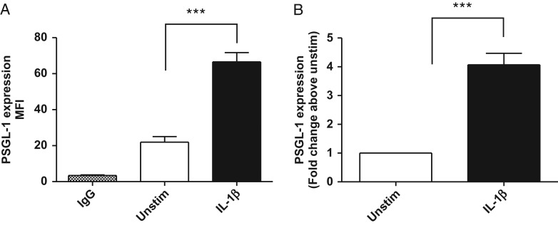
Fig. 3.
IL-1β-induced PSGL-1 cell surface levels on human monocytes. Human monocytes were isolated from healthy donors (n = 17) and incubated in the presence (black bar) and absence (white bar) of 10 ng/mL recombinant human IL-1β for 72 h. Cells were harvested and co-stained with PE CD162, FITC CD14 and isotype matched controls. (A) Represents mean fluorescent intensity (MFI) values in unstimulated and IL-1β-treated (10 ng/mL) cells and (B) fold change in PSGL-1 levels upon stimulation with IL-1β, relative to unstimulated (media alone) control values. IL-1β significantly induced a 4.0 ± 0.4 fold increase in PSGL-1 cell surface levels (***P < 0.0001, students paired t test, n = 21). Data represent mean ± SEM from 21 independent experiments using monocytes isolated from 17 human donors (10M/7F).

