Abstract
Background Scapholunate (SL) interosseus ligament injuries detected at an early stage could allow the surgeon to prevent progression through the spectrum of injury that leads to instability, and eventually osteoarthritis. We contend that early instability following injury can be detected by visualizing the relative motions and distances between the involved carpal bones (scaphoid and lunate) during wrist movement in vivo. The purpose of this study is to demonstrate the utility of dynamic CT (i.e., 4DCT) in diagnosing SL interosseus ligament injuries in two patients with clinical suspicion of SL interosseus ligament injury during flexion-extension (FE), radial-ulnar (RU) deviation, and dart thrower's (DT) motions.
Case Description 4DCT images obtained from two individual cases were analyzed to assess the proximity between the scaphoid and lunate during wrist motion using standard image processing techniques. Proximity maps representing the distances between the scaphoid and lunate bones during each phase of wrist motion were determined. These maps provide insight into the severity of diastasis (large separation) and location of diastasis at the SL joint. The patients' proximity maps indicated dorsal diastasis and subtle uniform diastasis.
Literature Review Complex musculoskeletal abnormalities, such as wrist joint instabilities, elude diagnosis during 2D fluoroscopy due to the 3D geometry of the anatomy and the inherent 3D nature of the bony kinematics. Even the most recent advances with MR arthrography lack good correlation with wrist arthroscopy. Wrist arthroscopy remains the gold standard for diagnosis to assess for intercarpal laxity. However, arthroscopy is an invasive procedure subjecting patients to the risk of infection, nerve injury, pain, and stiffness.
Clinical Relevance 4DCT allows noninvasive characterization of where ligament injuries likely occur; this may allow for a more selective surgical treatment directed at the specific location of the tear.
Keywords: scapholunate interosseus ligament, computed tomography, 4DCT
The scapholunate (SL) interosseus ligament is a ligament that provides intrinsic stability to the SL joint. The mechanism of its injury is typically impact loading in wrist dorsiflexion, ulnar deviation, and intercarpal supination. The spectrum of injury that ensues encompasses four main stages: predynamic, dynamic, static instability, and scapholunate advanced collapse (SLAC). Current treatment focuses on later-stage disease, where restoration of normal kinematics is difficult. Early diagnosis may be critical for less invasive treatment and to prevent persistent instability and degenerative arthritis; however, conventional diagnostic techniques may miss early-stage injury.
In predynamic or dynamic instability, the diagnosis of SL interosseus ligament injury may be difficult to make. Despite being tender over the SL, radiographs may be negative and magnetic resonance imaging (MRI) may lack the sensitivity and specificity for the injury.1 Other invasive methods are less frequently used and include arthrography, MRI with gadolinium contrast, and arthroscopy. Arthroscopy is the gold standard for the diagnosis of predynamic and dynamic SL instability; however, it is an invasive procedure that has related morbidity.
SL interosseus ligament injuries detected at an early stage could allow the surgeon to prevent progression through the spectrum of injury that leads to instability, and eventually osteoarthritis. We contend that early instability following injury can be detected by visualizing the relative motions and distances between the involved carpal bones (scaphoid and lunate) and the radius during wrist movement in vivo. We propose the use of dynamic CT (4DCT) for quantifying bony motion with high temporal and spatial resolution.2 3 The clinical utility of the volume rendered 4DCT movie sequences during wrist joint motion has been recognized by others.4 5 6 7 8 9 10 The purpose of this study is to demonstrate the utility of 4DCT in diagnosing SL interosseus ligament injuries in two patients with clinical suspicion of SL injury during flexion-extension (FE), radial-ulnar (RU) deviation, and dart thrower's (DT) motions.
Case Reports
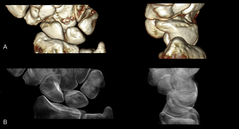
Two patients (53-year-old woman [Case 1], 44-year-old man [Case 2]) were recruited as part of an IRB-approved research protocol. The patients had clinical suspicion of unilateral SL interosseus ligament injury, including tenderness over the SL, negative radiographs, equivocal MRI, and no history of wrist injury. They underwent 4DCT imaging on each wrist separately during FE, RU, and DT motions. Each wrist was scanned for 2 seconds as the patients moved their wrists using an established nongated 4DCT technique.2 3 Patients lay prone with their forearm in neutral rotation. Dynamic scans during each of the motions resulted in 18 image volumes across the 2-second trials. Volume rendering technique (VRT) movies were generated from the 3D VRT images at the 18 time points (Fig. 1). The patients underwent diagnostic wrist arthroscopy and the presence of SL instability was confirmed. One patient (Case 1) had Geissler 3 instability; one (Case 2) had Geissler 411 findings. Both patients then proceeded with definitive SL interosseus ligament reconstruction.
Fig. 1.

Two volumes from an 18-volume 4DCT imaging session rendered using volume rendering technique (VRT) with more (A) and less (B) bone opacity.
The 4DCT technique generated high spatial (0.234 mm in-plane, 0.4 mm slice thickness) and high temporal (75 ms) resolution images of the moving wrist joint. The images can be viewed using different VRTs (Fig. 1), both statically and during dynamic motion to facilitate viewing wrist movements including individual carpal bone motions.
Two fellowship-trained hand surgeons independently reviewed 4DCT videos of both the injured and uninjured wrists for each motion cycle. Differences in carpal kinematics, such as altered SL diastasis and abnormal pronation of the scaphoid seen in SL injury, between the normal and injured wrists were visually examined and scored using a 5-point ordinal scale (1 = no difference to 5 = obvious difference). Values for the two surgeons' assessments of the two patients can be seen in Table 1; mean values for the two surgeons were 4.75, 4.0, and 3.0 for the FE, RU, and DT movement, respectively.
Table 1. Image interpretation of 4DCT images by two hand surgeons.
| Case 1 | Case 2 | Mean | |
|---|---|---|---|
| Flexion-extension Reader 1 Reader 2 |
4 5 |
5 5 |
4.75 |
| Radial-ulnar deviation Reader 1 Reader 2 |
2 4 |
5 5 |
4 |
| Dart thrower's Reader 1 Reader 2 |
2 1 |
4 5 |
3 |
Note: Data show that flexion-extension and radial-ulnar deviation may better delineate SL injury compared with dart thrower's motion.
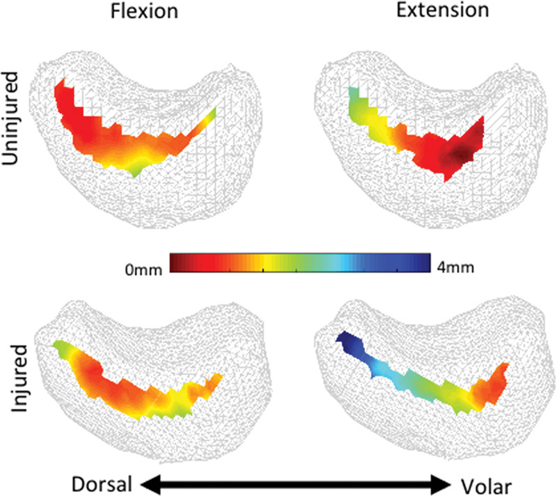
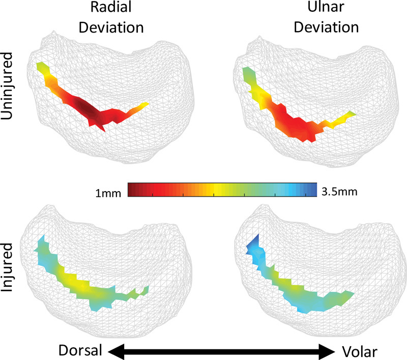
4DCT images obtained from the individual cases were further analyzed to assess the proximity between the scaphoid and lunate during the wrist motions2 3 using standard image processing techniques. Proximity maps are color representations of the distances calculated between the two bones. These maps provide insight into the magnitude and location of diastasis at the SL joint. Patients' proximity maps are shown, indicating dorsal diastasis (Fig. 2: Case 1) and subtle uniform diastasis (Fig. 3: Case 2).
Fig. 2.

(Case 1) Sagittal view of the articular surface of the lunate. Color corresponds to interosseous distances at flexion-extension extrema during dynamic CT imaging. The injured side (bottom row) exhibits considerable dorsal diastasis, indicating dorsal ligament disruption. Note: patient right-side lunate mirrored for comparison purposes.
Fig. 3.

(Case 2) Sagittal view of the articular surface of the lunate. Color corresponds to interosseous distances at radial-ulnar deviation extrema during dynamic CT imaging. The injured side (bottom row) exhibits uniform, though moderate diastasis. Note: patient right-side lunate mirrored for comparison purposes.
Discussion
The most common intrinsic ligament injury in the wrist involves the SL. If left untreated, this can lead to a predictable pattern of wrist instability, pain, and arthritis.12 Though the true incidence of SL injury has yet to be determined, it has been estimated to occur in up to 50% of distal radius fractures and 30% of scaphoid fractures.13 14 15 16 A spectrum of injury exists from partial disruption to a complete tear with altered carpal kinematics. The former can be easily missed with plain radiographs and MRI lacks specificity for the SL, presumably because both modalities are static and cannot detect alterations in carpal motion.17 Stress radiography can be used to detect SL injury but may lack sensitivity and specificity to detect injury.18 19 Fluoroscopy is useful for imaging the dynamic movement of the carpus when the movement occurs primarily about a single axis and when evaluating gross instabilities or abnormalities; however, because the images obtained with fluoroscopy are 2D projections of 3D objects, subtle bony movements in three dimensions are impossible to detect. In addition, joints with a complicated, multiarticulated structure suffer from superposition of the bony anatomy in the 2D projection. For these reasons, complex musculoskeletal abnormalities, such as wrist joint instabilities, elude diagnosis during clinical 2D fluoroscopy due to the 3D geometry of the anatomy and the inherent 3D nature of the bony kinematics. Even the most recent advances with MR arthrography lack good correlation with wrist arthroscopy.20 Wrist arthroscopy remains the gold standard for diagnosis to assess for intercarpal laxity. However, arthroscopy is an invasive procedure subjecting patients to the risk of infection, nerve injury, pain, and stiffness. An arthroscopic scoring system exists for grading SL instability but is prone to inter- and intraobserver error and may not adequately diagnose pathologic instability.11 21
Once the diagnosis has been determined, a myriad of surgical treatments exists for SL instability ranging from closed reduction and percutaneous pinning to formal ligament reconstruction. Results from studies reporting treatment of partial or acute injuries seem to be more favorable than those that are more chronic in nature or those requiring formal ligament reconstructions.22 23 24 25 Given this, early detection of SL injury will allow physicians to potentially intervene earlier.
The 4DCT technique generated high spatial and temporal resolution images of the moving wrist joint. The 4DCT VRT movie sequences allowed visual assessment of the differences in the carpal bone motion and joint spacing between the normal and injured wrists. Differences were observed by the surgeons in the study and the differences between the injured and uninjured wrists were more readily observed during RU and FE than during DT motion for the two patients. This discrepancy may be due to the lack of requisite scaphoid and lunate movement during the DT movement, thus providing less visual information to guide the surgeons' decision making. The abnormal motion between the scaphoid and lunate can be accurately measured using 4DCT and may allow earlier diagnosis of SL injury.
Finally, 4DCT may allow for a more selective surgical treatment directed at the specific location of the tear.26 27 4DCT allowed noninvasive characterization of where the ligament injury likely occurred. The injuries were validated intraoperatively demonstrating the ability of 4DCT to accurately diagnose SL injury, which were not present on standard radiographs or MRI.
Limitations of the study are primarily related to the small numbers of patients studied. This data, however, will serve as a pilot for a larger study in which we intend to quantify SL kinematics, precise injury mechanisms, and the effect of surgical reconstructions using 4DCT.
Acknowledgments
The project described was supported by award number R21 AR 057902 from the National Institute of Arthritis and Musculoskeletal and Skin Diseases, award number T32 HD 07447 from the National Institute of Child Health and Human Development, and the Mayo Clinic Center for Individualized Medicine (CIM). The study sponsors had no role in study design, collection, analysis or interpretation of the data, in the writing of the manuscript, or in the decision to submit this manuscript for publication.
Footnotes
Conflict of Interest Sanjeev Kakar is a consultant for Arthrex, Skeletal Dynamics, and AM Surgical. Cynthia H. McCollough has received funding from Siemens Healthcare.
References
- 1.Schmid M R, Schertler T, Pfirrmann C W. et al. Interosseous ligament tears of the wrist: comparison of multi-detector row CT arthrography and MR imaging. Radiology. 2005;237(3):1008–1013. doi: 10.1148/radiol.2373041450. [DOI] [PubMed] [Google Scholar]
- 2.Leng S, Zhao K, Qu M, An K N, Berger R, McCollough C H. Dynamic CT technique for assessment of wrist joint instabilities. Med Phys. 2011;38 01:S50. doi: 10.1118/1.3577759. [DOI] [PMC free article] [PubMed] [Google Scholar]
- 3.Zhao K, Breighner R, Holmes D III, Leng S, McCollough C, An K N. A technique for quantifying wrist motion using four-dimensional computed tomography: approach and validation. J Biomech Eng. 2015;137(7) doi: 10.1115/1.4030405. [DOI] [PMC free article] [PubMed] [Google Scholar]
- 4.Choi Y S, Lee Y H, Kim S, Cho H W, Song H T, Suh J S. Four-dimensional real-time cine images of wrist joint kinematics using dual source CT with minimal time increment scanning. Yonsei Med J. 2013;54(4):1026–1032. doi: 10.3349/ymj.2013.54.4.1026. [DOI] [PMC free article] [PubMed] [Google Scholar]
- 5.Demehri S, Wadhwa V, Thawait G K. et al. Dynamic evaluation of pisotriquetral instability using 4-dimensional computed tomography. J Comput Assist Tomogr. 2014;38(4):507–512. doi: 10.1097/RCT.0000000000000074. [DOI] [PubMed] [Google Scholar]
- 6.Edirisinghe Y, Troupis J M, Patel M, Smith J, Crossett M. Dynamic motion analysis of dart throwers motion visualized through computerized tomography and calculation of the axis of rotation. J Hand Surg Eur. 2014;39(4):364–372. doi: 10.1177/1753193413508709. [DOI] [PubMed] [Google Scholar]
- 7.Garcia-Elias M, Alomar S X, Monill S J. Dart-throwing motion in patients with scapholunate instability: a dynamic four-dimensional computed tomography study. J Hand Surg Eur. 2014;39(4):346–352. doi: 10.1177/1753193413484630. [DOI] [PubMed] [Google Scholar]
- 8.Goh Y P, Lau K K. Using the 320-multidetector computed tomography scanner for four-dimensional functional assessment of the elbow joint. Am J Orthop. 2012;41(2):E20–E24. [PubMed] [Google Scholar]
- 9.Halpenny D, Courtney K, Torreggiani W C. Dynamic four-dimensional 320 section CT and carpal bone injury—a description of a novel technique to diagnose scapholunate instability. Clin Radiol. 2012;67(2):185–187. doi: 10.1016/j.crad.2011.10.002. [DOI] [PubMed] [Google Scholar]
- 10.Shores J T, Demehri S, Chhabra A. Kinematic “4 dimensional” CT imaging in the assessment of wrist biomechanics before and after surgical repair. Eplasty. 2013;13:e9. [PMC free article] [PubMed] [Google Scholar]
- 11.Geissler W B, Freeland A E, Savoie F H, McIntyre L W, Whipple T L. Intracarpal soft-tissue lesions associated with an intra-articular fracture of the distal end of the radius. J Bone Joint Surg Am. 1996;78(3):357–365. doi: 10.2106/00004623-199603000-00006. [DOI] [PubMed] [Google Scholar]
- 12.Manuel J Moran S L The diagnosis and treatment of scapholunate instability Orthop Clin North Am 2007382261–277., vii [DOI] [PubMed] [Google Scholar]
- 13.Forward D P, Lindau T R, Melsom D S. Intercarpal ligament injuries associated with fractures of the distal part of the radius. J Bone Joint Surg Am. 2007;89(11):2334–2340. doi: 10.2106/JBJS.F.01537. [DOI] [PubMed] [Google Scholar]
- 14.Jørgsholm P, Thomsen N O, Björkman A, Besjakov J, Abrahamsson S O. The incidence of intrinsic and extrinsic ligament injuries in scaphoid waist fractures. J Hand Surg Am. 2010;35(3):368–374. doi: 10.1016/j.jhsa.2009.12.023. [DOI] [PubMed] [Google Scholar]
- 15.Laulan J, Bismuth J P. Intracarpal ligamentous lesions associated with fractures of the distal radius: outcome at one year. A prospective study of 95 cases. Acta Orthop Belg. 1999;65(4):418–423. [PubMed] [Google Scholar]
- 16.Wong T C, Yip T H, Wu W C. Carpal ligament injuries with acute scaphoid fractures—a combined wrist injury. J Hand Surg Br. 2005;30(4):415–418. doi: 10.1016/j.jhsb.2005.02.011. [DOI] [PubMed] [Google Scholar]
- 17.Hobby J L, Tom B D, Bearcroft P W, Dixon A K. Magnetic resonance imaging of the wrist: diagnostic performance statistics. Clin Radiol. 2001;56(1):50–57. doi: 10.1053/crad.2000.0571. [DOI] [PubMed] [Google Scholar]
- 18.Kuo C E, Wolfe S W. Scapholunate instability: current concepts in diagnosis and management. J Hand Surg Am. 2008;33(6):998–1013. doi: 10.1016/j.jhsa.2008.04.027. [DOI] [PubMed] [Google Scholar]
- 19.Linscheid R L, Dobyns J H, Beabout J W, Bryan R S. Traumatic instability of the wrist. Diagnosis, classification, and pathomechanics. J Bone Joint Surg Am. 1972;54(8):1612–1632. [PubMed] [Google Scholar]
- 20.Mak W H, Szabo R M, Myo G K. Assessment of volar radiocarpal ligaments: MR arthrographic and arthroscopic correlation. AJR Am J Roentgenol. 2012;198(2):423–427. doi: 10.2214/AJR.11.6919. [DOI] [PubMed] [Google Scholar]
- 21.Rimington T R, Edwards S G, Lynch T S, Pehlivanova M B. Intercarpal ligamentous laxity in cadaveric wrists. J Bone Joint Surg Br. 2010;92(11):1600–1605. doi: 10.1302/0301-620X.92B11.24798. [DOI] [PubMed] [Google Scholar]
- 22.Garcia-Elias M, Lluch A L, Stanley J K. Three-ligament tenodesis for the treatment of scapholunate dissociation: indications and surgical technique. J Hand Surg Am. 2006;31(1):125–134. doi: 10.1016/j.jhsa.2005.10.011. [DOI] [PubMed] [Google Scholar]
- 23.Hirsh L, Sodha S, Bozentka D, Monaghan B, Steinberg D, Beredjiklian P K. Arthroscopic electrothermal collagen shrinkage for symptomatic laxity of the scapholunate interosseous ligament. J Hand Surg Br. 2005;30(6):643–647. doi: 10.1016/j.jhsb.2005.07.011. [DOI] [PubMed] [Google Scholar]
- 24.Weiss A P, Sachar K, Glowacki K A. Arthroscopic debridement alone for intercarpal ligament tears. J Hand Surg Am. 1997;22(2):344–349. doi: 10.1016/S0363-5023(97)80176-1. [DOI] [PubMed] [Google Scholar]
- 25.Whipple T L. The role of arthroscopy in the treatment of scapholunate instability. Hand Clin. 1995;11(1):37–40. [PubMed] [Google Scholar]
- 26.Del Piñal F. Arthroscopic volar capsuloligamentous repair. J Wrist Surg. 2013;2(2):126–128. doi: 10.1055/s-0033-1343016. [DOI] [PMC free article] [PubMed] [Google Scholar]
- 27.Wahegaonkar A L, Mathoulin C L. Arthroscopic dorsal capsulo-ligamentous repair in the treatment of chronic scapho-lunate ligament tears. J Wrist Surg. 2013;2(2):141–148. doi: 10.1055/s-0033-1341582. [DOI] [PMC free article] [PubMed] [Google Scholar]


