

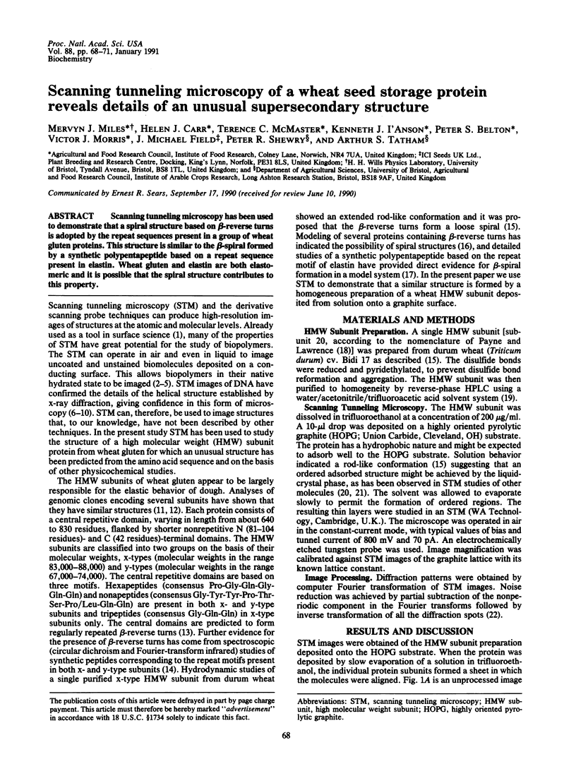
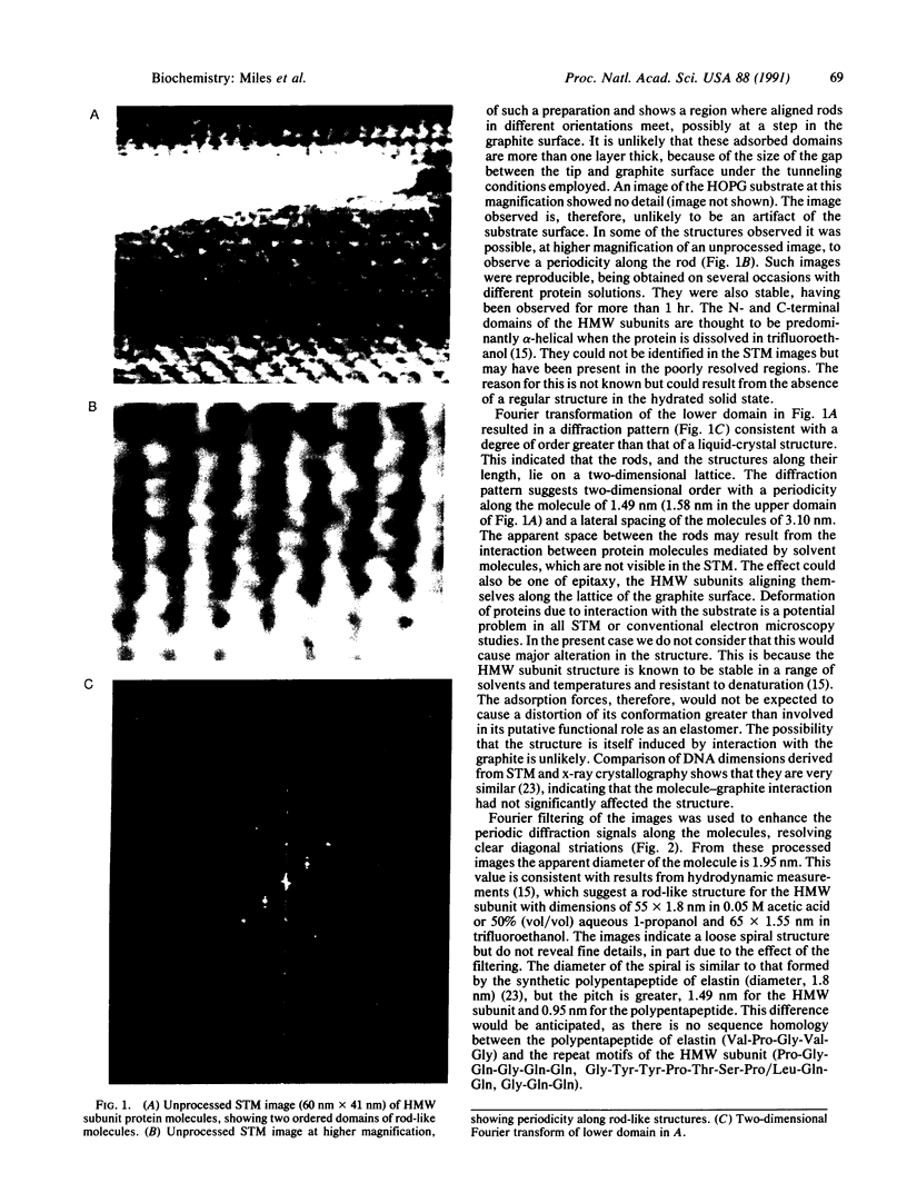
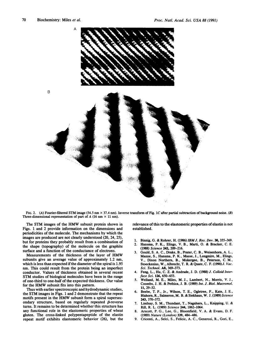
Abstract
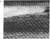
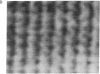
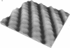
Scanning tunneling microscopy has been used to demonstrate that a spiral structure based on beta-reverse turns is adopted by the repeat sequences present in a group of wheat gluten proteins. This structure is similar to the beta-spiral formed by a synthetic polypentapeptide based on a repeat sequence present in elastin. Wheat gluten and elastin are both elastomeric and it is possible that the spiral structure contributes to this property.
Full text
PDF
Images in this article

Selected References
These references are in PubMed. This may not be the complete list of references from this article.
- Amrein M., Dürr R., Stasiak A., Gross H., Travaglini G. Scanning tunneling microscopy of uncoated recA-DNA complexes. Science. 1989 Mar 31;243(4899):1708–1711. doi: 10.1126/science.2928803. [DOI] [PubMed] [Google Scholar]
- Arscott P. G., Lee G., Bloomfield V. A., Evans D. F. Scanning tunnelling microscopy of Z-DNA. Nature. 1989 Jun 8;339(6224):484–486. doi: 10.1038/339484a0. [DOI] [PubMed] [Google Scholar]
- Beebe T. P., Jr, Wilson T. E., Ogletree D. F., Katz J. E., Balhorn R., Salmeron M. B., Siekhaus W. J. Direct observation of native DNA structures with the scanning tunneling microscope. Science. 1989 Jan 20;243(4889):370–372. doi: 10.1126/science.2911747. [DOI] [PubMed] [Google Scholar]
- Carr H. J., O'Brien E. J., Morris E. P. Structure of tropomyosin-troponin T cocrystals. J Muscle Res Cell Motil. 1988 Oct;9(5):384–392. doi: 10.1007/BF01774065. [DOI] [PubMed] [Google Scholar]
- Driscoll R. J., Youngquist M. G., Baldeschwieler J. D. Atomic-scale imaging of DNA using scanning tunnelling microscopy. Nature. 1990 Jul 19;346(6281):294–296. doi: 10.1038/346294a0. [DOI] [PubMed] [Google Scholar]
- Dunlap D. D., Bustamante C. Images of single-stranded nucleic acids by scanning tunnelling microscopy. Nature. 1989 Nov 9;342(6246):204–206. doi: 10.1038/342204a0. [DOI] [PubMed] [Google Scholar]
- Field J. M., Tatham A. S., Shewry P. R. The structure of a high-Mr subunit of durum-wheat (Triticum durum) gluten. Biochem J. 1987 Oct 1;247(1):215–221. doi: 10.1042/bj2470215. [DOI] [PMC free article] [PubMed] [Google Scholar]
- Hansma P. K., Elings V. B., Marti O., Bracker C. E. Scanning tunneling microscopy and atomic force microscopy: application to biology and technology. Science. 1988 Oct 14;242(4876):209–216. doi: 10.1126/science.3051380. [DOI] [PubMed] [Google Scholar]
- Lindsay S. M., Thundat T., Nagahara L., Knipping U., Rill R. L. Images of the DNA double helix in water. Science. 1989 Jun 2;244(4908):1063–1064. doi: 10.1126/science.2727694. [DOI] [PubMed] [Google Scholar]
- Matsushima N., Creutz C. E., Kretsinger R. H. Polyproline, beta-turn helices. Novel secondary structures proposed for the tandem repeats within rhodopsin, synaptophysin, synexin, gliadin, RNA polymerase II, hordein, and gluten. Proteins. 1990;7(2):125–155. doi: 10.1002/prot.340070204. [DOI] [PubMed] [Google Scholar]
- Urry D. W. Characterization of soluble peptides of elastin by physical techniques. Methods Enzymol. 1982;82(Pt A):673–716. doi: 10.1016/0076-6879(82)82096-x. [DOI] [PubMed] [Google Scholar]
- Welland M. E., Miles M. J., Lambert N., Morris V. J., Coombs J. H., Pethica J. B. Structure of the globular protein vicilin revealed by scanning tunnelling microscopy. Int J Biol Macromol. 1989 Feb;11(1):29–32. doi: 10.1016/0141-8130(89)90036-6. [DOI] [PubMed] [Google Scholar]