Abstract
Volatile sulfur compounds (VSCs) produced by oral anaerobes are the major compounds responsible for oral malodor. Enterococcus faecium WB2000 is recognized as an antiplaque probiotic bacterium. In this study, the effect of E. faecium WB2000 on VSC production by Porphyromonas gingivalis was evaluated, and the mechanism of inhibition of oral malodor was investigated. P. gingivalis ATCC 33277 was cultured in the presence of four lactic acid bacteria, including E. faecium WB2000. Subsequently, P. gingivalis ATCC 33277, W50, W83, and two clinical isolates were cultured in the presence or absence of E. faecium WB2000, and the emission of VSCs from spent culture medium was measured by gas chromatography. The number of P. gingivalis ATCC 33277 in mixed culture with E. faecium WB2000 decreased at 6 h, and the rate of decrease was higher than that in mixed cultures with the other lactic acid bacteria. The numbers of five P. gingivalis strains decreased at similar rates in mixed culture with E. faecium WB2000. The concentration of methyl mercaptan was lower in spent culture medium from P. gingivalis and E. faecium WB2000 cultures compared with that from P. gingivalis alone. Therefore, E. faecium WB2000 may reduce oral malodor by inhibiting the growth of P. gingivalis and neutralizing methyl mercaptan.
1. Introduction
Oral malodor is caused mainly by the metabolism of sulfur amino acids by anaerobic bacteria inhabiting the oral cavity [1]. The main compounds responsible for oral malodor are volatile sulfur compounds (VSCs), such as hydrogen sulfide (H2S), methyl mercaptan (CH3SH), and dimethyl sulfide; these compounds are produced by some periodontopathic bacteria. Indeed, Porphyromonas gingivalis, Treponema denticola, Tannerella forsythia, and Prevotella intermedia generate considerable amounts of H2S and CH3SH [2].
Probiotic bacteria, defined as live microorganisms that benefit the health of the host when administered in adequate amounts (FAO/WHO 2001), are thought to play a role in the maintenance of oral health [3]. Enterococci are facultatively anaerobic, Gram-positive cocci that form a part of the normal flora of the gastrointestinal tract of animals and humans. They are also frequently found in fermented food, such as cheese and meat [4]. Enterococcus faecium and Enterococcus faecalis are the most clinically relevant members of the genus Enterococcus. Traditionally, they are regarded as low-grade pathogens but have emerged as important causes of nosocomial infections [5]. Clinical use of E. faecium and E. faecalis during food fermentation and as probiotics requires a careful safety evaluation [6].
E. faecium strains have been reported to inhibit biofilm formation by cariogenic bacteria in vitro [7, 8]. In addition, a previous double-blind randomized trial in which the subjects cleaned their teeth using a dentifrice containing E. faecium WB2000 or placebo for 4 weeks revealed improvements in salivary flow, salivary buffering capacity, and plaque accumulation [9]. This study aimed to investigate the effect of E. faecium WB2000 on VSC production by P. gingivalis and the mechanism of inhibition of oral malodor by E. faecium WB2000.
2. Materials and Methods
2.1. Bacterial Strains and Culture Conditions
The bacterial strains used in the study are listed in Table 1. Enterococcus faecium WB2000, previously classified as Streptococcus faecalis [10], was provided by Wakamoto Pharmaceutical. The selective medium for P. gingivalis consisted of Brucella agar (Becton Dickinson, Le Pont de Claix, France) supplemented with 5% horse blood, hemin (5 μg/mL), vitamin K (1 μg/mL), and gentamicin (50 μg/mL). Lactic acid bacteria were cultivated on BL agar (Nissui, Tokyo, Japan). Bacterial strains were cultivated at 37°C anaerobically for 40 h and suspended in sterile physiological saline to an optical density at 560 nm (OD560) of 1.0 for P. gingivalis and an OD560 of 0.02 for lactic acid bacteria.
Table 1.
Bacterial strains used in the study.
| Species | Strain |
|---|---|
| Porphyromonas gingivalis | ATCC 33277 |
| ATCC 53978 (W50) | |
| ATCC BAA-308 (W83) | |
| 2-1 (clinical isolate) | |
| 7-1 (clinical isolate) | |
| Enterococcus faecium | WB2000 |
| Lactobacillus salivarius | CIP 103140 |
| Lactobacillus reuteri | JCM 1112 |
| Streptococcus salivarius | JCM 5707 |
2.2. Cocultivation of P. gingivalis and Lactic Acid Bacteria
Bacterial cocultivation was carried out using 100 μL of P. gingivalis suspension, 100 μL of lactic acid bacterial suspension, and 10 mL fresh GAM broth (Nissui, Tokyo, Japan) supplemented with 0.7% glucose, hemin (5 μg/mL), and vitamin K (1 μg/mL) at 37°C anaerobically. Viable bacterial counts in the culture medium were determined at 6, 12, 24, and 48 h on the appropriate agar medium at 37°C anaerobically for 48 h.
2.3. Measurement of Volatile Sulfur Compounds
The VSC concentration in spent medium from P. gingivalis cultured in the presence or absence of E. faecium WB2000 was measured after 24 and 48 h. Aliquots (0.2 mL) of spent culture medium were added to 5 mL conical tubes, which were sealed and incubated at room temperature for 5 min. Then, 0.5 mL of the gas phase was collected and measured by gas chromatography (model GC2014, Shimadzu Works, Kyoto, Japan).
3. Results
3.1. Effect of Lactic Acid Bacteria on the Growth of P. gingivalis ATCC 33277
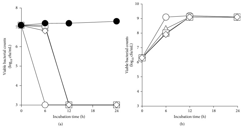
The number of viable P. gingivalis ATCC 33277 decreased to less than the detection limit after 6 h in mixed culture with E. faecium WB2000 (Figure 1(a)). In mixed cultures with the other three lactic acid bacteria (S. salivarius JCM 5707, L. salivarius CIP 103140, and L. reuteri JCM 1112), the number of viable P. gingivalis ATCC 33277 decreased to less than the detection limit at 12 h. In mixed culture with P. gingivalis ATCC 33277, E. faecium WB2000 grew more rapidly than it did in mixed cultures with the other three lactic acid bacteria, and its growth plateaued at 6 h (Figure 1(b)).
Figure 1.
Viable counts of P. gingivalis ATCC 33277 in the presence or absence of each of the four lactic acid bacteria (a) and viable counts of lactic acid bacteria in mixed culture with P. gingivalis ATCC 33277 (b). ●: number of P. gingivalis ATCC 33277 in monoculture, ○: number of P. gingivalis ATCC 33277 or E. faecium WB2000 in mixed cultures, △: number of P. gingivalis ATCC 33277 or L. salivarius CIP 103140 in mixed cultures, □: number of P. gingivalis ATCC 33277 or L. reuteri JCM 1112 in mixed cultures, and ⋄: number of P. gingivalis ATCC 33277 or S. salivarius JCM 5707 in mixed cultures.
3.2. Effect of E. faecium WB2000 on the Growth of Various P. gingivalis Strains
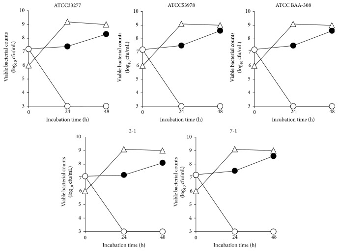
The effect of E. faecium WB2000 on the growth of five P. gingivalis strains (ATCC 33277, ATCC 53978, ATCC BAA-308, 2-1, and 7-1) was evaluated. The numbers of all P. gingivalis strains decreased to less than the detection limit at 24 and 48 h in mixed cultures with E. faecium WB2000 (Figure 2). In contrast, the number of E. faecium WB2000 reached a plateau at 24 h.
Figure 2.
Viable counts of five strains of P. gingivalis (ATCC 33277, ATCC 53978, ATCC BAA-308, 2-1, and 7-1) and E. faecium WB2000 in mixed cultures. ●: number of P. gingivalis in monoculture, ○: number of P. gingivalis in mixed cultures with E. faecium WB2000, and △: number of E. faecium WB2000 in mixed cultures with P. gingivalis.
3.3. Effect of E. faecium WB2000 on VSC Production by P. gingivalis Strains
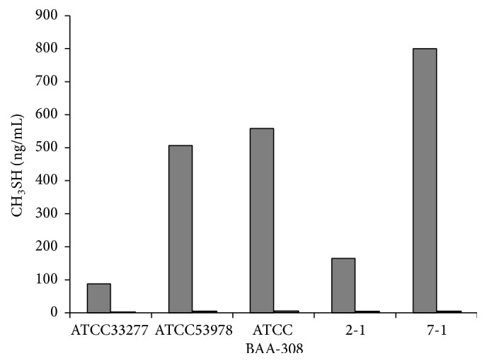
The concentrations of VSCs in spent culture medium were measured by gas chromatography (Table 2). The levels of H2S produced by P. gingivalis strains were lower than those of CH3SH. The concentrations of H2S in mixed culture media were higher than that in medium in which only P. gingivalis was cultured, with the exception of medium from mixed cultures of two P. gingivalis clinical isolates (2-1 and 7-1) and E. faecium WB2000 after 48 h. In contrast, the CH3SH concentration in mixed culture medium was lower than that in medium from culture of P. gingivalis alone, with the exception of medium from mixed cultures of two P. gingivalis strains (W50 and 2-1) and E. faecium WB2000 after 24 h. The levels of CH3SH in spent medium from P. gingivalis cultured for 48 h in the presence or absence of E. faecium WB2000 are shown in Figure 3. Although CH3SH production by P. gingivalis was strain dependent, the CH3SH concentration was markedly lower in spent medium from mixed cultures of all P. gingivalis strains and E. faecium WB2000 than in medium from culture of the latter microorganism only.
Table 2.
The hydrogen sulfide and methyl mercaptan concentrations in spent culture medium (ng/mL).
| Pg strain | Culture medium | Hydrogen sulfide | Methyl mercaptan | ||
|---|---|---|---|---|---|
| 24 h | 48 h | 24 h | 48 h | ||
| ATCC 33277 | Pg | 0.45 | 0.86 | 11.45 | 87.34 |
| Pg + Ef WB2000 | 0.94 | 1.49 | 2.38 | 1.53 | |
| ATCC 53978 (W50) | Pg | 0.87 | 2.20 | 3.40 | 507.27 |
| Pg + Ef WB2000 | 1.44 | 2.91 | 4.36 | 2.92 | |
| ATCC BAA-308 (W83) | Pg | 0.68 | 2.46 | 62.53 | 558.22 |
| Pg + Ef WB2000 | 1.75 | 2.83 | 6.61 | 4.32 | |
| 2-1 | Pg | 0.43 | 2.18 | 1.70 | 164.44 |
| Pg + Ef WB2000 | 1.11 | 2.03 | 3.26 | 2.81 | |
| 7-1 | Pg | 0.86 | 9.24 | 58.18 | 799.95 |
| Pg + Ef WB2000 | 2.06 | 2.44 | 6.03 | 4.05 | |
Pg: Porphyromonas gingivalis; Ef: Enterococcus faecium.
Figure 3.

The levels of CH3SH in spent medium from P. gingivalis cultured for 48 h in the presence or absence of E. faecium WB2000 (ng/mL). Grey bars: single culture; white bars: dual culture with E. faecium WB2000.
4. Discussion
The hypothetical mechanisms of probiotic action in the oral cavity are (1) involvement in binding of oral microorganisms to proteins, (2) effects on plaque formation and its complex ecosystem by competing and interfering with interbacterial attachment, (3) involvement in the metabolism of substrates, and (4) production of compounds that inhibit oral bacteria [11]. E. faecium has been reported to inhibit biofilm formation by cariogenic bacteria [7, 8]. Kumada et al. [8] reported a protein that inhibited biofilm formation by streptococci. In addition, our previous study suggested that E. faecium inhibits the growth of some mutans streptococci [7]. In this study, E. faecium WB2000 inhibited the growth of, as well as reduced CH3SH production by, P. gingivalis. A study on L. salivarius TI 2711 reported that a low pH (≤6.0) and the presence of lactic acid (40–50 mmol/L) induced P. gingivalis death [12]. The pH of E. faecium WB2000 culture medium was 4.4 after 24 h of incubation (data not shown). The growth rate of E. faecium WB2000 was higher than that of the other lactic acid bacteria, suggesting that this organism rapidly inhibited the growth of P. gingivalis.
P. gingivalis strains produced CH3SH and a low level of H2S, as reported previously [13]. E. faecium WB2000 suppressed CH3SH production by P. gingivalis but did not inhibit production of H2S in the current study. Some probiotic bacteria produce VSCs in the presence of cysteine or methionine [14]. The GAM broth used in the current study contains cysteine, and thus E. faecium WB2000 might have produced H2S. Streptococcus thermophilus inhibited the growth and H2S, CH3SH, and CH3SCH3 production of P. gingivalis [13]. To determine whether E. faecium WB2000 specifically inhibits production of CH3SH, different media and culture conditions should be used. Furthermore, future studies are needed to evaluate other P. gingivalis strains producing high levels of H2S.
In addition to E. faecium and S. thermophilus, other organisms have been reported to inhibit oral malodor. Streptococcus salivarius K12 suppressed the growth of Solobacterium moorei, which produces H2S and is involved in halitosis [15, 16]. Lactobacillus salivarius WB21 and L. reuteri have been reported to inhibit oral malodor in clinical trials [17–19]. L. salivarius WB21 reduced the number of Fusobacterium nucleatum and ubiquitous bacteria in saliva [19].
E. faecium WB2000 has been used in traditional Japanese medicine (Strong Wakamoto®) to treat gastrointestinal discomfort, and its effect on dry eye was reported recently [20]. Moreover, the effect of dentifrice containing E. faecium WB2000 on plaque control has been investigated [9]. The findings of this study suggest that E. faecium WB2000 may reduce oral malodor by inhibiting the growth of P. gingivalis and neutralizing CH3SH. The effect of E. faecium WB2000 on oral malodor will be investigated in a future clinical trial involving the use of dentifrice.
Acknowledgments
This study was supported in part by a Grant-in-Aid for Young Scientists (no. 16K20707) and by Grants-in-Aid for Scientific Research (nos. 26463203 and 26463175) from the Ministry of Education, Culture, Sports, Science and Technology, Japan.
Competing Interests
The authors declare that they have no competing interests.
References
- 1.Scully C., Porter S., Greenman J. What to do about halitosis. British Medical Journal. 1994;308(6923):217–218. doi: 10.1136/bmj.308.6923.217. [DOI] [PMC free article] [PubMed] [Google Scholar]
- 2.Persson S., Edlund M. B., Claesson R., Carlsson J. The formation of hydrogen sulfide and methyl mercaptan by oral bacteria. Oral Microbiology and Immunology. 1990;5(4):195–201. doi: 10.1111/j.1399-302x.1990.tb00645.x. [DOI] [PubMed] [Google Scholar]
- 3.Stamatova I., Meurman J. H. Probiotics: health benefits in the mouth. American Journal of Dentistry. 2009;22(6):329–338. [PubMed] [Google Scholar]
- 4.Pesavento G., Calonico C., Ducci B., Magnanini A., Lo Nostro A. Prevalence and antibiotic resistance of Enterococcus spp. isolated from retail cheese, ready-to-eat salads, ham, and raw meat. Food Microbiology. 2014;41:1–7. doi: 10.1016/j.fm.2014.01.008. [DOI] [PubMed] [Google Scholar]
- 5.Guzman Prieto A. M., van Schaik W., Rogers M. R., et al. Global emergence and dissemination of Enterococci as nosocomial pathogens: attack of the clones? Frontiers in Microbiology. 2016;7, article 788 doi: 10.3389/fmicb.2016.00788. [DOI] [PMC free article] [PubMed] [Google Scholar]
- 6.Eaton T. J., Gasson M. J. Molecular screening of Enterococcus virulence determinants and potential for genetic exchange between food and medical isolates. Applied and Environmental Microbiology. 2001;67(4):1628–1635. doi: 10.1128/aem.67.4.1628-1635.2001. [DOI] [PMC free article] [PubMed] [Google Scholar]
- 7.Suzuki N., Yoneda M., Hatano Y., Iwamoto T., Masuo Y., Hirofuji T. Enterococcus faecium WB2000 inhibits biofilm formation by oral cariogenic streptococci. International Journal of Dentistry. 2011;2011:5. doi: 10.1155/2011/834151.834151 [DOI] [PMC free article] [PubMed] [Google Scholar]
- 8.Kumada M., Motegi M., Nakao R., et al. Inhibiting effects of Enterococcus faecium non-biofilm strain on Streptococcus mutans biofilm formation. Journal of Microbiology, Immunology and Infection. 2009;42(3):188–196. [PubMed] [Google Scholar]
- 9.Hatano Y., Suzuki N., Yoneda M., Hirofuji T. Clinical study on the improvement effect of a lactic acid bacterium-contianing dentifrice (Avantbise) on oral hygiene. The Japanese Journal of Conservative Dentistry. 2012;55:219–225. [Google Scholar]
- 10.Schleifer K. H., Kilpper-Bälz R. Transfer of Streptococcus faecalis and Streptococcus faecium to the genus Enterococcus nom. rev. as Enterococcus faecalis comb. nov. and Enterococcus faecium comb. nov. International Journal of Systematic Bacteriology. 1984;34(1):31–34. doi: 10.1099/00207713-34-1-31. [DOI] [Google Scholar]
- 11.Meurman J. H. Probiotics: do they have a role in oral medicine and dentistry? European Journal of Oral Sciences. 2005;113(3):188–196. doi: 10.1111/j.1600-0722.2005.00191.x. [DOI] [PubMed] [Google Scholar]
- 12.Matsuoka T., Nakanishi M., Aiba Y., Koga Y. Mechanism of Porphyromonas gingivalis killing by Lactobacillus salivarius TI 2711. Journal of the Japanese Society of Periodontology. 2004;46(2):118–126. [Google Scholar]
- 13.Lee S.-H., Baek D.-H. Effects of Streptococcus thermophilus on volatile sulfur compounds produced by Porphyromonas gingivalis . Archives of Oral Biology. 2014;59(11):1205–1210. doi: 10.1016/j.archoralbio.2014.07.006. [DOI] [PubMed] [Google Scholar]
- 14.Sreekumar R., Al-Attabi Z., Deeth H. C., Turner M. S. Volatile sulfur compounds produced by probiotic bacteria in the presence of cysteine or methionine. Letters in Applied Microbiology. 2009;48(6):777–782. doi: 10.1111/j.1472-765x.2009.02610.x. [DOI] [PubMed] [Google Scholar]
- 15.Stephen A. S., Naughton D. P., Pizzey R. L., Bradshaw D. J., Burnett G. R. In vitro growth characteristics and volatile sulfur compound production of Solobacterium moorei . Anaerobe. 2014;26:53–57. doi: 10.1016/j.anaerobe.2014.01.007. [DOI] [PubMed] [Google Scholar]
- 16.Masdea L., Kulik E. M., Hauser-Gerspach I., Ramseier A. M., Filippi A., Waltimo T. Antimicrobial activity of Streptococcus salivarius K12 on bacteria involved in oral malodour. Archives of Oral Biology. 2012;57(8):1041–1047. doi: 10.1016/j.archoralbio.2012.02.011. [DOI] [PubMed] [Google Scholar]
- 17.Iwamoto T., Suzuki N., Tanabe K., Takeshita T., Hirofuji T. Effects of probiotic Lactobacillus salivarius WB21 on halitosis and oral health: an open-label pilot trial. Oral Surgery, Oral Medicine, Oral Pathology, Oral Radiology and Endodontology. 2010;110(2):201–208. doi: 10.1016/j.tripleo.2010.03.032. [DOI] [PubMed] [Google Scholar]
- 18.Keller M. K., Bardow A., Jensdottir T., Lykkeaa J., Twetman S. Effect of chewing gums containing the probiotic bacterium Lactobacillus reuteri on oral malodour. Acta Odontologica Scandinavica. 2012;70(3):246–250. doi: 10.3109/00016357.2011.640281. [DOI] [PubMed] [Google Scholar]
- 19.Suzuki N., Yoneda M., Tanabe K., et al. Lactobacillus salivarius WB21-containing tablets for the treatment of oral malodor: a double-blind, randomized, placebo-controlled crossover trial. Oral Surgery, Oral Medicine, Oral Pathology and Oral Radiology. 2014;117(4):462–470. doi: 10.1016/j.oooo.2013.12.400. [DOI] [PubMed] [Google Scholar]
- 20.Kawashima M., Nakamura S., Izuta Y., Inoue S., Tsubota K. Dietary supplementation with a combination of lactoferrin, fish oil, and Enterococcus faecium WB2000 for treating dry eye: a rat model and human clinical study. The Ocular Surface. 2016;14(2):255–263. doi: 10.1016/j.jtos.2015.12.005. [DOI] [PubMed] [Google Scholar]


