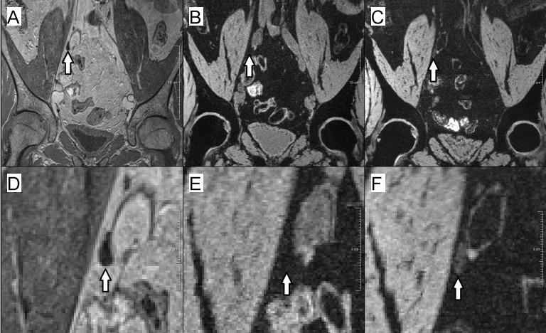
Figure 2. Example of a normal lymph node in ferumoxtran-10 MRL and ferumoxytol MRL.
(A–C) Overviews; (D–F) zoomed-in images. (A & D) USPIO-insensitive 3D T1-weighted (VIBE) sequence. The lymph node is visible as a hypointense structure. (B & E) 3D T2*-weighted (MEDIC) sequence, enhanced with ferumoxtran-10. The normal lymph node is as dark as the fat-suppressed fat and is thus indistinguishable from the background. (C & F) 3D T2*-weighted sequence, enhanced with ferumoxytol. Due to contrast uptake, the normal lymph node is darker than it would have been in non contrast-enhanced MRI, but it is not as dark as the background, and thus may be scored as metastatic.

