Abstract
Traditional Sicilian cheese productions are carried out employing traditional wooden vats, called tina. Many studies have highlighted the beneficial role of wooden dairy equipment by contributing to enriching the milk microflora and improving the acidification processes. The present work was undertaken to evaluate the safety of the wooden vats used to coagulate milk. To this purpose, the different microbial populations hosted onto the internal surfaces of the vats used to produce two different stretched cheeses, namely Caciocavallo Palermitano and Vastedda della valle del Belìce DOP, were investigated for the presence of spoilage and pathogenic microorganisms as well as for bacteria with inhibitory effect in vitro against pathogenic microorganisms. A wide biodiversity of protechnological lactic acid bacteria (LAB), in terms of species, was revealed. Several LAB inhibited the growth of Listeria monocytogenes ATCC 7644. The wooden vats analysed resulted safe for three main findings: absence of the main pathogenic species, presence of high levels of LAB, anti-Listeria activity of many LAB.
Key words: Wooden vat, Biofilm, Sicilian cheese, Inhibitory activity, Food safety
Introduction
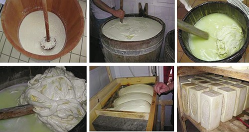
Bovine and ovine Sicilian farms have traditionally produced raw milk cheeses, using different wooden equipment consisting of: tina, a truncated conical flat base 200-400 L container for milk coagulation; rotula, an about 20 cm long stick with a terminal disc used for breaking the curd; piddiaturi, an about 50 L washtub where the stretching is performed; and tavuleri, a rectangular container where Caciocavallo Palermitano assumes its characteristic parallelepiped shape (Figure 1).
Figure 1.

Wooden equipment used for Caciocavallo Palermitano cheese production.
The possibility offered by the 92/46 CEE Directive, implemented by the 54/97 DPR, to derogate the hygiene requirements of traditional cheeses production equipment, induced the Sicilian Region to identify the traditional dairy products by the 4492/98 Decree of the Sicilian Region. To allow the derogation application, the decree briefly describes each cheese production processes and the corresponding wooden equipment, which normally could not be used because of the porous material that does not allow an effective cleaning and sanitation.
The current Regulation 852/2004 of the European Community (European Commission, 2004) on food hygiene allows the use of any type of equipment as long as food business operators are able to demonstrate they are made with materials ensuring the safety of the final product. Sicilian cheese makers have traditionally managed wooden vat hygiene through washing of this container with the hot deprotenised whey resulting from the production of ricotta cheese and/or hot water, sometimes with a careful brushing, leaving the wooden vat full with the whey for about 12 h. These operations tend to empirically regulate, also according to the environmental temperatures, the so-called tina acidity, influencing the microbial composition on the vat inner surface, that is in direct contact with the milk.
Some studies have shown that the wooden vats used for raw milk cheese production allows the development of a biofilm on the inner surface which actively participates in the production process (Lortal et al., 2008; Didienne et al., 2012; Settanni et al., 2012).
In this study, we evaluated the wooden vats used for the production processes of Caciocavallo Palermitano cheese, a bovine raw milk pasta filata cheese with 3 to 18 months ripening, and Vastedda della valle del Belìce DOP, a fresh sheep raw milk pasta filata cheese. The aim of the study was to determine the presence of spoilage and pathogenic microorganisms, to evaluate the species composition of the lactic acid bacteria (LAB) hosted onto the inner surfaces of the wooden vats, and to investigate on their in vitro antibacterial effect.
Materials and Methods
In this study, the wooden vats of 12 dairy factories of western Sicily were microbiologically investigated. Eight factories, all located within the Palermo province, produce Caciocavallo Palermitano (CP) cheese, while the other 4 factories produce Vastedda della valle del Belìce DOP (VB) cheese in the production area of the provinces of Agrigento and Trapani. The analyses were carried out during two consecutive production years. A total of 20 wooden vats (13 used for CP cheese production and made of chestnut wood and 7 used for VB cheese and made of Douglas fir) were sampled.
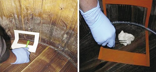
The biofilms were collected for the investigation of total mesophilic count (TMC), lactic acid populations and hygiene indicator bacteria from the inside of the vats by brushing a 100 cm2 area, delimited with a 10×10 cm sterile plastic mask (80 cm2 from the bottom and 20 cm2 from the side surface) with a sterile brush moistened with a transport medium (buffered peptone water, BPW). The same surface, was then cleaned with a sterile gauze (Figure 2). Both brush and gauze were aseptically transferred in BPW, kept refrigerated during transport by a portable fridge. The search of Listeria monocytogenes and Salmonella spp. was performed on a larger surface area, 400 cm2, using a 20×20 cm mask.
Figure 2.

Sampling on the wooden vat surfaces.
All the samples were analysed to determine the following microbiological counts: TMC at 30°C on Plate Count Agar (PCA) - Oxoid (Basingstoke, UK) incubated at 30°C for 72 h (ISO 4833:2003; ISO, 2003b), sulphite reducing anaerobes on Iron Sulphite Agar - Oxoid, incubated at 37°C for 24 h (ISO 15213:2003; ISO, 2003c), coliforms on Violet Red Bile agar (VRBA) - Oxoid, incubated at 37°C for 24 h (ISO 4832:2006; ISO, 2006), enterococci on Rapid Enterococcus Agar (REA) - Biorad (Hercules, CA, USA) incubated at 44°C for 48 h and confirmation of suspected colonies by microscopic examination, catalase testing (Fluka, St. Gallen, Switzerland) and detection on Bile esculina agar (Oxoid), Escherichia coli on Tryptone Bile Glucuronide Agar-Oxoid incubated at 44°C for 24h (ISO 16649-2:2001; ISO, 2001) and coagulase positive staphylococci on Baird Parker (BP) with added RPF supplement-Oxoid, incubated at 37°C for 24 h (ISO 6888-2: 1999 Amend 1:2003; ISO, 2003a).
Listeria monocytogenes investigation was performed by using the enzyme immunoassay ELFA (Enzyme Linked Fluorescent Assay) instrument VIDAS (bioMerieux, Marcy-l’Etoile, France). Salmonella spp was detected using the AFNOR BIO 12/23-05/07 method with a preenrichment in Buffered Peptone Water incubated at 37°C for for 16-20 h followed by subculture in Xylose Lysine Deoxycholate (XLD) Agar (Oxoid) and Brillant Green Agar (Oxoid) at 37°C for 20-24 h. Listeria monocytogenes was detected using the AFNOR BIO 12/11-03/04 with a pre-enrichment in Half Fraser broth (Oxoid) incubated at 30°C for 24-26 h and subsequent culture on Fraser broth (FB) (Oxoid) incubated at 37°C for 24-26 h. An aliquot of FB is used to perform the VIDAS LMO2 test (bioMerieux).
Lactic acid bacteria were counted and isolated by using the ISTISAN 08/36 Reports: mesophilic and thermophilic cocci were plated on M17 agar incubated at 30°C for 72 h and 44°C for 48 h, respectively; mesophilic lactobacilli were isolated by using MRS agar (pH 5.4) incubated anaerobically at 37°C for 72 h.
Lactic acid bacteria isolates were genetically identified by 16S rRNA gene sequencing. The DNA was extracted by the Instagene Matrix (Biorad) according to the manufcturer’s instructions. PCRs were performed as described by Weisburg et al. (1991) using the primers rD1 (5′-AAGGAGGTGATCCAGCC-3′) and fD1 (5′-AGAGTTTGATCCTGGCTCAG-3′) and the AmpliTaq Gold® 360 DNA Polymerase (Life Technologies, Carlsbad, CA, USA). DNA sequences were determinated by using an ABI PRISM 3130 Genetic Analyzer (Applied Biosystems, Carlsbad, CA, USA) and compared using a BLAST search in the GenBank/EMBL/DDBJ database (http://www.ncbi.nlm.nih.gov). The isolates were considered to represent a given species when a 97% or higher similarity was detected.
For the in vitro antibacterial activity assay, LAB isolates were tested using the spot on the lawn method, which shows the inhibitory activity against target microorganisms, by the detection of an inhibition halo around the colony strain tested (Patriarca et al., 2012). The following bacteria sensitive to the inhibitory activity of LAB were used as indicators: L. monocytogenes ATCC 7644, E. coli ATCC 25922, Salmonella enteritidis ATCC 13076 and Staphylococcus aureus ATCC 25923.
The strains cryopreserved at -80°C were revitalised in 5 mL of MRS broth at 37°C for 72h in the presence of CO2. Petri plates containing Tryptone Soya Agar (TSA) plus 0.5% of yeast extract were spotted with 2 µL of each culture broth and incubated anaerobically overnight at 30°C. Brain Heart Infusion (BHI; Difco) containing 1% agar was tempered to 45°C and seeded with 105-106 CFU/mL of each pathogen. The spotted plates were overlaid with 8 mL of the seeded BHI agar and then incubated at 30°C in anaerobiosis for 24 h. Inhibition was detected by a zone of clearing (>3 mm) around the producer colony (Patriarca et al., 2012).
Results
The data obtained, expressed as mean values, are reported in Tables 1 and 2. Total mesophilic count and mesophilic LAB cocci were between 3.62 and 6.95 Log CFU/cm2, while thermophilic LAB cocci were slightly higher (3.57 - 7.43 Log CFU/cm2), especially in CP vats. Total coliforms were detected in 16 wooden vats: 5 of these showed concentrations <1 Log CFU/cm2, 10 between 1.04 and 3.18 Log CFU/cm2 and only one 6.04 Log CFU/cm2. Five wooden vats showed E. coli concentrations between 0.30 and 2.46 Log CFU/cm2; only in the farm where the coliforms were highly present, E. coli resulted 3.18 Log CFU/cm2. In 11 wooden vats, enterococci ranged from 0.70 Log and 3.40 Log CFU/cm2. In all the samples coagulase-positive staphylococci, L. monocytogenes and Salmonella spp. were absent.
Table 1.
Microbial groups detected in the vats used for Caciocavallo Palermitano cheese production (Log CFU/cm2).
| Mean | Min | Max | SD | |
|---|---|---|---|---|
| TMC | 5.26 | 3.62 | 6.95 | 0.91 |
| Mesophilic LAB cocci | 5.38 | 4.00 | 6.48 | 0.63 |
| Thermophilic LAB cocci | 5.81 | 5.15 | 7.43 | 0.71 |
| LAB rods | 4.52 | 3.48 | 5.96 | 0.82 |
| Enterococci | 1.95 | 0.70 | 3.15 | 0.75 |
| Total coliforms | 1.77 | 0.30 | 3.18 | 1.07 |
| E. coli | 1.66 | 0.30 | 2.46 | 0.94 |
TMC, total mesophilic count; LAB, lactic acid bacteria.
Table 2.
Microbial groups detected in the vats used for Vastedda della valle del Belice cheese production (Log CFU/cm2).
| Mean | Min | Max | SD | |
|---|---|---|---|---|
| TMC | 5.20 | 3.60 | 6.85 | 1.17 |
| Mesophilic LAB cocci | 5.02 | 4.15 | 6.60 | 0.91 |
| Thermophilic LAB cocci | 4.57 | 3.57 | 6.64 | 1.06 |
| LAB rods | 3.77 | 2.60 | 6.30 | 1.39 |
| Enterococci | 2.40 | 1.65 | 3.40 | 0.80 |
| Total coliforms | 2.30 | 0.60 | 6.04 | 2.04 |
| E. coli | 1.75 | 0.30 | 3.18 | 1.44 |
TMC, total mesophilic count; LAB, lactic acid bacteria.
The LAB isolated from the CP vats showed a predominance of Lactobacillus casei, Enterococcus faecium, Lactobacillus rhamnosus, Streptococcus thermophilus and Pediococcus acidilactici (Table 3). The LAB isolated from the VB vats revealed the predominance of two species: Enterococcus faecium and Lactobacillus casei. The percentage of lactic acid strains able to inhibit the growth of L. monocytogenes ATCC 7644 was 66.7% for the LAB isolated from the VB vats and of 55.7% for those isolated from the CP vats. The LAB did not show inhibition against the other bacteriocin-sensitive bacteria tested.
Table 3.
Lactic acid bacteria strains isolated from the wooden vats and strains with inhibitory activity vs L. monocytogenes ATCC 7644.
| LAB | CP vats | VB vats | ||
|---|---|---|---|---|
| Isolates (n) | Inhibitory substance producer (n) | Isolates (n) | Inhibitory substance producer (n) | |
| Enterococcus faecalis | 5 | 3 | 1 | 0 |
| Enterococcus faecium | 12 | 10 | 15 | 14 |
| Enterococcus hirae | 0 | 0 | 2 | 1 |
| Enterococcus spp. | 3 | 0 | 1 | 1 |
| Lactobacillus brevis | 1 | 1 | 4 | 3 |
| Lactobacillus casei | 13 | 2 | 10 | 2 |
| Lactobacillus delbrueckii | 1 | 0 | 2 | 2 |
| Lactobacillus fermentum | 0 | 0 | 2 | 2 |
| Lactobacillus rhamnosus | 11 | 9 | 1 | 0 |
| Lactococcus lactis | 2 | 2 | 1 | 0 |
| Lactococcus lactis subsp. cremoris | 4 | 3 | 0 | 0 |
| Leuconostoc lactis | 1 | 1 | 0 | 0 |
| Leuconostoc mesenteroides | 1 | 0 | 0 | 0 |
| Leuconostoc pseudomesenteroides | 1 | 1 | 0 | 0 |
| Pediococcus acidilactici | 7 | 3 | 4 | 4 |
| Pediococcus lolii | 5 | 2 | 0 | 0 |
| Pediococcus pentosaceus | 2 | 1 | 0 | 0 |
| Streptococcus macedonicus | 2 | 1 | 4 | 4 |
| Streptococcus thermophilus | 8 | 5 | 4 | 1 |
| Total | 79 | 44 (55.7%) | 51 | 34 (66.7%) |
CP, Caciocavallo Palermitano; VB, Vastedda della valle del Belìce DOP; LAB, lactic acid bacteria.
Discussion
The data obtained from this study confirmed the presence of a certain species diversity on the inner surface of the wooden vats used for the typical Sicilian cheese productions (Licitra et al., 2007; Lortal et al., 2008; Settanni et al., 2012). The LAB isolated from the CP vats showed a predominance of Lactobacillus casei, Enterococcus faecium, Lactobacillus rhamnosus, Streptococcus thermophilus and Pediococcus acidilactici, while in those used for the production of a similar cheese, the Ragusano, a predominance of Streptococcus thermophilus and the absence of Enterococcus faecium was detected. Data on indicator microorganisms (coliforms and E. coli) showed the efficacy of the sanitation procedures applied during cheese production. Only in one case, the presence of high total coliforms loads (6.04 Log CFU/cm2) and E. coli (3.18 Log CFU/cm2) indicated the need of revising this process.
Probably the biofilm composition might be affected by the characteristics of the milk used, the specific production processes of the two cheeses studied and the wooden vat sanitation procedures. The presence of more than 50% of the LAB isolates with inhibitory activity vs. L. monocytogenes ATCC 7644 constitutes an additional positive feature, although it must be evaluated in relation to the type of the cheese produced, because traditional Sicilian raw milk cheeses are not commonly subjected to L. monocytogenes contamination, as observed in previous studies (Scatassa et al., 2009).
Conclusions
The correct maintenance of wooden vats, as part of good manufacturing practices of the Caciocavallo Palermitano and the Vastedda della valle del Belìce DOP cheese production, promotes the selection of a microbial flora able to play an active role in the achievement of the food safety objectives through the biocompetitive activity of LAB and the inhibitory activity against pathogenic bacteria, particularly L. monocytogenes.
Acknowledgments
The authors would like to thank A. Carrozzo and B. Ducato for their precious collaboration.
Funding Statement
Funding: this work was supported by the Italian Ministry of Health Research Project RC 06-2011 Prodotti lattiero caseari tradizionali Siciliani: tecniche di produzione e rischio microbiologico.
References
- Didienne R, Defargues C, Callon C, Meylheuc T, Hulin S, Montel MC, 2012. Characteristics of microbial biofilm on wooden vats (‘gerles’) in PDO Salers cheese. Int J Food Microbiol 156:91-101. [DOI] [PubMed] [Google Scholar]
- European Commission, 2004. Regulation of the European Parliament and of the council of 29 April 2004 on the hygiene of foodstuffs, 852/2004/EC. In: Official Journal, L 139/1, 30/04/2004. [Google Scholar]
- ISO, 2001. Microbiology of food and animal feeding stuffs. Horizontal method for the enumeration of beta-glucuronidase-positive Escherichia coli. Part 2: Colony-count technique at 44 degrees C using 5-bromo-4-chloro-3-indolyl beta-D-glucuronide. ISO Norm 16649-2:2001. International Standardization Organization ed., Geneva, Switzerland. [Google Scholar]
- ISO, 2003a. Microbiology of food and animal feeding stuffs. Horizontal method for the enumeration of coagulase-positive staphylococci (Staphylococcus aureus and other species). Part 2: Technique using rabbit plasma fibrinogen agar medium AMENDMENT 1: Inclusion of precision data [Google Scholar]
- ISO, 2003b. Microbiology of food and animal feeding stuffs. Horizontal method for the enumeration of microorganisms. Colony-count technique at 30 degrees C. ISO Norm 4833:2003. International Standardization Organization ed., Geneva, Switzerland. [Google Scholar]
- ISO, 2003c. Microbiology of food and animal feeding stuffs. Horizontal method for the enumeration of sulfite-reducing bacteria growing under anaerobic conditions. ISO Norm 15213:2003. International Standardization Organization ed., Geneva, Switzerland. [Google Scholar]
- ISO, 2006. Microbiology of food and animal feeding stuffs. Horizontal method for the enumeration of coliforms. Colony-count technique. ISO Norm 4832:2006. International Standardization Organization ed., Geneva, Switzerland. [Google Scholar]
- Licitra G, Ogier JC, Parayre S, Pediliggieri C, Carnemolla TM, Falentin H, Madec MN, Carpino S, Lortal S, 2007. Variability of bacteria biofilms of the “Tina” wood vats used in the Ragusano cheese-making process. Appl Environ Microb 73:6980-87. [DOI] [PMC free article] [PubMed] [Google Scholar]
- Lortal S, Di Blasi A, Madec MN, Pediliggieri C, Tuminello L, Tanguy G, Fauguant J, Lecuona Y, Campo P, Carpino S, Licitra G, 2009. Tina wooden vat biofilm: a safe and highly efficient lactic acid bacteria delivering system in PDO Ragusano cheese making. Int J Food Microbiol 132:1-8. [DOI] [PubMed] [Google Scholar]
- Patriarca V, Di Bartolo I, Tozzoli R, Agrimi U, 2012. Valutazione dell’attività antibatterica delle batteriocine nei confronti di patogeni alimentari. Fiore A, Vilmercati A, Anniballi F, De Medici D, Argomenti di Sanità Pubblica Veterinaria e Sicurezza Alimentare. Seminari dipartimentali 2011. Istituto Superiore di Sanità, Rome, Italy, pp 28-35. [Google Scholar]
- Scatassa ML, Di Noto AM, Cardamone C, Sciortino S, Todaro M, Caracappa S, 2009. Vastedda della valle del Belìce cheese: experimental contamination with Salmonella and Listeria spp. Proceedings of XVII FeMESPRum International Congress, pp 298-99. [Google Scholar]
- Settanni L, Di Grigoli A, Tornambè G, Bellina V, Francesca N, Moschetti G, Bonanno A, 2012. Persistence of wild Streptococcus thermophilus strains on wooden vat and during the manufacture of a traditional Caciocavallo type cheese. Int J Food Microbiol 155:73-81. [DOI] [PubMed] [Google Scholar]
- Weisburg W, Barns SM, Pelletier DA, Lane DJ, 1991. 16S ribosomal DNA amplification for phylogenetic study. J Bacteriol 173:697-703. [DOI] [PMC free article] [PubMed] [Google Scholar]


