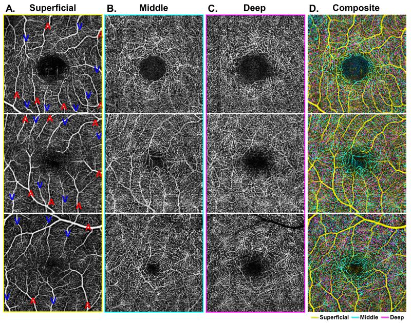
Figure 3. Characterization of the foveal avascular zone (FAZ) at the level of the three capillary plexuses.
A-C: Demonstrate the three capillary plexuses of three different eyes from healthy control patients (each row). Arterioles and veins are labeled with a red A or blue V, respectively, in the superficial capillary plexus (SCP) (Column A). In the middle capillary plexus (MCP) (Column B), the border of the foveal avascular zone (FAZ) is best observed as a well-demarcated and clearly circumscribed area, with a border that is composed of branches from both arterioles and venules in the MCP. This regular FAZ border is not observed in the deep capillary plexus (DCP) (Column C). The composite image (Column D) shows an overlay of all three capillary plexus layers, with the SCP in yellow, MCP in cyan, and DCP in magenta. In the composite image, there is significant overlap between the capillary networks, but a distinct area of cyan is observed in the FAZ region, while the magenta DCP does not approach the FAZ region to the same extent. Consequently, the FAZ is appreciably larger in the DCP compared to the MCP.

