
Figure 3. QKI5 suppresses GC cell growth and metastasis in vivo.
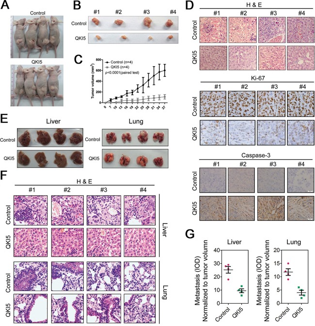
A. Xenograft model in nude mice. QKI5-and control-transfected MGC-803 cells were injected s.c. into the posterior flank of nude mice. The graph is representative of tumors in mice at seven weeks after inoculation. B. The graph is representative of excised tumors from killed mice. C. Tumor volume was calculated and all data are shown as the mean ± SD. D. Pathology analysis of tumor sections from xenograft mice. H&E staining and labeling with anti-Ki-67 and anti-caspase3 was performed. Bars: 20 μm. E. Nude mice were injected with MGC-803 cells infected with QKI5 or control lentivirus through the lateral tail vein. Five weeks after the injection, the mice were sacrificed, and the lungs and livers were dissected for microscopic histology. F. Histological analysis of sections from livers (the upper panel) and lungs (the lower panel) of mice as described in E. G. Quantification of visible liver (the left panel) and lung (the right panel) metastases counted using a microscope.