Abstract
Chondromyxoid fibroma (CMF) is a rare benign cartilaginous tumor with a predilection for the bones of lower extremities and about one fourth of the tumors involve the foot. Radiologically, an eccentric lytic lesion with well-defined margins is seen in the metaphysis of the bone. We hereby, report an 18 yr old young male who presented to Orthopedic Outpatient Department, JN Medical College, Aligarh Muslim University, India diagnosed with giant cell tumor of the third metatarsal bone of right foot on radiography but on fine needle aspiration cytology (FNAC) the diagnosis of CMF was made. Preoperative diagnosis of this benign condition helped in doing minimum surgical intervention in the form of curettage along with bone grafting. Histopathology further confirmed the diagnosis of CMF. The case is being discussed to highlight the importance of FNAC to diagnose these uncommon benign bone lesions.
Key Words: Chondromyxoid fibroma (CMF), Cytodiagnosis, Metatarsal bone
Introduction
Fine-needle aspiration cytology (FNAC) is a reliable and cost-effective means of diagnosing lesions of the skeletal system in conjunction with clinical and radiological findings (1). There are several studies on FNAC of bone tumors, but there are only very few case reports regarding cytological diagnosis of Chondromyxoid fibroma (CMF) (2, 3).
CMF is the rarest benign cartilaginous tumor, constituting around 0.5% of all bone tumors characterized by incomplete cartilage differentiation (4). It usually presents in second and third decade of life. Jaffe and Lichtenstein described this tumor for the first time in 1948 (5). CMF is usually an intra-medullary eccentric lesion located in the metaphyseal region of the distal femur and proximal tibia but sometimes it can also be seen at unusual sites like flat bones of the skull, pelvis, ribs, hands and feet where it can cause diagnostic difficulties (6).
Owing to the paucity of published literature about the cytodiagnosis of chondromyxoid fibroma which is one of the rarest primary bone tumor, this case is being reported here.
Case Report
An 18 yr old young male of average build presented to Orthopedic Outpatient Department, JN Medical College, Aligarh Muslim University, India with complaints of pain, tenderness and swelling over the dorsum of right foot for the past 1 year. There was no significant past medical history or any history of trauma.Written informed consent was obtained from the patient for publication of this case report and accompanying images.
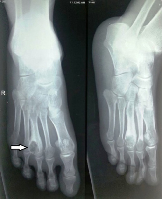
Radiograph of the right foot was suggestive of a lytic lesion with well-defined margins in the region of head of the third metatarsal (Fig. 1). A presumptive diagnosis of giant cell tumor of the bone was made. FNAC was done and smears were prepared and stained with hematoxylin and eosin (H & E).
Fig. 1.
Radiograph of the right foot suggestive of a lytic lesion in the region of head of third metatarsal
Cytological examination:
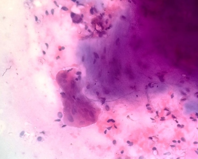
On cytological examination of the stained smears there was moderate cellularity with round to ovoid cells having moderate amount of cytoplasm, benign appearing nuclei and bland chromatin. Many scattered dense, fibrillary chondromyxoid tissue fragments were appreciated but there was no mature cartilaginous tissue or foci of calcification. Stellate cells and spindle- shaped fibrocytoid cells along with fewosteoclastic giant cells were also seen on a myxoidstroma. The overall cytological picture favored the diagnosis of CMF (Fig. 2).
Fig. 2.
FNAC smear showing chondromyxoid tissue fragments, fibrocytes and osteoclastic giant cells (hematoxylin & eosin 400X
Preoperative diagnosis of this benign condition helped in doing minimum surgical intervention in the form of curettage and grafting.
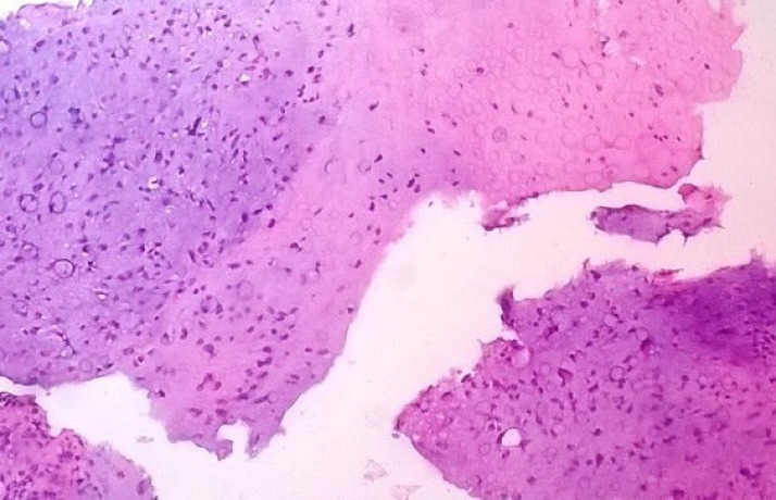
Histopathological examination of the resected specimen showed hypocellular lobules of myxochondroid appearance separated by more cellular tissue composed of fibroblast like spindle cells having ovoid nuclei and moderate cytoplasm along with osteoclastic giant cells in places (Fig.3). There was mild nuclear atypia. No mitoses, necrosis or calcifications were seen. The findings were consistent with the diagnosis of CMF.
Fig. 3 .
Section showing hypocellular lobules of myxochondroid appearance separated by cellular tissue composed of fibroblast like spindle cells (hematoxylin & eosin 400X
The post-operative period was uneventful. Currently, the patient is in follow up and doing well 6 months after the surgery. The differential diagnosis includes giant cell tumor, aneurysmal bone cyst, chondroma, chondroblastoma and nonossifying fibroma. It may be confused at times with chondrosarcoma on histology. The zonal and lobular pattern of CMF with nodules of chondroid tissue separated by fibromyxoid areas along with osteoclastic giant cells differentiates it from other probable diagnoses.
Discussion
CMF is the rarest benign chondroid neoplasm and represents less than 0.5% of all bone tumors. It usually presents in patients during their second and third decade and has a slightly higher incidence in males (7). The tumor has a predilection for the metaphyseal region of the distal femur and proximal tibia with some unusual locations like metatarsals as in our case. Patients usually complain of a slow growing painful swelling. Pathological fracture can alsooccur in about 5% of cases of chondromyxoid fibroma (8). Radio- logically CMF shows an expansile radiolucent lesion with well-defined sclerotic margins (9).
Feit et al. reported a case of chondromyxoid fibroma of fourth metatarsal and Goldenharet al. reported a similar case in first metatarsal and agnosed on histopathological examination after resection of the tumor (10, 11). Pre-operative diagnosis is very important so as to avoid unnecessary radical surgical procedures which increase the morbidity of the patient. The cytological features of CMF are distinctive enough to make a pre-operative diagnosis on FNAC after clinic-radiological correlation (12, 13).
Our patient was a young male who presented with a painful swelling in his right foot diagnosed as giant cell tumor of the third metatarsal of the right foot on radiological examination. However, FNAC was done which revealed scattered dense, fibrillary chondromyxoid tissue fragments, stellate cells and spindle- shaped fibrocytoid cells along with few osteoclastic giant cells in a myxoid stroma which favored the diagnosis of CMF.
On cytological study of CMF, there are varying combinations of chondroid, myxoid and fibroid elements (2). The chondroid and myxoid elements form the irregular fragments of matrix in the background. The cellular elements comprise a variable mixture of spindle cells, stellate cells and chondroblasts with nuclear pleomorphism but with uniformly bland, smudgy chromatin and no mitotic figures (6, 12). Cytologically it may be confused with benign lesions like chondroblastoma or at times even with myxoid chondrosarcoma but cellular pleomorphism, nuclear atypia and mitoses seen in the latter help to distinguish the two. Chondroblastoma shows chondroblasts, giant cells and chondroid matrix (4, 7).
The histopathological picture of CMF is characteristic with lobular arrangement of myxoid and chondroid tissue surrounded by zones of hypercellular fibrous tissue. No well-formed cartilage or bone is seen (2). Some cases of malignant transformation of CMF have been reported (8). In our patient histopathological examination showed hypocellular lobules of myxochondroid tissue separated by fibroblast like spindle cells along with osteoclastic giant cells. There was mild nuclear atypia and no mitoses, necrosis or calcifications were seen. These findings were consistent with the diagnosis of CMF.
The ideal treatment for CMF is curettage combined with bone grafting, and hence a correct pre-operative diagnosis is very helpful in selecting this line of management (13).
CMF is the rarest benign tumor of cartilaginous origin which sometimes poses a great di- agnostic challenge especially when it is located at rare sites. Preoperative diagnosis is also very important so as to avoid any radical surgical procedures owing to the benign nature of the condition. Radiological investigations may sometimes be misleading, as in our case therefore cytology plays an important role here.
Acknowledgements
The authors declare that there is no conflict of interests.
References
- 1.Handa U, Bal A, Mohan H, Bhardwaj S. Fine needle aspiration cytology in the diagnosis of bone lesions. Cytopathology . 2005;16:59–64. doi: 10.1111/j.1365-2303.2004.00200.x. [DOI] [PubMed] [Google Scholar]
- 2.Walke VA, Nayak SP, Munshi MM, Bobhate SK. Cytodiagnosis of chondromyxoid fibroma. J Cytol. 2010;27:96–8. doi: 10.4103/0970-9371.71873. [DOI] [PMC free article] [PubMed] [Google Scholar]
- 3.Layfield LJ, Ferreiro JA. Fine needle aspiration cytology of chondromyxoid fibroma: a case report. DiagnCytopathol . 1988;4:148–51. doi: 10.1002/dc.2840040215. [DOI] [PubMed] [Google Scholar]
- 4.Nielsen GP, Layfield LJ, Rosenberg AE. 4th ed. Philadelphia: Churchill Livingstone; 2006. Neoplastic and tumor like lesion of bone. In: Silverberg SG, editor. Silverbergs principles and practice of surgical pathology and cytopathology; pp. 701–83. [Google Scholar]
- 5.Jaffe HL, Linchtenstein L. Chondromyxoid fibroma of bone; a distinctive benign tumor likely to be mistaken especially for chondrosarcoma. Arch Pathol (Chic) . 1948;45:541–51. [PubMed] [Google Scholar]
- 6.Daneshbod Y, Khademi B. Chondromyxoid fibroma of the mandible: A diagnostic pitfall on aspiration cytology of Parotid. ActaCytol . 2008;52:636–8. doi: 10.1159/000325612. [DOI] [PubMed] [Google Scholar]
- 7.Wolf DA, Chaljub G, Maggo WG, Gelman BB. Intracranial chondromyxoid fibroma Report of a case and review of literature. Arch Pathol Lab Med. 1997;121:626–30. [PubMed] [Google Scholar]
- 8.Dürr HR, Lienemann A, Nerlich A, Stumpenhausen B, Refior HJ. Chondromyxoid fibroma of bone. Arch Orthop Trauma Surg. 2000;120(1-2):42–7. doi: 10.1007/pl00021214. [DOI] [PubMed] [Google Scholar]
- 9.Greenspan A. Tumors of cartilage origin. OrthopClin North Am. 1989;20:347–366. [PubMed] [Google Scholar]
- 10.Feit EM, Dobbs BM. Chondromyxoid fibroma of the fourth metatarsal. J Am Podiatr Med Assoc. 2000 Apr;90(4):211–6. doi: 10.7547/87507315-90-4-211. [DOI] [PubMed] [Google Scholar]
- 11.Goldenhar AS, Neil J, Whittaker S. Chondromyxoid fibroma of a metatarsal and cuneiform. J Am Podiatr Med Assoc. 1994;84(8):413–5. doi: 10.7547/87507315-84-8-413. [DOI] [PubMed] [Google Scholar]
- 12.Hazarika D, Kumar RV, Ramarao C, Mukherjee G, Pattabhirama V, ChadraShekhar M. Fine needle aspiration cytology of chondroblastoma and chondromyxoid fibroma: A report of 2 cases. Acta Cytol. 1995;38:592–6. [PubMed] [Google Scholar]
- 13.Gupta S, Dev G, Marya S. Chondromyxoid fibroma: A fine- needle aspiration diagnosis. Diagn Cytopathol. 1993;9:63–5. doi: 10.1002/dc.2840090113. [DOI] [PubMed] [Google Scholar]
- 14.De Las Casas LE, Singh HK, Halliday BE, Xuf , Strausbauch PH, Silverman JF. Myxoid chondrosarcoma of sphenoid sinus and chondromyxoid fibroma of iliac bone: cytomorphologic findings of two distinct and uncommon myxoid lesion. Diagn Cytopathol. 2000;22:383–9. doi: 10.1002/(sici)1097-0339(200006)22:6<383::aid-dc11>3.0.co;2-h. [DOI] [PubMed] [Google Scholar]
- 15.Bhamra JS, Al-Khateeb H, Dhinsa BS, Gikas PD, Tirabosco R, Pollock RC, et al. Chondromyxoid fibroma management: a single institution experience of 22 cases. World J Surg Oncol . 2014 Sep;12:283. doi: 10.1186/1477-7819-12-283. [DOI] [PMC free article] [PubMed] [Google Scholar]


