Lymphangioma circumscriptum (LC) is an abnormal saccular dilatation of lymph ducts in the skin, subcutaneous tissue and deep dermal layer. Proximal extremities, trunk, axilla, buttocks and oral cavity are some common sites of involvement. Vulval involvement is extremely uncommon. It can be developmental or acquired [1]. We describe a very rare case of lymphangioma circumscriptum involving the vulva.
Case Report
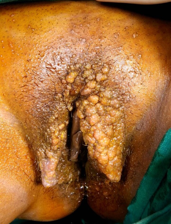
A 65-year-old postmenopausal woman presented with bilateral pedal edema for past 20 years and progressively increasing vulval mass for past 15 years. She had consulted a private practitioner for genital lesions in 2008 and had undergone a biopsy. Histopathology showed lymphangioma circumscriptum, following which she underwent endovenous laser ablation of the lesion. Her vulval swelling recurred in 2013 for which she went to another private hospital. Repeat biopsy showed a benign inflammatory polyp, negative for granuloma or neoplasia. She came to our institute in June 2014. She had history of ulcerative colitis since 2000. There was no prior history of fever, vulvar pruritus, pain, lymphorrhea, lymphedema of leg or lymphadenopathy. Also, there was no history of sexually transmitted disease, tuberculosis or filariasis. On examination of vulva, there was a large irregular warty growth involving both labia majora, resembling frog spawn (Fig. 1). Growth was around 10 × 10 cm, non-tender, firm, with minimal serosanguinous discharge. Clitoris and labia minora were free from lesion. Per speculum and per vaginal examination did not reveal any abnormality. There were no palpable lymph nodes or dilated veins. There were also multiple small satellite lesions on the mons pubis. Lesions were painful vesicles which gradually increased in number and were associated with occasional blood tinged discharge. She also had bilateral pedal edema which was gradually progressive with no associated pain, redness or fever. Her Pap smear was normal. MRI pelvis was done which showed few tortuous and serpiginous foci in left labia majora associated with diffuse soft tissue thickening involving vulva suggestive of lymphomatous malformation of vulva. Lymphangiography was done which showed normal lymphatic drainage in both lower limbs. Her blood and biochemical investigations were normal. Her Mantoux test, filarial antigen, chlamydia antibody, HPV DNA testing, VDRL, HIV test were all negative. Biopsy was done which showed irregular papillary and warty covering stratified squamous epithelium, hyperkeratosis and congestion of rete ridges, large dilated lymphatic spaces present in upper dermis suggestive of lymphangioma circumscriptum. Patient was planned for wide local excision of mass. Mass was excised with wide normal margins and liberal use of electrocautery at the depth of excision. Primary closure of skin was done. Histopathology of the specimen confirmed the lesion as lymphangioma circumscriptum. Her postoperative course was uneventful. Wound healed well without complications. Her pedal edema resolved slowly over a period of 3 months. It has been 1 year since the surgery and patient is fine; there is no recurrence.
Fig. 1.

Clinical picture showing large irregular warty growth involving both labia majora, with hyperkeratosis and small lesions over mons pubis, resembling frog spawn
Discussion
Lymphangioma circumscriptum (LC) can be of two types—congenital and acquired form. Congenital LC occurs due to local malformation of lymphatics and manifests at birth or before 5 years of age. Obstruction of lymphatics due to any cause results in acquired forms of LC which can manifest at any age. The most common causes of acquired forms include pelvic surgery, radiation therapy, infection such as tuberculosis and Crohn’s disease [2]. Differentiation between congenital and acquired lymphangiomas with respect to localization within the skin has been made: The former results from a hamartomatous malformation of lymphatic vessels, and the latter from acquired obstruction of lymph vessels, e.g., after surgical or radiation treatment of malignancies of the breast or uterus. The diagnosis is usually made by biopsy [3]. The basic pathology in LC is the presence of dilated muscle-coated lymphatic cistern in the subcutaneous plane with communication to the large dermal lymphatics finally forming multiple clustered vesicles with clear, yellow, pink or hemorrhagic fluid over the skin surface as blow-out phenomenon. These abnormal lymphatics are not connected to the normal lymphatics. The basic developmental defect is sequestration of cisterns in the plane of subcutaneous tissue [4]. Clinical presentation of LC may vary from pseudovesicles to nodules or wart-like lesions; therefore, correct clinical diagnosis is difficult to make. Most common differential diagnosis includes genital warts, herpes zoster, molluscum contagiosum and even leiomyoma [2]. Vulvar LC can be asymptomatic, pruritic, burning or painful. The LC is asymptomatic in its localized form; the most common symptom is oozing of clear fluid mixed with blood, spontaneously or after minor trauma. Complications are rare in LC. The most common complications are bleeding, pain and infection by Staphylococcus aureus causing cellulitis. Although chances of malignancy are rare, few cases of lymphangiosarcoma have been reported in patients previously treated by radiation therapy [4]. Apart from above complications, LC may also cause psychosexual dysfunction [2]. There is no standard therapy for LC. As there is no medical treatment, surgical excision is the main modality of treatment, although recurrence is common. Other treatment options include sclerotherapy, cryotherapy, superficial radiotherapy, pulsed dye laser, intense pulse light, electrocautery, vaporization with CO2 laser. Electrodessication of the lesion is a new treatment option which has been tried following failure of pulsed dye laser with good results [2]. The main aim of the treatment is to remove or destroy the diseased lymphatics and subcutaneous components which act as a nidus for recurrence. Follow-up is necessary to treat recurrence and detect the occasional case of lymphangiosarcoma.
Varnit Toshyan
obtained his MBBS degree from the M. L. B. Medical College, Jhansi, U.P., and his MD in Obstetrics and Gynecology in 2015 from the All India Institute of Medical Sciences, New Delhi.
Compliance with Ethical Standards
Conflict of interest
Authors declare that they have no conflict of interest.
Footnotes
Dr Varnit Toshyan obtained his MBBS degree from the M.L.B. Medical College, Jhansi, U.P. and his MD in obstetrics and gynaecology in 2015 from the All India Institute of Medical Sciences; Dr Latika Chawla is a Senior Resident at the Department of Obstetrics and Gynecology; Dr Swati Verma is Junior Resident at the Department of Obstetrics and Gynecology; Dr Juhi Bharti is Senior Resident at the Department of Obstetrics and Gynecology; Dr Venus Dalal is a Senior Resident at the Department of Obstetrics and Gynecology; Dr K. K. Roy is a Professor at the Department of Obstetrics and Gynecology; Dr Sunesh Kumar is a Professor and Head of Unit at the Department of Obstetrics and Gynecology
References
- 1.Basak S, De A, Bag T. Surgery as the treatment of choice in vulvar lymphangioma circumscriptum: case report and review of other management options. Eur J Obstet Gynecol Reprod Biol. 2010;152(2):225–226. doi: 10.1016/j.ejogrb.2010.05.028. [DOI] [PubMed] [Google Scholar]
- 2.Sinha A, Phukan JP, Jalan S, Pal S. Lymphangioma circumscriptum of the vulva: report of a rare case. J Life Health. 2015;6(2):91–93. doi: 10.4103/0976-7800.158968. [DOI] [PMC free article] [PubMed] [Google Scholar]
- 3.Mehta V, Nayak S, Balachandran C, Monga P, Rao R. Extensive congenital vulvar lymphangioma mimicking genital warts. Indian J Dermatol. 2010;55(1):121–122. doi: 10.4103/0019-5154.60372. [DOI] [PMC free article] [PubMed] [Google Scholar]
- 4.Kudur MH, Hulmani M. Extensive and invasive lymphangioma circumscriptum in a young female: a rare case report and review of the literature. Indian Dermatol Online J. 2013;4(3):199–201. doi: 10.4103/2229-5178.115516. [DOI] [PMC free article] [PubMed] [Google Scholar]


