. 2016 Oct 19;5:e19375. doi: 10.7554/eLife.19375

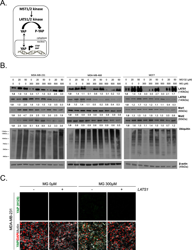
Figure 6. MG induces LATS1 kinase decrease in breast cancer cells.

(A) Schematic representation of the Hippo pathway focused on MST1/2 and LATS1/2 kinases control of nuclear-cytoplasmic shuttling of YAP co-transcription factor. (B) LATS1, LATS2, MST1 and MST2 expression in MDA-MB-231, MDA-MB468 and MCF7 cells treated with MG (300 and 500 µM) in presence of increasing concentrations of MG132 proteasome inhibitor during 6 hr using Western blot. Ubiquitin immunoblot were performed to validate proteasome inhibition by MG132. Immunoblot data were quantified by densitometric analysis and normalized for β-actin. Numbers represent fold increase relative to the condition shown with bold number. (C) YAP immunofluorescence (Santa Cruz antibody, H125) in MDA-MB-231 cells transiently transfected with LATS1 expression vector (+) or empty vector used as control (-) and then treated with MG (300 µM) until confluence. All data are representative of three independent experiments.

DOI: http://dx.doi.org/10.7554/eLife.19375.018

Figure 6.

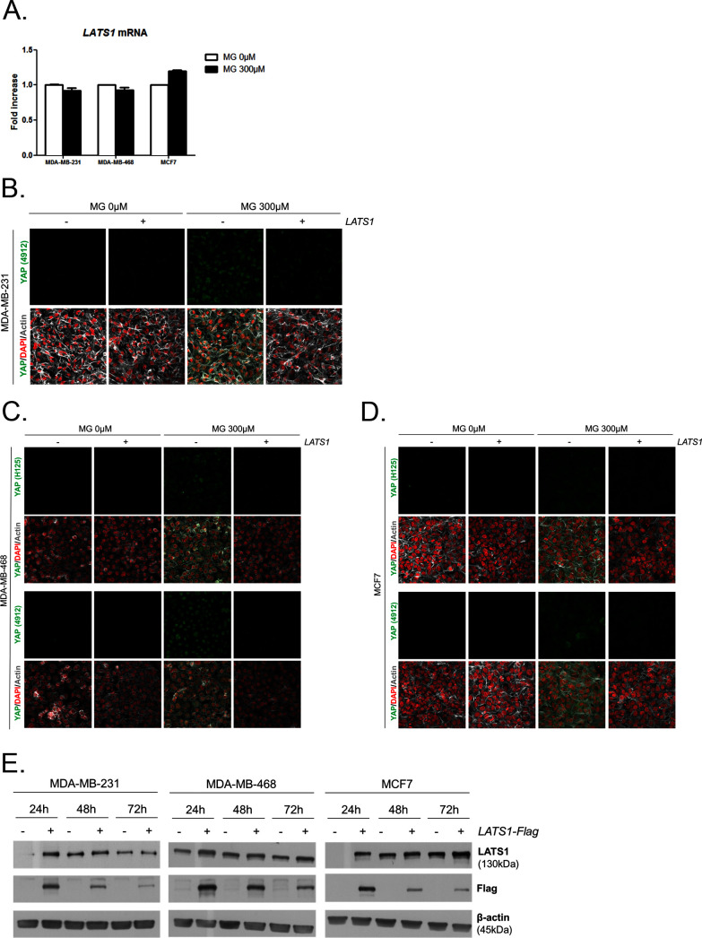
Figure 6—figure supplement 1. MG leads to YAP cellular accumulation through LATS1 expression decrease.

Figure 6—figure supplement 1.

(A) MG treatment from low to high density did not affect LATS1 mRNA levels as assessed by qRT-PCR in the indicated cell lines. Data were analyzed using unpaired student’s t test and shown as the mean values ± SEM of three independent experiments. (B) YAP immunofluorescence (Cell Signaling antibody, 4912) in MDA-MB-231 cells transiently transfected with LATS1 (+) or empty vector (-) and then treated with MG (300 µM) until confluence. Magnification 630x. (C and D) YAP immunofluorescence detection (Santa Cruz antibody, H125 and Cell Signaling antibody, 4912) in MDA-MB-468 and MCF7 cells, respectively, transiently transfected with LATS1 (+) or empty vector (-) and then treated with MG (300 µM) until confluence. Magnification 630x. (E) Validation of LATS1 overexpression in breast cancer cells by western blot using LATS1 and Flag antibodies. β-actin is used for normalization. All data are representative of three independent experiments.