Abstract
We report on 2 patients (3½ year-old-male and 6-year-old female) with the ring 15 chromosome syndrome and speech delays and review 25 cases from the literature. The main characteristics of this syndrome include growth retardation (100%), variable mental retardation (95%), microcephaly (88%), hypertelorism (46%), and triangular facies (42%). Other frequent findings include delayed bone age (75%), brachydactyly (44%), speech delay (39%), frontal bossing (36%), anomalous ears (30%), café-au-lait spots (30%), cryptorchidism (30%), and cardiac abnormalities (30%). The average age at diagnosis was 8.1 years. The average maternal and paternal age at the time of birth was 28 and 31 years, respectively.
Keywords: short stature, microcephaly, triangular facies, hypertelorism, brachydactyly, mental retardation, speech delay, r(15) chromosome
INTRODUCTION
Ring 15 chromosome syndrome, a rare disorder first described by Jacobsen [1966], is characterized by growth retardation, microcephaly, triangular facies and variable mental retardation. Since that time at least 25 cases have been reported [Jacobsen, 1966; Emberger et al., 1971; Forabosco et al., 1972; Stoll et al., 1975; Kawasaki et al., 1975; Rumenic et al., 1976; Ferrante et al., 1977; Pfeiffer et al., 1977; Fujita and Matsumoto, 1978; Scheibenreiter and Frisch, 1978; Schmid et al., 1978; Wisniewski et al., 1979; Fryns et al., 1979; Ledbetter et al., 1980; Gardner et al., 1980; Meinecke and Koske-Westphal, 1980; Kousseff, 1980; Yunis et al., 1981; Fryns et al., 1981; Moreau and Teyssier, 1982; Neri et al., 1983; Otto et al., 1984; Wilson et al., 1985; Kosztolanyi and Pap, 1986]. Herein we report on 2 additional patients and review the literature.
CLINICAL REPORTS
Patient 1
This white male was the product of a term gestation ta a 27-year-old-white primagravida female and 33-year-old white father. Intrauterine growth failure was detected by ultrasound during the fifth month of pregnancy. The infant was delivered by C-section because of breech presentation. The birth weight was 1930 g and length was 42.5 cm. The APGAR scores were 9 and 9 at one and five minutes, respectively. On physical examination, the infant had microcephaly, bilateral hip dislocations, bilateral club feet, bilateral cryptorchidism, and mild first degree hypospadias.
Developmentally, the patient rolled over at 3 months, babbled using syllables (eg, b, d, m) at 4½ months, supported weight on his legs at 5½ months, spoke in consonant-vowel syllables at 7 months, had his first tooth at 9 months, and sat up at 10 months. At 15 months, he could scoot with the use of his arms and had a five to seven word vocabulary. By 18 months, his vocabulary leveled off at 10–15 words, and he was not able to put words together.
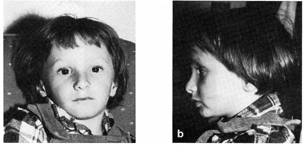
Physical examination at 20 months showed a weight of 7.7 kg (<5th centile), height of 72.4 cm (<5th centile) and OFC of 42.5 cm (<5th centile). He was a small but symmetrically proportionate child with a triangular shaped face, micrognathia, borderline high palate, sharp tapering ears, and postsurgical foot abnormalities (Fig. 1). No other abnormalities were found on physical examination except for hypospadias. His bone age was below one year at a chronological age of 20 months.
Fig. 1.
a, b. Patient 1 at 43 months of age.
Chromosome preparations with Giemsa-trypsin banding and AgNOR staining methods were undertaken from peripheral blood lymphocytes of the patient and his parents. Chromosomal analysis of 22 cells showed a 46,XY,r(15)(p11q26) chromosome constitution in 19 cells and 45,XY, -15 in 3 cells. A large ring which probably represents a double ring was seen in 20% of cells while the remaining cells contined only the single r(15) chromosome. Parental chromosomes were normal.
At age 26 months, audiometric testing showed mild to moderate hearing loss which may have been secondary to chronic otitis media. The Sequenced Inventory of Communication Development (SICD) was used to evaluate age appropriate task mastery. While his mental abilities clustered at the 15 to 16 month level, his motor abilities clustered at the 11 month level. His expressive skills were markedly delayed at approximately the 12 month level, although his receptive language skill was at a level of 16 to 20 months. The Receptive-Expressive Emergent Level Language Scale was used to further investigate language skills, and he achieved a receptive language quotient of 81, corresponding to an achievement age of 22 months.
An oral and speech examination showed several abnormalities. He was often found to suck his food prior to chewing and swallowing. When he did chew, he predominantly used an immature up-and-down chewing patttern. His teeth were found to occlude at the incisors but the lateral teeth did not close. His consonant repertoire appeared to be limited to the earliest developing sounds and he had difficulty with rapid lateralization of his tongue. Because of language difficulties, his parents have taught him sign language which he has learned easily and used spontaneously in new situations.
Patient 2
This white female was the 2.1 kg, 44.4 cm product of an uncomplicated term pregnancy to a 28-year-old primagravida white female and 29-year-old father. The mother denied smoking or medications, and had only used alcohol occasionally during pregnancy. There was no consanguinity.
During infancy, she was a poor feeder and height-weight parameters were less than the 3rd centile for age, although her growth curve paralleled normal. Endocrine evaluation at this time included SMA, CBC, thyroid studies, and serum amino acid screen, all of which were normal. The bone age was 6 months at a chronological age of 11 months.
Developmentally, she had normal motor milestones as an infant, but later had developmental delay and speech articulation difficulties. She was formally evaluated at age years, with the following results: 5 month delay in expressive language skills and difficulty with oral motor skills, including elevation and rapid lateralization of the tongue and oral diadochokinesis. There was also concern with her performance of fine motor skills and rapid alternating movements. Mild hypotonia was also noted.
At age 5 years she was re-evaluated for an endocrine disorder but normal values were obtained for prolactin, thyroxine and somatomedin C. Her bone age was less than two standard deviations below the mean for her chronological age. Although she was thought to have the Russell-Silver syndrome, she was referred at age 6½ years for further genetic evaluation.
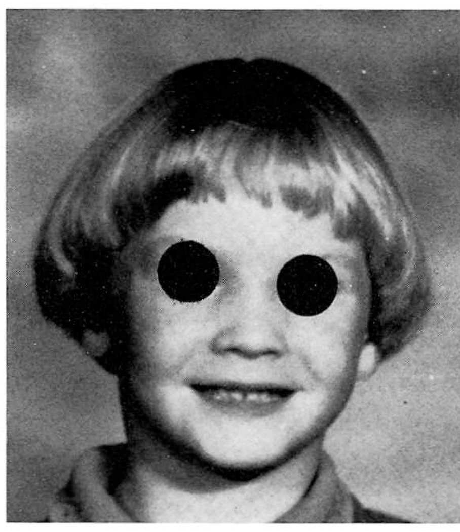
On physical examination she was a pleasant, white girl who weighed 18.2 kg (10th centile), height 107 cm (<3rd centile) and OFC 46.2 cm (<3rd centile) (Fig. 2). Her ears were well-formed and her nasal bridge was broad while the palate was moderately high. The inner canthal, outer canthal and interpupillary distances were normal. The cardiovascular, abdominal, and neurological examinations were normal except for hyporeflexia. The limbs showed bilateral 5th finger clinodactyly, whorls on 8 of 10 digits and distal axial triradii. Café-au-lait spots were found on the back and left thigh. The rest of the examination was normal. Peripheral blood was obtained and chromosome analysis on 20 cells showed that the majority of cells (>80%) had a 46,XX,r(15)(p11q26) chromosome constitution while the remaining cells were 45,XX,-15. No large ring chromosomes representing double rings were observed. Parental chromosomes were normal.
Fig. 2.
Patient 2 at 6 years of age.
DISCUSSION
We reviewed 25 previously reported cases of ring chromosome 15 syndrome and present 2 additional cases (Table I). Similar phenotypic abnormalities that occur in these patients include short stature, microcephaly, mental retardation, and frequently triangular facies with hypertelorism. Other visceral and musculoskeletal malformations include brachydactyly (44%), cardiac abnormalities of various types (30%), cryptorchidism (30%), fifth finger clinodactyly (26%), and talipes equinovarus (15%).
TABLE I.
Summary of Clinical Manifestations in Ring Chromosome 15 Patients
| Clinical Features | Patient 1 | Patient 2 | Literature | Total | % |
|---|---|---|---|---|---|
| Cranio-Facial | |||||
| Microcephaly (<3rd centile) | + | + | 19/22 | 21/24 | 88 |
| Hypertelorism | + | − | 10/22 | 11/24 | 46 |
| Triangular facies | + | + | 8/22 | 10/24 | 42 |
| Broad nasal bridge | − | + | 9/25 | 10/27 | 37 |
| Frontal bossing | − | − | 8/20 | 8/22 | 36 |
| Anomalous ears | − | − | 8/25 | 8/27 | 30 |
| Micrognathism | + | − | 6/22 | 7/24 | 29 |
| High arched palate | + | + | 4/25 | 6/27 | 22 |
| Skin | |||||
| Café-au-lait spots | + | + | 5/21 | 7/23 | 30 |
| Achromic areas | − | − | 3/21 | 3/23 | 13 |
| Simian crease | − | − | 3/25 | 3/27 | 11 |
| Uro-Genital | |||||
| Cryptorchidism | + | n/a | 2/9 | 3/10 | 30 |
| Hypospadias | + | n/a | 1/9 | 2/10 | 20 |
| Renal malformations | − | − | 2/25 | 2/27 | 7 |
| Musculo-Skeletal | |||||
| Short Stature | + | + | 25/25 | 27/27 | 100 |
| Birth length (<46 cm) | + | + | 12/15 | 14/17 | 82 |
| Delayed bone age | + | + | 4/6 | 6/8 | 75 |
| Birth weight (<2.5 kg) | + | + | 17/24 | 19/26 | 73 |
| Brachydactyly | − | − | 12/25 | 12/27 | 44 |
| Fifth finger clinodactyly | + | + | 5/25 | 7/27 | 26 |
| Small hands | − | + | 6/24 | 6/26 | 23 |
| Talipes equinovarus | + | − | 3/25 | 4/27 | 15 |
| Second and third toe syndactyly | + | − | 2/25 | 3/27 | 11 |
| Scoliosis | − | − | 2/25 | 2/27 | 7 |
| Cardiovascular | |||||
| Cardiac abnormalities | − | − | 8/25 | 8/27 | 30 |
| CNS Function | |||||
| Mental retardation | + | + | 18/19 | 20/21 | 95 |
| Speech delay | + | + | 7/21 | 9/23 | 39 |
| Hypotonia | − | + | 6/25 | 7/27 | 26 |
n/a = not applicable.
The average age at diagnosis for all cases was 8.1 years for the 17 females and 10 males, including our patients. The average maternal and paternal age at the time of birth was 28 years and 31 years, respectively. Developmental milestones were somewhat delayed with an average age of 11 months for sitting (N = 8); 22 months for walking (N = 10); and 20 months for talking (N = 6).
The growth in our patients paralleled standardized curves but fell consistently below the 3rd centile although growth hormone studies in patient 2 and in several previously reported ring chromosome 15 patients have been normal [Wilson et al., 1985].
Of special importance in our two cases is the delay in psychomotor development, especially pertaining to speech. Both patients have expressive speech skills significantly delayed compared with receptive speech abilities. A similar result was found in one previous report with formal speech testing [Ferrante et al., 1977] and 6 other patients were described as having delays in speech. Formal testing in our patients showed difficulties in lateralization of the tongue that may account for the speech difficulty. Eating and swallowing problems may also be due to this motor deficiency. The speech problems in our patients suggest that a significant part of the difficulty in communication may not be due to mental retardation. Although mental retardation is present in nearly all patients (95% of patients from the literature reported with at least mild mental retardation), there is a wide variation in mental capacity.
A program of manual communications was instituted with our first patient. Presently, at age 3½ years he has mastered over 250 signs, and can combine signs. For those patients with receptive speech capability this approach may be of importance until oral communication improves.
In summary, there is a wide spectrum of anomalies associated with ring chromosome 15 syndrome. An index of suspicion should allow diagnosis when a number of the presented phenotypic abnormalities are identified. Most characteristic are intrauterine and post-natal growth delay, microcephaly with a triangular face and varying degrees of mental retardation. In some patients specific tongue motor abnormalities may account for speech difficulties and the appearance of more severe mental retardation. Therefore, speech delay may be an under-recognized feature of ring chromosome 15 patients. Further study of the specific speech impediments combined with specific therapy and manual communications may allow a significant improvement of mental development in some ring chromosome 15 patients.
Contributor Information
Merlin G. Butler, Division of Genetics, Department of Pediatrics, Vanderbilt University School of Medicine, Nashville, Tennessee
Agnes B. Fogo, Department of Pathology, Vanderbilt University School of Medicine, Nashville, Tennessee
David A. Fuchs, Division of Genetics, Department of Pediatrics, Vanderbilt University School of Medicine, Nashville, Tennessee
Francis S. Collins, Division of Medical Genetics, Department of Internal Medicine, University of Michigan; Ann Arbor, Michigan
Viathilingam G. Dev, Division of Genetics, Department of Pediatrics, Vanderbilt University School of Medicine, Nashville, Tennessee
John A. Phillips, III, Division of Genetics, Department of Pediatrics, Vanderbilt University School of Medicine, Nashville, Tennessee.
REFERENCES
- Emberger JM, Rossi D, Jean R, Bonnet H, Dumas R. Etude d’une observation de chromosome du groupe 13–15 en anneau (46,XY,15r) Humangenetik. 1971;11:295–299. doi: 10.1007/BF00278656. [DOI] [PubMed] [Google Scholar]
- Ferrante E, Boscherini B, Bruni B, Vignetti P, Finocchi G. La sindrome r(5) (cromosoma 15 ad anello). Descripzione di un caso. Minerva Pediatr. 1977;29:2163–2168. [PubMed] [Google Scholar]
- Forabosco A, Dutrillaux B, Vazzoler G, Lejeune J. Chromosome 15 en anneau: r(15). Identification par denaturation menagee. Ann Génét (Paris) 1972;15:267–270. [PubMed] [Google Scholar]
- Fryns JP, Jaeken J, Devlieger H, Debucquoy P, Eggermont E, van den Berghe H. Ring chromosome 15 syndrome. Acta Paediatr Belg. 1981;34:47–49. [PubMed] [Google Scholar]
- Fryns JP, Timmermans J, D’Hondt F, Francois B, Emmery L, van den Berghe H. Ring chromosome 15 syndrome. Hum Genet. 1979;51:43–48. doi: 10.1007/BF00278290. [DOI] [PubMed] [Google Scholar]
- Fujita H, Matsumoto H. Ring chromosome 15;46,XX,r(15)(p11q26) in a girl. Jap J Hum Genet. 1978;23:233–237. doi: 10.1007/BF01872473. [DOI] [PubMed] [Google Scholar]
- Gardner RJM, Chewings WE, Holdaway MD. A ring 15 chromosome in a girl with minor abnormalities. N Z Med J. 1980;91:173–174. [PubMed] [Google Scholar]
- Jacobsen P. A ring chromosome in the 13–15 group associated with microcephalic dwarfism, mental retardation and emotional immaturity. Hereditas. 1966;55:188–191. [Google Scholar]
- Kawaski T, Kamimae A, Maeda J, Shonohara T, Tomita H. A case with No 15 ring chromosome. Jap J Pediatr Pract. 1975;38:544. [Google Scholar]
- Kosztolanyi G, Pap M. Severe growth failure associated with atrophic intestinal mucosa and ring chromosome 15. Acta Paediatr Scand. 1986;75:326–331. doi: 10.1111/j.1651-2227.1986.tb10209.x. [DOI] [PubMed] [Google Scholar]
- Kousseff BG. Ring chromosome 15 and failure to thrive. Am J Dis Child. 1980;134:798–799. doi: 10.1001/archpedi.1980.02130200066022. [DOI] [PubMed] [Google Scholar]
- Ledbetter DH, Riccardi VM, Au WW, Wilson DP, Holmquist GP. Ring chromosome 15: Phenotype, Ag-NOR analysis, secondary aneuploidy and associated chromosome instability. Cytogenet Cell Genet. 1980;27:111–122. doi: 10.1159/000131472. [DOI] [PubMed] [Google Scholar]
- Meinecke P, Koske-Westphal T. Ring chromosome 15 in a male adult with radial defects. Evaluation of the phenotype. Clin Genet. 1980;18:428–433. doi: 10.1111/j.1399-0004.1980.tb01788.x. [DOI] [PubMed] [Google Scholar]
- Moreau N, Teyssier M. Ring chromosome 15: Report of a case in an infertile man. Clin Genet. 1982;21:272–279. doi: 10.1111/j.1399-0004.1982.tb00763.x. [DOI] [PubMed] [Google Scholar]
- Neri G, Ricci R, Pelino A, Bova R, Tedeschi B, Serra A. A boy with ring chromosome 15 derived from a t(15q;15q) Robertsonian translocation in the mother. Cytogenetic and biochemical findings. Am J Med Genet. 1983;14:307–314. doi: 10.1002/ajmg.1320140211. [DOI] [PubMed] [Google Scholar]
- Otto J, Back E, Furste HO, Abel M, Bohm N, Pringsheim W. Dysplastic features, growth retardation, malrotation of the gut and fatal ventricular septal defect in a 4-month-old girl with ring chromosome 15. Eur J Pediatr. 1984;142:229–231. doi: 10.1007/BF00442457. [DOI] [PubMed] [Google Scholar]
- Pfeiffer RA, Dhadial R, Lenz W. 46,XX/46,XX,r(15) mosaicism. Report of a case. J Med Genet. 1977;14:63–65. doi: 10.1136/jmg.14.1.63. [DOI] [PMC free article] [PubMed] [Google Scholar]
- Rumenic LJ, Joksimovic I, Anaf M. Ring chromosome 15 in a child with a minor dysmorphism of phenotype. Hum Genet. 1976;33:187–188. doi: 10.1007/BF00281895. [DOI] [PubMed] [Google Scholar]
- Scheibenreiter S, Frisch H. Ein Kind mit Ringchromosom 15. Wien Klin Wochenschr. 1978;90:22–25. [PubMed] [Google Scholar]
- Schmid H, Henrichs I, Nestler H, Knorr-Gartner H, Teller WM, Krone W. Analysis of banding patterns and mosaic configurations in a case of ring chromosome 15. Hum Genet. 1978;41:289–299. doi: 10.1007/BF00284763. [DOI] [PubMed] [Google Scholar]
- Stoll C, Juif TG, Luckel JC, Lausecker C. Ring chromosome 15:r(15). Identification by R-banding. Hum Genet. 1975;27:259–262. doi: 10.1007/BF00278356. [DOI] [PMC free article] [PubMed] [Google Scholar]
- Wilson GN, Sauder SE, Bush M, Beitins IZ. Phenotypic delineation of ring chromosome 15 and Russell-Silver syndromes. J Med Genet. 1985;22:233–236. doi: 10.1136/jmg.22.3.233. [DOI] [PMC free article] [PubMed] [Google Scholar]
- Wisniewski L, Pronicka E, Lech H, Niezabitowska A. The child with chromosome ring 15. Klin Padiatr. 1979;191:429–432. [PubMed] [Google Scholar]
- Yunis E, Leibovici M, Quintero L. Ring (15) chromosome. Hum Genet. 1981;57:207–209. doi: 10.1007/BF00282025. [DOI] [PubMed] [Google Scholar]


