

Abstract
The osteomeatal complex plays an important role in the development of Chronic rhinosinusitis. The ethmoidal infundibulum is bordered medially by the uncinate process, and the anatomic relationship between the ethmoidal infundibulum and the frontal recess may depend upon the types of attachment of the uncinate process. The osteomeatal complex is the main area targeted in chronic rhinosinusitis and within it uncinate process is the first anatomical structure encountered. The aim of this study was to evaluate the types of attachment of the uncinate process and its implications in the development of sinus inflammation. The significance of anatomical variations of uncinate process in chronic sinusitis were evaluated. A prospective CT scan study on 64 patients of chronic sinusitis (128 uncinate processes) was done. The results were tabulated and analyzed using Statistical Package for Social Science 16.0. Type I superior attachment of uncinate process into the lamina papyracea was the most common variety in all ages and both sexes and a statistically significant association between Type 1 Uncinate process and frontal sinusitis was found. (P < 0.05). The superior attachment of uncinate process alters the frontal sinus drainage and causes the frontal sinusitis.
Keywords: Uncinate process, Anatomic variations, Frontal sinusitis
Introduction
The osteomeatal complex (OMC)—is the small compartment located in the region between the middle turbinate and the lateral nasal wall in the middle meatus—represents the region for drainage of anterior ethmoid, maxillary and frontal sinuses. The variation in any one of the component of ostiomeatal complex causes improper drainage of these sinuses causing chronic sinusitis. Uncinate process is a latin term, meaning hooked out growth. It is a thin, bony leaflet almost sagitally oriented which runs in sickle shaped curve from anterosuperior to posteroinferior, parallel to anterior surface of ethmoid bulla. The ascending convex anterior margin may be attached superiorly to skull base, laterally to lamina papyracea, or may fuse medially with the insertion of middle turbinate. Zinreich et al. [1]
The anatomic variations of uncinate process were defined as Stammberger and Bolger [2]
Superior attachment of uncinate could be to a. Type I—the uncinate process is inserted into the lamina papyracea, b. Type II—the uncinate process extends superiorly to the roof of the ethmoid, that is the skull base, c. Type III—the uncinate process is attached to the middle turbinate
Medially bent uncinate process
Laterally bent uncinate process
Hypertrophied uncinate process
Pneumatized uncinate process
Surgically, the uncinate must be removed to gain access to the ethmoid infundibulum and maxillary sinus ostium. Once the uncinate has been resected, the ostium of the maxillary sinus can be visualized and enlarged by a maxillary antrostomy. These steps, uncinectomy and maxillary antrostomies, form the basis of the FESS procedure and are essential for optimal surgical outcome (Fig. 1).
Fig. 1.

Gender distribution of disease
Materials and Method
The prospective and retrospective study was carried out in the Department of otorhinolaryngology, SIMS, Hapur from July 2013 to august 2015. A prospective CT scan study on 64 patients of chronic sinusitis (128 uncinate processes) was done. Patients with clinical evidence of chronic rhinosinusitis with or without nasal polyposis not responding to 3 week of medical treatment were evaluated with computed tomographic scanning of Paranasal Sinuses Coronal view (axial when needed). To support a clinical diagnosis of chronic rhinosinusitis, in accordance with the American Academy of Otolaryngology and Head and Neck Surgery (AAO-HNS) criteria, at least two major or one major and two minor symptoms are required. Lanza and Kennedy [3].
Chronic rhinosinusitis was defined as representing a state of persistent sinus disease associated with at least one of the following; nasal congestion, hyposmia, facial pain, or nasal discharge.
Patients with acute rhinosinusitis or those who had previously undergone nasal or sinus surgery, either open or endoscopic, patients with facial trauma, pregnant females, patients with fungal sinusitis were excluded from the study (Fig. 2).
Fig. 2.

Age group distribution the the disease
All patients with identifiable disease were subjected to detailed clinical examination, and CT scan PNS coronal section (axial section when required). Coronal planes were selected, as it is the plane in which anatomy and pathology are examined almost identical to view of endoscopist and it best displays both the osteomeatal changes.
Statistical Analysis
The comparison was conducted using the Chi square statistical test with Statistical Program for Social Science (SPSS) version 16.0. A P value <0.05 was considered statistically significant.
Observations
In the present study carried out in the Department of Otorhinolaryngology, SIMS, Hapur, a total of 64 patients (128 sides) having symptoms and signs of chronic sinusitis were included.
Out of 64 patients; 33 were male and 31 were female with M:F ratio = 1.06:1. The patients were in the range of 16–65 years. The age group of 16–30 (37.5 %) was the most commonly affected.
Computed Tomography Evaluation: Uncinate Process Variation
Out of 64 patients (128 uncinate process), 24 cases showed medially bent uncinate process, 3 cases showed laterally bent uncinate process, 2 cases showed pneumatized uncinate process and 3 cases showed hypertrophied uncinate process. Over all the abnormal uncinate process found in 32 cases (25.0 %).
Out of 128 sides superior attachment of uncinate process was identified for 108 sides. Type I uncinate process was seen in 74 (57.8 %). Type II uncinate process was seen in 26 (20.3 %) and Type III was seen in 8 (6.2 %).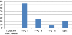
Medially bent was the most frequent variation of uncinate process seen in 24 cases (18.8 %) followed by laterally bent and hypertrophied, seen in 3 cases(2.3 %) each, followed by pneumatised uncinate process seen in 2 cases(1.6 %) observed in all the age groups and was significant on Chi square test.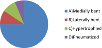
Discussion
Computerized tomographic imaging of sinonasal region has become the gold standard in the evaluation of patients with chronic rhinosinusitis. Its ability to accurately map out the bony and soft tissue anatomy of the paranasal sinus has proven invaluable to the endoscopic surgeon in the diagnostic workup of the surgical candidate.
Endoscopic examination in conjunction with computed tomography (CT) has proven to be ideal combination in recent years and are already accepted as the “Standard of Care” for sinus diseases.
Stammberger and Wolf [4] has documented the preeminent role of the ostiomeatal complex as a gateway to sinus disease. As a consequence of various anatomic and functional relationships immediately adjacent to the OMC, proliferation of disease into the anterior sinuses is the natural corollary.
In this study,the incidence of anatomical variations in the ostiomeatal complex and distribution of the mucosal abnormalities in each paranasal sinuses was investigated by coronal plane CT Scanning. The study population consisted of 64 patients. Age of the patients included in this study ranged from 16 to 60 years. Males were predominantly affected.
Computed Tomographic Detection of Anatomical Variations and Mucosal Abnormalities
Coronal CT Scan paranasal sinus focuses mainly attention to the area of the ostiomeatal unit, now has emerged as the standard in presurgical evaluation of patients under consideration for possible F.E.S.S (Zinreich) [5]. CT Scan coronal section can provide specific diagnosis underlying causes of sinusitis. It can clarify the anatomical relationships and varitions that may play a role in sinusitis and also road map for F.E.S.S.
Uncinate Process Variations
The uncinate process may show a large number of anatomic variations. In the radiologic assessment of the uncinate process in coronal sections, its anatomic course must always be kept in mind. As a result of its arc-shaped course, the most anterior coronal sections will show the uncinate process at its widest. In the middle third of its course, the uncinate process appears to lie immediately adjacent to the posterior wall of the nasolacrimal duct. In the more posterior sections it appears increasingly narrower because of its arc from anterosuperior to posteroinferior.
The free edge of the Uncinate process may deviate medially, laterally or anteriorly (Stammberger and Wolf) [4]. The most frequent, pathologically significant variation of the uncinate process is a marked medial curvature or bend. A markedly medially bent or folded uncinate process with a corresponding area of extensive contact with the middle turbinate is one of the most frequent pathological findings in patients with chronic rhinosinusitis (Stammberger and Wolf) [4]. The exact prevalence of these variations and their relation to sinus disease is not determined. “In our study we observed medially bent Uncinate process in 18.8 % of the cases. Laterally bent Uncinate process was seen in 2.3 % cases, hypertrophied uncinate in 2.3 % and pneumatised uncinate in 1.6 % cases”.
Pneumatization of the uncinate process, also referred to as the Uncinate bulla, has been implicated as an anatomic variation that can impair sinus ventilation, specifically in the anterior ethmoid, frontal recess and infundibular regions. Although this entity was noted many years ago during gross anatomic studies, early investigators failed to describe the mechanism by which Uncinate pneumatization occurred and did not provide data regarding the prevalence of this anatomic variation. Zinreich [5] noted uncinate bulla in one patient in a series of 230 patients (0.4 %) with chronic sinus complaints evaluated by coronal CT scan.
Variations of uncinate process in our series were found in only 32 % cases. This was similar to that of 30 % reported by Tuli et al. [6] and Aiyer et al. but higher than that of 2.5 % reported by Bolger et al. [7] and 2 % by Asruddin et al. [8] and lower than that of 45 % reported by Wanamaker [9] and 65 % reported by Mamatha et al. [10].
| Study | Our Study | Tuli et al. | Aiver et al. | Bolger et al. | Asrudin et al. | Wanamaker | Mamatha et al. |
|---|---|---|---|---|---|---|---|
| Variations | 32 % | 30 % | 30 % | 2.5 % | 2 % | 45 % | 65 % |
The most commonly seen superior attachment in our series was Type I uncinate process in 57.8 % cases as compared to 79.8 % cases reported by Tuli et al. [6] This was higher than that reported by Krzeski et al. (17.83 %), Min et al. (54 %) and Landsberg and Friedman (52 %). Type II uncinate process was the second most common seen in 20.3 % cases and Type III uncinate process was seen in 6.2 %. These were lower than that reported by Krzeski et al. of Type II being 33.12 % and Type III 14.33 % and Min et al. who found that the superior attachment of the uncinate was to skull base in 24.5 % sides (Type II) and to middle turbinate in 21.5 % (Type III) Tuli et al. [6].
| Study | Our study (%) | Tuli et al. (%) | Krzeski et al. (%) | Min et al. (%) | Landsberg and Friedman |
|---|---|---|---|---|---|
| Type I | 57.8 | 67 | 17.83 | 54 | 52 % |
| Type II | 20.3 | 14 | 33.12 | 24.5 | – |
| Type III | 6.2 | 3 | 14.33 | 21.5 | – |
A detailed analysis of coronal plane CT scan of paranasal sinuses was done and various structures and anatomical variations of lateral nasal wall were evaluated as a with special attention towards the ostiomeatal complex region. Coronal CT Scan was found to be a useful preoperative evaluation tool because of the advent of FESS, CT of the paranasal sinuses has gained considerable importance.
The extent of sinonasal inflammatory disease, important anatomic landmarks and their variations can be easily depicted on CT scans thereby providing a reliable “road map” to endoscopic surgery aimed more at functional restoration and preservation. One of the keys to comprehending sinus disease and preventing surgical complications during sinus surgery is an understanding of the complex ostiomeatal unit anatomy on coronal CT images PNS. Dua et al. [11]
Anatomic variations and mucosal abnormalities were carefully analysed in each C.T scan and their prevalence was found to correlate well with the observations of previous investigators.
Conclusion
From this study we concluded that Chronic rhinosinusitis is fairly a common disease condition affecting most commonly the age group between 16 and 45 years. In our present study, it was found that the superior part of uncinate process commonly attaches with the lamina papyracea (Type 1) and its attachment alters the frontal sinus drainage thus showing statistically significant association with frontal sinusitis.
Removal of disease in Ostiomeatal complex region is the basic principle of FESS which is best appreciated on CT Scan. Anatomical variation in any one of the component of Ostiomeatal complex causes improper drainage of paranasal sinuses causing sinusitis. CT scan must be done prior to any functional endoscopic sinus surgery. They help in assessing the extent of sinus disease and to know the anatomical variations
Compliance with Ethical Standards
Funding
This study was funded by Saraswathi Institute of Medical Sciences, Hapur.
Conflict of interest
Author A declares that he has no conflict of interest. Author B declares that he has no conflict of interest.
Ethical Approval
This article does not contain any studies with human participants or animals performed by any of the authors.
Informed Consent
Informed consent was obtained from all individual participants included in the study.
References
- 1.Zinreich SJ, Kennedy DW, Rosenbaum AE, Gayler BW, Kumar AJ, Stammberger H. Paranasal sinuses: CT imaging requirements for endoscopic surgery. Radiology. 1987;163:769–775. doi: 10.1148/radiology.163.3.3575731. [DOI] [PubMed] [Google Scholar]
- 2.Stammberger HR, Bolger WE. Paranasal sinuses: anatomic terminology and nomenclature. The Anatomic Terminology Group. Ann Otol Rhinol Laryngol Suppl. 1995;167:7–16. [PubMed] [Google Scholar]
- 3.Lanza DC, Kennedy DW. Adult rhinosinusitis defined. Otolaryngol Head Neck Surg. 1997;117:S1–S7. doi: 10.1016/S0194-5998(97)70001-9. [DOI] [PubMed] [Google Scholar]
- 4.Stammberger H, Wolf G. Headaches and sinus disease: the endoscopic approach. Ann Otol Rhinol Laryngol Suppl. 1988;134:3–23. doi: 10.1177/00034894880970s501. [DOI] [PubMed] [Google Scholar]
- 5.Zinreich SJ. Paranasal sinus imaging. Otolaryngol Head Neck Surg. 1990;103(5):863–868. doi: 10.1177/01945998901030S505. [DOI] [PubMed] [Google Scholar]
- 6.Tuli IP, Sengupta S, Munjal S, Kesari SP. Anatomical variations of uncinate process observed in chronic sinusitis. Indian J Otolaryngol Head Neck Surg. 2013;65(2):157–161. doi: 10.1007/s12070-012-0612-8. [DOI] [PMC free article] [PubMed] [Google Scholar]
- 7.Bolger WE, Butzin CA, Parson DS. Paranasal sinus bony anatomic variations and mucosal abnormalities; CT analysis for endoscopic sinus surgery. Laryngoscope. 1991;101:56–64. doi: 10.1288/00005537-199101000-00010. [DOI] [PubMed] [Google Scholar]
- 8.Asruddin, Yadav RK, Singh J. Low dose CT in chronic sinusitis. Indian J Otolaryngol Head Neck Surg. 1999;52(1):17–22. doi: 10.1007/BF02996425. [DOI] [PMC free article] [PubMed] [Google Scholar]
- 9.Wanamaker HH. Role of Haller’s cell in headache and sinus disease: a case report. Otolaryngol Head Neck Surg. 1996;114(2):324–327. doi: 10.1016/S0194-5998(96)70196-1. [DOI] [PubMed] [Google Scholar]
- 10.Mamatha H, Shamasundar NM, Bharathi MB, Prasanna LC. Variations of ostiomeatal complex and its applied anatomy: a CT scan study. Indian J Sci Technol. 2010;3(8):904–907. [Google Scholar]
- 11.Dua K, Chopra H, Khurana AS, Munjal M(2005) CT scan Variations in chronic sinusitis. Indian J Radiol Imaging [serial online] [cited 2015 sep 14]. http://www.ijri.org/text.asp?2005/15/3/315/29144