Abstract
Temporal bone dissection has important role in educating, and training the surgeons. Temporal bone has complicated three dimensional anatomy and it is challenging for young surgeons to understand and operate. Not knowing the anatomy may cause serious consequences to patient due to injury to vital structures. It is important to learn temporal bone harvesting techniques, preservation of specimens, fixation and to reduce the health hazards posed by these specimens by taking safety measures. Spending more time in temporal bone laboratory and repeated dissection of temporal bones provides the skills necessary in the operating room for future generation. All training institutes should establish temporal bone laboratory in their department to provide the necessary expertise to future generation while maintaining safe and secure environment.
Keywords: Temporal bone dissection, Resident curriculum, Transmastoid approach, Intra cranial approach
Introduction
Temporal bone dissection plays an important role in the education, research, training of residents and young surgeons in Otorhinolaryngology. The three dimensional anatomy of temporal bone makes it challenging for young surgeons to operate and serious morbidity and mortality can occur from injuring vital structures in the temporal bone while performing surgery [1–8]. Also the dissection of temporal bone is essential to develop innovative surgical techniques in middle ear, mastoid and perform transmastoid approach to clear intracranial lesions or develop an approach to cerebello-pontine angle lesions [1–4]. It is universally accepted that repeated dissection of temporal bone is essential to understand the core anatomy of temporal bone and to develop safe otological skills [2, 7, 8].
This creates demand to attain and harvest temporal bone from cadaver. Usually temporal bones are removed while performing post-mortem [1–4, 9]. The great demand of dissection of temporal bone by the otologists made it essential to establish a temporal bone laboratory in institutes and hospitals [3, 4, 10].
The main objective of this article is to enumerate the requirements in establishing the temporal bone laboratory, legal aspects in harvesting the temporal bone from cadaver, preservation techniques of specimen and safety measurements.
Temporal Bone Lab Facility at AIIMS
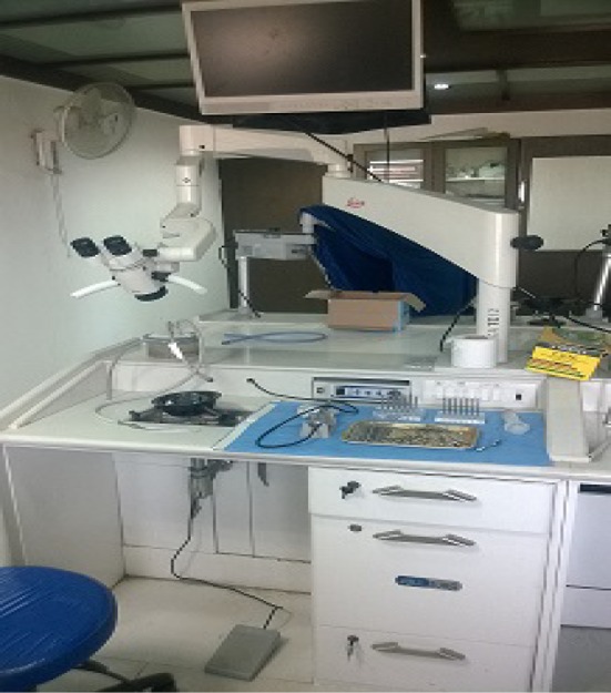
The department of Otorhinolaryngology and Head & Neck surgery in All India Institute of Medical Sciences, New Delhi, India has established a permanent and fixed temporal bone laboratory 5 years ago which is air conditioned. The laboratory has eight fixed working stations and each station equipped with a Leica operating microscope with monitor attachment, temporal bone holder, irrigation and suction, electrical micro drill with hand piece, surgical instruments and drill burrs (Figs. 1, 2a–c).
Fig. 1.

Working station with monitor attached microscope, suction and irrigation, electric drill with foot pedal, instruments and temporal bone holder
Fig. 2.
a Consists micro drill with hand piece, burrs, micro instruments with micro suction cannulas, b straight and angled hand pieces c left to right syringe, suction cannulas, micro suction cannulas and adaptor
Usually temporal bones are harvested during the post-mortem from unclaimed cadavers under Indian anatomy act for medical education and research purpose. There are three types of harvesting methods to remove the temporal bone from cadaver and soon after removal it should be preserved to sterilize and to reduce bio-hazards.
Under the guidance of faculty in the department of Otorhinolaryngology and Head & Neck surgery, All India Institute of Medical Sciences, New Delhi, the trainee’s and postgraduates attend the temporal bone laboratory for approximately 12 h in a week. With help of dissection manuals and prepared DVDs, each trainee is able to identify/grasp the normal and abnormal anatomy of temporal bone and later on practice detailed procedures. The HOD made compulsory rule to utilise the temporal bone laboratory by trainees to produce skilled future generation and to reach the operating standards of the Institution.
Discussion
A good temporal bone laboratory with adequate equipment gives boost to trainees to learn anatomy of temporal bone. This fixed type laboratory providing round the clock accessibility to trainees and dissectors is not available in many other institutes in the country.
- 1. Requirements
-
A. Microscope It is the basic requirement for dissection. A good illuminating microscope is needed to visualise the millimetre vital structures in the temporal bone [1, 4]. However, the microscope should have following specifications [11], (1). High quality optics/high resolution, (2). Variable magnification, (3). Objectives with appropriate focal distances, (4). Adjustable eye pieces, (5). Inbuilt camera for photos/video demonstration, (6). Suitable stand, (7). Objective ocular eye piece or monitor attachment, (8). Illuminated by either a halogen or xenon light source provides good magnification and visualization.The scope shall be fixed or mobile type; fixed type causes less damage to scope rather than mobile type because frequent movement of scope can cause dislodgment of lenses. Our institute has fixed Leica microscope with light source.
- B. Motor A drastic development in medical technology changed the trends of otological surgeries from goose and hammer dissection to compact micro motor dissection. It dramatically reduces the morbidity and duration of surgery. The motors are gas driven and electric operated [11]. The advantages of gas driven drills has more power and speed which helps in rapid removal of bone, the disadvantage of this drills are need high pressure lines in the operating room and need compressed gas cylinders. The electric drill quite contrast to gas driven drills, these are all compact models which are easy to arrange, lower noise levels, easy to handle and the limiting factor is lack of speed [11].
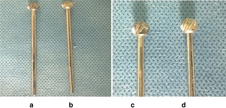
- C. Drill Burrs Have key role in dissection and they are cutting and diamond varieties. These can be stainless steel and TC burrs are available in the market. However the durability of the burr depends on the material used and utilization in dissection. The diamond burrs are available in the following sizes: 7.0, 6.0, 5.0, 4.0, 3.1, 2.3, 1.4, 1.0, 0.8 mm. The selection of cutting burrs is very difficult and one has to find the number of cutting edges on the cutting burrs. The burrs with fewer cutting teeth are dangerous because they catch unwanted bone edges. The advisable cutting edges in each burr are as following 7.0–16, 6.0–16, 5.0–12, 4.5–12, 2.3–10, 1.8–8, 1.4–8, 1.0–6 [12, 13] (Fig. 3a–d).
- D. Suction and Irrigation High vacuum suction and water irrigation is needed to remove the bone fragments from the dissection field continuously [11]. Mucus sucker can be used manually to attain clear field during the dissection. Where facilities are available electric suction and auto-irrigation can be used which reduce the dissection time and give comfort to the dissector.
- E. Others Other requirements such as room with good ventilation and space; table with comfortable height which gives good leg room and comfort to the surgeon; chair with back rest or revolving chair with easily adjustable height; proper drainage and sink to clear sludge at the end of the procedure; and waste bin to separate the bio-tissue and normal wastage for further disposal.
-
F. Maintenance The protocol of laboratory is to maintain clean surroundings and integrity of the lab. In view of the above, the dissector shall himself clean the platform after dissection. In addition to that, the dissector should clean the bone dust which falls on the microscope including the lenses and cover the microscope at the end of dissection.The micro motor hand piece should be lubricated with appropriate lubricant to reduce the rusting of bearings and to provide smooth movements. Finally clean the drill burrs and make them free from bone dust.
-
- 2. Harvesting Temporal Bones
-
A. Legal Aspects The requirement for temporal bones is increasing day by day and at the same time availability of bones is decreasing due to legal and traditional reasons in harvesting the bones from cadavers. In United Kingdom, the Human tissue act (2004) made amendments in the law to utilization of human tissues for academic, research, medical education and training and now requires informed consent [4]. This causes impact on harvesting of temporal bones for both temporal bone laboratory and dissection courses [4]. USA has its own National Temporal Bone Hearing and Balance Pathology Resource Registry and the activity of this organization is to encourage the individuals with ear disorders to donate temporal bone for scientific research. However trainees are not getting enough of temporal bones for dissection.In India, most of the temporal bones are removed at time of post-mortem in the Forensic Medicine Department or in the Department of Anatomy under the Anatomy act [4]. The Anatomy act is a state act which is published in the state Government Gazette [4]. The Bombay anatomy act 1949—An act to provide unclaimed bodies of deceased persons, donation of body or any part of the body after his death made by a person while he was alive to hospitals and medical and teaching institutions for therapeutic purposes or for the purpose of medical education or research including anatomical examination and dissection-adapted from Bombay government gazette publication, part IV, on the 22nd April 1949 [4, 14]. Most of states in India have their own legal acts to harvest human tissue for academic and research activities. According to these legal acts the unclaimed or unknown cadaver bodies can be utilised for medical education and research purpose.Several researchers have developed 3-dimensional models and computer aided software for temporal bone dissections but nothing replaces the original temporal bone dissection [5].
- B. Techniques/Methods The procured temporal bones are fixed in a 10 % neutral buffered formalin solution for a period of 2 weeks at 4 °C temperature or in a high concentration of alcohol [1]. Hypertonic saline may be used but it is not documented in literature. Frozen temporal bones offer better colour, consistency but have the risk of viral transmission (e.g., hepatitis B). The buffered solution decreases the oxidation effect of formalin. When exposed to air it forms formic acid which is a caustic substance hazardous to health. The recommended volume ratio of fixative to specimen is at least three parts to one part (3:1). The container holding the specimen should be labelled with an ID number and care should be taken to retain all tissues from each specimen together for future crimination. Some of the dissectors fix the specimens in hypertonic saline which makes the tissue soft and can be removed easily. However, it is more hazardous to health as it spread air-borne infections (like viral infections).
-
-
3. Preservation techniques
For research purpose the bone should be harvested within 24 h of death to avoid post-mortem reaction of tissues. However, in reality there is a delay in procuring the specimen due to unavoidable and unexplainable reasons. There are three types of harvesting methods from cadaver [1, 3],- Intra cranial method (removal of all the ear structures)
- Extended intra cranial technique (in addition to the ear structures, Eustachian tube and part of palate also removed)
- Extended cranial method (when complete autopsy with brain removal is not done).
It is important to avoid damages to internal structures and land marks of temporal bone, such as dura should be intact to avoid damage to endolymphatic sac, seventh and eighth nerves, porus acousticus and traumatic avulsion of internal auditory canal.
Instruments
Conventional method employs use of hammer and chisel which can cause undesirable damage to bone and vital structures. However using electric saw can take ideal cut margins, preserve the vital structures. However many centres don’t have the facility.
-
4. Safety measurements
Care should be taken regarding biological and chemical hazards which come from temporal bone laboratory. Some of the pathogens (especially viruses) are not destroyed by the fixation. So while performing dissection, the dissector should wear gowns, disposal masks, gloves and protection to eyes. Eating and drinking in the laboratory should be avoided as the alcohol is inflammable; ethyl ether is explosive while HCl is caustic and formaldehyde is carcinogenic. Healthy and safety measurements are important to limit all the above hazards.
The bones are usually preserved in the formaldehyde or high concentration alcohol or frozen, but this carries the risk of viral transmission of hepatitis B and prions (means proteinaceous infectious organisms) [1, 2, 4]. The prions can cause sub acute encephalopathy namely Creutzfeldt–Jacob disease (CJD) [4]. During mastoid surgery and bone dissection there is scattering of bone dust which consists of neural tissue and it causes aerosol inoculation into the surgeon’s conjunctiva. To reduce this risk, the surgeon should take precautionary measures to protect his eyes [1, 2, 4].
Having the temporal bone laboratory in the department allows preparing the specimen for dissection without breaching the legislation acts and health hazards. And also it provides security to collected and labelled specimen for further crimination or burial.
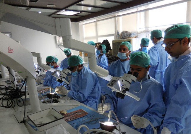
To overcome the challenges of temporal bone surgery, it is essential for trainees to develop hand-eye coordination and fine hand movements during the surgery and to attain anatomical knowledge under the microscopic vision. The combination of surgical experience and laboratory cadaveric dissection gives good quality results to the society (Fig. 4).
Fig. 3.
Cutting burrs selection (a) has more catchment gap between teeth’s (b) has less catchment gap between teeth’s (c, d are zoomed pictures)
Fig. 4.

Workshop-training to young surgeons
Conclusion
Establishing the temporal bone laboratory with equipment in the department gives boost to young otorhinolaryngologists and postgraduates to learn the anatomy of temporal bone. It increases/hikes the quality of surgical skills and develops new innovative techniques. It can also be utilized to conduct courses and workshops to train the new generation of surgeons. All the department heads must insist upon the trainees to perform the dissection before performing ear surgeries. “The important way of learning temporal bone anatomy and surgical skills is through repeated dissection of temporal bone in a cadaver”.
Compliance with Ethical Standards
Conflict of interest
All the authors have no conflict of interest.
Ethical Standard
This article does not contain any studies with human participants or animals performed by any of the authors.
References
- 1.Fennessy BG, O’Sullivan P. Establishing a temporal bone laboratory: considerations for ENT specialist training. Ir J Med Sci. 2009;178:393–395. doi: 10.1007/s11845-009-0373-x. [DOI] [PubMed] [Google Scholar]
- 2.George AP, De R. Review of temporal bone dissection teaching: how it was, is and will be. J Laryngol Otol. 2010;124:119–125. doi: 10.1017/S0022215109991617. [DOI] [PubMed] [Google Scholar]
- 3.Walvekar RR, Harless LD, Loehn BC, Swartz W. Block method of human temporal bone removal: a technical modification to permit rapid removal. Laryngoscope. 2010;120:1998–2001. doi: 10.1002/lary.21052. [DOI] [PubMed] [Google Scholar]
- 4.Naik SM, Naik MS, Bains NK. Cadaveric temporal bone dissection: Is it obsolete today? Int Arch Otorhinolaryngol. 2014;18:63–67. doi: 10.1055/s-0033-1351681. [DOI] [PMC free article] [PubMed] [Google Scholar]
- 5.Wang H, Northrop C, Burgess B, Liberman MC, Merchant SN. Three-dimensional virtual model of the human temporal bone: a stand-alone, downloadable teaching tool. Otol Neurotol. 2006;27:452–457. doi: 10.1097/01.mao.0000188353.97795.c5. [DOI] [PMC free article] [PubMed] [Google Scholar]
- 6.García JMM, Da Costa ASA, Schmitz TR, Estomba CMC, Zavarce MIH. Commentary temporal bone dissection practice using a chicken egg. Otol Neurotol. 2014;35:941–943. doi: 10.1097/MAO.0000000000000390. [DOI] [PubMed] [Google Scholar]
- 7.Francis HW, Malik MU, Varela DADV, Barffour MA, Chien WW, Carey JP, Niparko JK, Bhatti NI. Technical skills improve after practice on virtual-reality temporal bone simulator. Laryngoscope. 2012;122:1385–1391. doi: 10.1002/lary.22378. [DOI] [PubMed] [Google Scholar]
- 8.Qiu MG, Zhang SX, Liu ZJ, Tan LW, Li QY, Li K, Wang YS, Deng JH, Tang Z-S. Visualization of the temporal bone of the Chinese visible human. Surg Radiol Anat. 2004;26:149–152. doi: 10.1007/s00276-003-0188-9. [DOI] [PubMed] [Google Scholar]
- 9.Manolis E, Filippou D, Theocharis S, Panagiotaropoulos T, Lappas D, Mompheratou E. Anatomical landmarks: dimensions of the mastoid air cell system in the Mediterranean population. Our experience from the anatomy of 298 temporal bones. Anat Sci Int. 2007;82:139–146. doi: 10.1111/j.1447-073X.2007.00175.x. [DOI] [PubMed] [Google Scholar]
- 10.Zirkle M, Taplin MA, Anthony R, Dubrowski A. Objective assessment of temporal bone drilling skills. Ann Otol Rhinol Laryngol. 2007;116(11):793–798. doi: 10.1177/000348940711601101. [DOI] [PubMed] [Google Scholar]
- 11.Gulya AJ, Minor LB, Poe DS (2010) Glasscock-Shambaugh Surgery of the ear. 6th edn. CBS Publishers & Distributors, New Delhi, pp 779–780
- 12.Fisch U and Mattox D (1988) Atlas on Microsurgery of the skull base. Thieme Medical Publisher, New York, p 10
- 13.Kylen P, Stjernvall J-E, Arlinger S. Variables affecting the drill-generated noise levels in ear surgery. Acta Otolaryngol. 1977;84:252–259. doi: 10.3109/00016487709123964. [DOI] [PubMed] [Google Scholar]
- 14.Supplements The Bombay Anatomy Act, 1949.[As modified up to 10th November 1997]. J Indian Acad Forensic Med 31(2). medind.nic.in/jal/t09/i2/jalt09i2p176


