Abstract
Severe gastrointestinal bleeding (GIB) secondary to jejunal diverticulosis (JD) is very rare. Delay in establishing a diagnosis is common and GIB from JD is associated with significant morbidity and mortality. We report an illustrative case diagnosed by push enteroscopy and managed with surgery.
Introduction
Gastrointestinal bleeding (GIB) is a cause of significant morbidity and mortality.1 Severe GIB secondary to jejunal diverticulosis (JD) is rare, with approximately 50 documented cases in the literature. The majority of patients with JD are asymptomatic; however, patients may present with complications such as bleeding, perforation, or abscess.2 Delay in establishing a diagnosis is common. Consequently, JD is associated with significant morbidity and mortality.2,3 The relative inaccessibility of the small bowel to endoscopy makes hemorrhage originating in the jejunum difficult to localize. However, video capsule endoscopy (VCE) or deep enteroscopy using a single or double balloon enteroscope (DBE) has been shown to detect small bowel bleeding in hemodynamically stable patients and may serve as a diagnostic, definitive or a temporizing intervention prior to definitive surgical management.1 Radionuclide techniques, computed tomography (CT) angiography, CT enterography, and conventional angiography can also be used to delineate obscure sources of GIB.1
Case Report
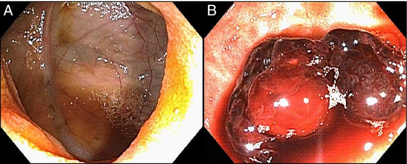
A 63-year old woman was referred to our institution from a community hospital with a 3-day history of hematochezia. Her medical history was significant for hypertension, Type II diabetes, cholecystectomy, colonic diverticulosis, and 2 episodes of severe GIB in the 9 years prior to presentation. She required about 8 U of packed red blood cells on both prior episodes of GIB; radiologic, nuclear medicine, and endoscopic evaluation were reportedly nondiagnostic, and GIB eventually stopped. During this presentation, upper and lower endoscopy were negative for a source of active bleeding at the referring hospital, and she received 9 U of packed red blood cells prior to transfer. On admission, she was actively bleeding but hemodynamically stable. Blood pressure was 118/74 mm Hg, heart rate was 115/min, respiratory rate was 20/min, and oxygen saturation was 98% on room air. Laboratory tests were significant for hemoglobin of 6.8 g/dL. Physical examination revealed severe pallor of her skin and mucous membranes and right lower quadrant abdominal tenderness with guarding and no rebound tenderness. There was no organomegaly, and her bowel sounds were hyperactive. Rectal examination revealed maroon-colored stool. Radionuclide scan was unrevealing, and she underwent an urgent colonoscopy, which revealed altered blood throughout her entire colon and terminal ileum. Therefore, a second-look push enteroscopy using a pediatric colonoscope was performed. Push enteroscopy revealed fresh blood in the proximal jejunum, which prompted deeper exploration. Three large diverticula were visualized, and 1 diverticulum had an adherent clot (Figure 1). Endoscopic intervention was not attempted because the pediatric colonoscope had reached maximum insertion with significant loss of tip control, which made definitive endotherapy less feasible.
Figure 1.
Push enteroscopy revealed (A) fresh blood in the proximal jejunum, (B) 3 large diverticula, and 1 adherent clot.
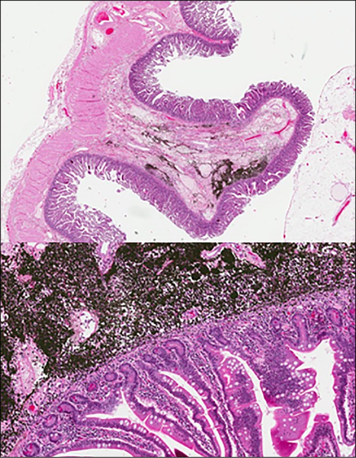
The jejunal mucosa around the diverticulum was tattooed with SpotTM ink (GI Supply, Camp Hill, PA). She was referred for surgery, and exploratory laparotomy confirmed the endoscopic findings and tattoo. The affected segment of jejunum was resected with primary reanastomosis. Gross and microscopic examination of the resected segment of her jejunum confirmed the diagnosis of jejunal diverticula with abundant submucosal black pigmentation from tattooing process (Figure 2 and 3). There has been no recurrent GIB in the first postoperative year.
Figure 2.
The resected segment of the jejunum confirmed the diagnosis of jejunal diverticula.
Figure 3.

Microscopic examination of the resected segment of the jejunum showed jejunal diverticula and abundant submucosal black pigmentation from tattooing process.
Discussion
The true incidence of JD is unknown; however, autopsy studies have reported an incidence of 0.4%-4.6%, whereas radiologic studies report an incidence ranging from 0.002% to 2.3% by enteroclysis.2 Jejunal diverticulosis was first described by Somerling in 1794 and later by Sir Astley Cooper in 1807.2 The diverticula arise on the mesenteric border of the bowel and are classified as pseudo-diverticula as they lack a muscularis propria layer.2 They are thought to be acquired pulsion lesions secondary to increased intraluminal pressure.2 They are seen more often in elderly patients, and some studies report a slight male predominance.3 Much rarer findings are a congenital (true) diverticulum in jejunum or small bowel duplication (mimics small bowel diverticulum).2,4 Eighty percent of small bowel diverticulosis occurs in the jejunum, 15% in the ileum, and 5% in both.4,5 The condition is associated with colonic diverticular lesions in 50%-75% of cases, with duodenal diverticula in 25.9% of cases, and with esophageal lesions in 2.3% of cases.2 Jejunal diverticula are typically asymptomatic; however, severe complications are seen in 10%-30% of patients.5 These complications include hemorrhage, small bowel perforation, malabsorption, volvulus, enterolith formation, abscess, diverticulitis, and intestinal obstruction.2-6
Bleeding from JD is seen in 3.4%-8.1% and can result in massive GIB.5 In 1923, Dr. L. R. Braithwaite, a British surgeon, was the first to report a case of bleeding from JD.6 The prevalence of small bowel lesions has been estimated to be ∼5%-10% in patients presenting with GIB.1 Evaluation of GIB should include upper and lower endoscopy, and, if overt bleeding persists with no identifiable source, VCE or angiography (for massive bleeding) should be obtained.1 A technetium-99m red blood cell scan (which detects bleeding rates of 0.1-0.5 mL/min), CT angiography or CT enterography may be considered for overt, nonmassive GIB if VCE is not available or contraindicated.1 Treatment of GIB from JD is typically surgical resection of the involved segment.2,5,6 However, in recent years, DBE has been used to evaluate and treat bleeding from JD.1,2,7,8 The most commonly used therapeutic modalities during this procedure are hemoclipping or argon plasma coagulation.7 Endoscopic band ligation during DBE has been used successfully for treatment in at least 1 case reported from Japan.8 Double balloon enteroscope is not available at most centers, therefore early surgical consultation is recommended, especially for massive or refractory small bowel bleeding after preoperative localization of the site of bleeding by marking the lesion with a tattoo.1
In this case, push enteroscopy eventually identified the source of bleeding after negative radiographic, nuclear medicine, and endoscopic evaluations. The patient underwent a successful resection of the affected segment of her jejunum with no further bleeding postoperatively. Previously, patients without an identified source of GIB after negative upper and lower endoscopy were classified as obscure GIB.1 The nomenclature for classification of GIB has recently been revised in light of advances in small bowel imaging with VCE, deep enteroscopy, and radiographic imaging, which has enabled the identification of a source of small bowel bleeding in most patients.1 Consequently, the term small bowel bleeding has been proposed as a replacement for the previous classification of obscure GIB. The term obscure GIB is now reserved for patients with no source of bleeding after evaluation with VCE and deep enteroscopy.1
Disclosures
Author contributions: AT Abegunde and E. Christman wrote and revised the manuscript. LA Hassell edited the manuscript and provided the pathological images. D. Kastens edited the manuscript and is the article guarantor.
Financial disclosure: None to report.
Informed consent was obtained for this case report.
References
- 1.Gerson LB, Fidler JL, Cave DR, Leighton JA. ACG clinical guideline: Diagnosis and management of small bowel bleeding. Am J Gastroenterol. 2015;110(9):1265–87. [DOI] [PubMed] [Google Scholar]
- 2.Yang CW, Chen YY, Yen H-H, et al. Successful double balloon enteroscopy treatment for bleeding jejunal diverticulum: A case report and review of the literature. J Laparoendosc Adv Surg Tech A. 2009;19(5):637–40. [DOI] [PubMed] [Google Scholar]
- 3.Levack MM, Madariaga ML, Kaafarani HM. Non-operative successful management of a perforated small bowel diverticulum. World J Gastroenterol. 2014;20(48):18477–9. [DOI] [PMC free article] [PubMed] [Google Scholar]
- 4.Patel VA, Jefferis H, Spiegelberg B, Iqbal Q, Prabhudesai A, Harris S. Jejunal diverticulosis is not always a silent spectator: A report of 4 cases and review of the literature. World J Gastroenterol. 2008;14(38):5916–9. [DOI] [PMC free article] [PubMed] [Google Scholar]
- 5.Yaqub S, Evensen BV, Kjellevold K. Massive rectal bleeding from acquired jejunal diverticula. World J Emerg Surg. 2011;6:17.. [DOI] [PMC free article] [PubMed] [Google Scholar]
- 6.Longo WE, Vernava AM. Clinical implications of jejuno-ileal diverticular disease. Dis Colon Rectum. 1992;35:381–8. [DOI] [PubMed] [Google Scholar]
- 7.Yen H-H, Chen Y-Y, Yang C-W, Soon M-S. The clinical significance of jejunal diverticular disease diagnosed by double balloon enteroscopy for obscure gastrointestinal bleeding. Dig Dis Sci. 2010;55:3473–8. [DOI] [PubMed] [Google Scholar]
- 8.Ikeya T, Ishii N, Shimamura Y, et al. Endoscopic band ligation for bleeding lesions in the small bowel. World J Gastrointest Endosc. 2014;6(10):488–92. [DOI] [PMC free article] [PubMed] [Google Scholar]


