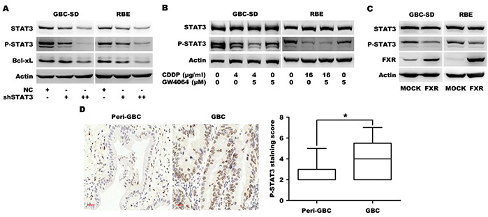
Figure 4. FXR agonist GW4064/CDDP co-treatment additively inhibits STAT3 phosphorylation.

A. Protein level of Bcl-xL in GBC-SD and RBE cells transfected with MOCK or shSTAT3 plasmid for 72 h. B. The STAT3 phosphorylation levels in GBC-SD and RBE cells that were treated with CDDP alone, GW4064 alone and CDDP/GW4064 combination for 24 h. C. Protein levels of FXR, P-STAT3 and STAT3 in GBC-SD and RBE cells transfected with MOCK or FXR plasmid for 72 h. D. The representative fields of patients' tissues of peri-gallbladder cancer (Peri-GBC, n=16) and gallbladder cancer (GBC, n=69) that were examined by P-STAT3 IHC. The nucleus brown staining represented positive signal for P-STAT3, and the cytoplasm was stained by hematoxylin. Scale bar=30 μm. The chart was the quantification of expression staining scores. *P <0.05, comparison between tumor and non-tumor tissues.
