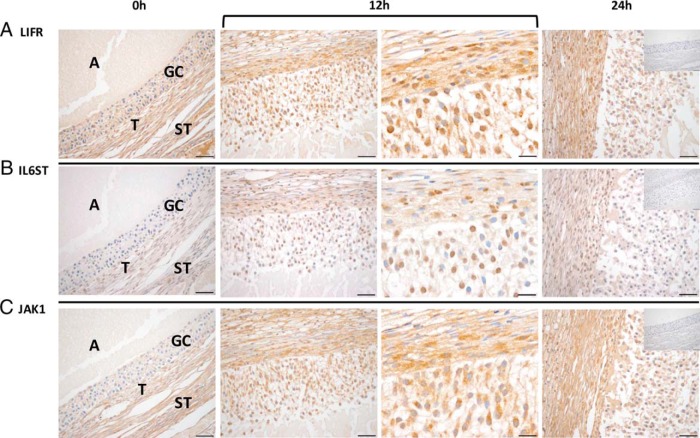
Figure 3.
LIFR, IL6ST, and JAK1 proteins are expressed in the rhesus macaque follicle. LIFR (A) immunostaining was evident before (0 h; ×40 objective) as well as 12 hours (bracket, left and right panels: ×40 and ×100 objectives, respectively) and 24 hours (×40 objective) after the injection of an ovulatory bolus of hCG. In contrast, both IL6ST (B) and JAK1 (C) immunostaining intensity increased primarily within the granulosa cells (GCs) 12 hours after hCG administration. The insets located in the 24-hour panel are representative negative controls for each primary antibody used, which consisted of a species and isotype matched irrelevant antibody. The various ovarian cell types and compartments are indicated in the 0-hour panels and include antrum (A), GCs, theca cells (T), and stroma (ST). Images are representative of ovaries obtained at each time point (0, 12, and 24 h after hCG) from 2–3 animals undergoing COv protocols. Scale bar, 50 and 20 μm for ×40 and ×100 magnification, respectively.

