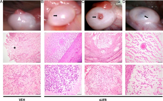
Figure 6.
Histological analysis confirms the presence of a trapped oocyte in follicles treated with sLIFR but not in those receiving vehicle. Rhesus macaque ovaries removed 52 hours after vehicle (PBS) (A) or sLIFR (10 μg/follicle) (B–D) intrafollicular injection were serially sectioned through the entire follicle. The top panels include images taken of the ovary before its removal by laparoscopy. A white arrowhead and asterisk indicate an ovulatory stigma and the site of follicle rupture, respectively (vehicle injection, n = 2; see also Ref. 15) (A), whereas black arrows denote unruptured follicles (sLIFR injection; n = 3). The middle panel revealed a discontinuous stroma indicative of a rupture site that was observed in the vehicle treated follicle (asterisk) (A), whereas a trapped oocyte was observed in follicles from all sLIFR treated animals (B–D). The bottom panels include a higher magnification of the follicle wall from vehicle (A)- and sLIFR (B–D)-injected follicles. Scale bars, 50 μm (middle panels) and 20 μm (bottom panels).

