Abstract
The degradation routes of poly(vinyl pyrrolidone) (PVP) exposed to sodium hypochlorite (bleach) have been previously investigated using chemical analyses such as infrared spectroscopy. So far, no reports have proposed mass spectrometry (MS) as an alternative tool despite its capability to provide molecular and structural information using its single stage electrospray (ESI) or matrix assisted laser desorption ionization (MALDI) and multi stage (MSn) configurations, respectively. The present study thus reports on the characterization of PVP after its exposure to bleach by high resolution MALDI spiralTOF-MS and Kendrick mass defect analysis providing clues as to the formation of a vinyl pyrrolidone/vinyl succinimide copolymeric degradation product. A thorough investigation of the fragmentation pathways of PVP adducted with sodium and proton allows one main route to be described—namely the release of the pyrrolidone pendant group in a charge remote and charge driven mechanism, respectively. Extrapolating this fragmentation pathway, the oxidation of vinyl pyrrolidone into vinyl succinimide hypothesized from the single stage MS is validated by the detection of an alternative succinimide neutral loss in lieu of the pyrrolidone release in the ESI-MSn spectra of the aged PVP sample. It constitutes an example of application of multi-stage mass spectrometry for the characterization of the degradation of polymeric samples at a molecular level.
Keywords: tandem mass spectrometry, fragmentation pathway, electrospray ionization, bleach, polymer degradation
INTRODUCTION
Poly(vinyl pyrrolidone) (PVP) designates an important class of water-soluble polymers used in various applications ranging from pharmaceuticals tablets (as binder) to nanoparticles and surface modification to membranes for water purification.1–3) In this last usage, PVP is often exposed to strong oxidizing agents such as sodium hypochlorite (bleach) during the cleaning of the membrane to remove the biofilms which readily forms at the surface.4) Such chemical cleaning is not inconsequential and turns out to be deleterious for the membrane, ultimately leading to its degradation. Numerous studies have dealt with the ageing of PVP exposed to sodium hypochlorite5–8) under various conditions (e.g. pH, concentration, exposure time…) and proposed several degradation routes and degradation products based on physico-chemical characterization such as infrared spectroscopy or electrochemistry.6) The two main degradation pathways reported in the literature concern 1) the vinyl pyrrolidone repeating unit which is modified in carboxylate/carboxylic acid or succinimide5) and 2) the polymeric backbone which undergoes chain scission.4) In other words, the chemical ageing of PVP leads to a changing of the repeating unit or of the end-groups of the polymer, two main pieces of information readily collected using mass spectrometry (MS).9,10) MS has become a powerful analytical tool for the investigation of polymeric samples,11–14) especially when coupled to soft ion sources such as electrospray (ESI) and matrix assisted laser desorption ionization (MALDI) to preserve the integrity of the chains.15) MS is indeed capable of measuring the mass of each individual chain, highlighting the occurrence of different distributions and allowing an accurate description of the samples in terms of repeating units, degrees of polymerization or end-groups. MS has logically been used in several publications for the characterization of PVP samples16–22) but—to the best of our knowledge—not for the analysis of aged PVP. Beyond the single stage experiment, tandem mass spectrometry (MS/MS) and its extended concept of multi stage mass spectrometry (MSn, n>2) (i.e., the isolation of an ion from the MS or any MSn−1 mass spectrum and its activation to observe its constituting fragments) further increases the knowledge of a polymeric chain by gaining information about its individual end-groups23,24) or its architecture.25) Nevertheless, tandem mass spectrometry—or multi stage mass spectrometry generally speaking—becomes an efficient tool if the fragmentation pathways of some standards belonging to the same class of polymers have been thoroughly investigated.26) Surprisingly, no reports have been found in the literature dealing with the tandem mass spectrometry of PVP. Echoing and capitalizing on the recent studies dealing with the characterization of copolymers27) or degraded polymers28) using tandem mass spectrometry, the present article thus reports on 1) the characterization of a PVP following its exposure to a bleach solution using high resolution MALDI-MS analysis to highlight the changes in the molecular composition of the sample and hypothesize the structure of an aged PVP and 2) the validation of this hypothesis by using multi-stage ESI-MSn mass spectrometry. For the latter, the fragmentation pathways of a pristine PVP sample used as standard will be first thoroughly investigated with sodium and proton adduction prior to the application of the so-established dissociation rules for the analysis of the degradation products.
MATERIALS AND METHOD
Chemicals
Poly(vinyl pyrrolidone) 2,500 g mol−1 (abbreviated as PVP) was purchased from Polyscience Asia Pacific, Inc. (Taipei, Taiwan). A solution of concentrated sodium hypochlorite has been prepared from a commercial bleach (composition: sodium hypochlorite and sodium hydroxide) diluted in milliQ water (dilution factor: 1 : 2, pH ∼11 with no buffer). Trans-2-[3-(4-tert-butylphenyl)-2-methyl-2-propenylidene]-malononitrile (known as DCTB) was from Sigma-Aldrich (St Louis, MO). Ammonium chloride (NH4Cl), sodium chloride (NaCl), sodium trifluoroacetate (NaTFA), chloroform (CHCl3) and methanol (MeOH) were from Wako Pure Chemical Industries, Ltd. (Osaka, Japan). All chemicals were used as received with no purification step.
MALDI spiralTOF high resolution MS and Kendrick mass defect analysis (KMD)
A solvent free preparation was used for both pristine PVP (powder) and aged PVP (aqueous solution). A few grains of matrix (DCTB), one grain of salt (NaTFA) and either a touch of pristine PVP or 5 μL of the aqueous solution of aged PVP were ground for a few minutes to produce a paste (until the water almost fully evaporates in the second case, known as evaporation-grinding (E-G) method.29)). A thin film was then formed on a non-hydrophobic surface (384 circles) from Hudson Surface Technology (HST Inc., Old Trappan, NJ) using a spatula. MALDI mass spectra were recorded using a JMS-S3000 SpiralTOF mass spectrometer (JEOL, Tokyo, Japan) equipped with a Nd:YLF laser irradiating the deposits. The so-generated ions were accelerated by a 20 kV high voltage and went through the spiralTOF analyzer along a spiral trajectory (approximate path length: 17 m) before their detection.30) The delay time was set at 350 ns to keep the peak width ΔM<0.03 Da at FWHM over the mass range of interest. Calibration was performed externally and internally using the sodium adducts of a poly(methyl methacrylate) 1,310 g mol−1 and a hydroxyl-ended polydimethylsiloxane 550 g mol−1 standards (DCTB, no salt added). MSTornado control/analysis (JEOL) was used for data recording and preliminary data treatment while mMass 5.5.0.0 was used for data treatment and artworks.31) For the mass defect analysis, the accurate mass measurements of ions on the IUPAC scale were converted to Kendrick masses (KM) according to KM(ion)=m/z (ion)×111/111.0684 (vinyl pyrrolidone as the base unit) and to Kendrick mass defects (KMD) according to KMD(ion)=NKM(ion)−KM(ion) with NKM (nominal Kendrick mass) the rounded KM to the next integer. The KMD plot displays the KMD of the detected oligomeric adducts as a function of their NKM using a “bubble chart” where each disk expresses a data triplet (NKM, KMD, abundance).32–34)
ESI multi-stage MSn
ESI-MS and ESI-MSn spectra (n=2–10) were recorded using an amaZon SL-STT2 ion trap (Bruker, Bremen, Germany) equipped with an electrospray ion source in infusion mode. The sample solution was introduced into the ionization source at a flow rate of 5 μL min−1 using a syringe pump. The apparatus was operated using the so-called “enhanced resolution mode” (mass range: 50–2,200 m/z, scanning rate: 8,100 m/z per second). The capillary voltage was set at −4,500 V and the endplate offset at −500 V. In MSn experiments, the width of the selection window was set at 2 Da and the amplification of the excitation was set according to the experiment (from 0.2 to 1.5 V). Instrument control, data acquisition and data processing of all experiments were achieved using Compass 1.3 SR2 software provided by Bruker while mMass 5.5.0.0 was used for data treatment and artworks.31)
RESULTS AND DISCUSSION
MALDI spiralTOF-MS of pristine and aged PVP
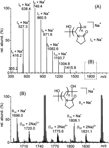
A first high resolution MALDI spiralTOF mass spectrum of a commercial PVP sample was recorded to properly describe its constituting polymeric distributions (Fig. 1A) prior to the analysis of its aged counterpart by multi-stage mass analysis. For the sake of simplicity and better comparison with the ESI-MS data in infusion mode, a direct MALDI-MS analysis has been conducted without any preliminary fractionation by SEC. The average molecular weight is consequently underestimated owing to the dispersity of the sample (Mn=760 g mol−1 as compared to 2,500 g mol−1 as mentioned by the supplier) but this bias has no incidence on the results presented in this paper. A main distribution of sodiated PVP is detected at a generic m/z 60.0575+n×111.0684+Na+ (n=2–18, accurate mass measurements listed in Table S1A in the Supporting Information) and assigned to a 2-hydroxyisopropyl/H-ended PVP series of elemental composition C3H7O–(C6H9NO)n–H (further noted In). It is in accordance with the end-groups proposed by Luo et al.35) (inset in Fig. 1A for its structure).
Fig. 1. (A) MALDI spiralTOF-MS spectrum of the pristine PVP. (B) Magnification of the spectrum from m/z 1690 to m/z 1860. The structures of the main distribution (2-hydroxyisopropyl/H-ended, noted I) and secondary distribution (2-hydroxyisopropyl/2-hydroxyisopropyl-ended, noted II) are depicted in insets of (A) and (B), respectively.
Looking closer to the mass spectrum (Fig. 1B), a secondary distribution (noted II) is also slightly detected and suspected to carry two 2-hydroxyisopropyl terminations (C3H7O–(C6H9NO)n–C3H7O, sodium adduction) based on the accurate mass measurements (Table S1B in the Supporting Information and inset in Fig. 1B for its proposed structure). Doubly charged sodiated oligomers of the distribution I are finally seen in the background (noted [In+2Na]2+). This propensity of PVP for multiply charge states will be advantageously used for the ESI-MS and MSn analyses of large oligomers in the next section.
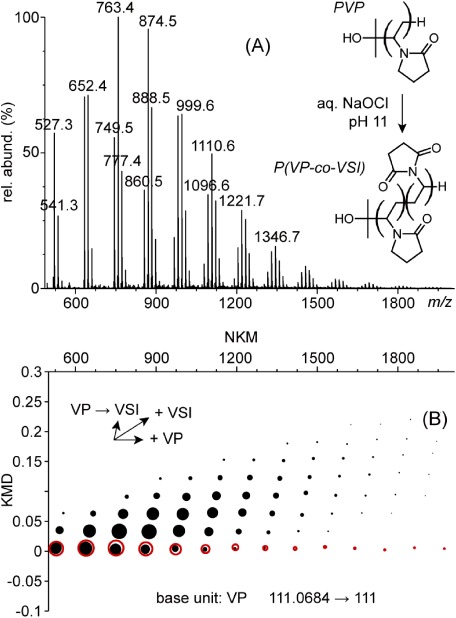
PVP has then been submitted to an accelerated chemical ageing by its exposure to a concentrated bleach solution for 6 days at pH 11. Such pH is not the most effective for the degradation of PVP (pH 8 leads to the fastest kinetics6)) but is comparable to the pH of the cleaning solutions used for membranes (pH 11 is the pH of a commercial bleach/water 50/50 solution, pH 8 is reached by voluntarily adding HCl). The MALDI spiralTOF-MS high resolution mass spectrum of the degraded PVP is depicted in Fig. 2A. Contrary to the MALDI-MS spectrum of the pristine PVP where a single distribution of peaks spaced by 111.1 Da was observed (Fig. 1), convoluted patterns of peaks spaced by 111.1 Da and 14.0 Da are readily seen and constitute a typical fingerprint of a copolymer. The degradation of PVP in basic pH is known to produce succinimide groups5) (abbreviated as SI36,37)), i.e., a newly formed vinyl succinimide repeating unit (abbreviated as VSI38,39)) of elemental composition C6H7NO2 (125.0 Da, +14.0 Da as compared to the VP repeating unit of elemental composition C6H9NO). The so-formed copolymeric skeleton will be noted P(VP-co-VSI) (inset in Fig. 2A).
Fig. 2. (A) MALDI spiralTOF-MS high resolution mass spectrum of a PVP sample in contact with an aqueous bleach solution at pH 11 for 6 days and (B) the associated KMD plot using VP as the base unit (black dots: aged sample; red dots: pristine sample). The structure of the poly(vinyl pyrrolidone-co-vinyl succinimide) copolymer formed upon ageing (noted P(VP-co-VSI) is depicted in inset of (A)).
To facilitate the visualization of the mass spectrum, a Kendrick mass defect analysis (KMD) has been conducted using VP as the base unit for the KM calculations (the mass of a VP unit at m=111.0684 in the IUPAC scale is now arbitrarily fixed at m=111.0000 and all the other masses are re-calculated accordingly). The resulting KMD plot is depicted in Fig. 2B (black filled circles). For sake of comparison the KMD plot for the pristine PVP is superimposed (red empty circles). From the horizontally aligned PVP pristine oligomers each point lining up in an oblique direction highlights the oxidation of a VP unit into a VSI unit, producing a scatter plot archetypal of a copolymer sample.32–34) The KMD plot of the aged sample displays a typical triangle shape: the more the VP units in the pristine backbone, the more the VSI units to be formed under ageing. It is worth mentioning the absence of isobaric interferences despite the high resolving power of the spiralTOF analyzer (6-mer at m/z 749.5: R=54,000; 15-mer at m/z 1749.1: R=75,000) for both the pristine and aged PVP samples. The fragmentation pathways of the PVP oligomers could thus be confidently investigated using an ion trap (no need for high resolution, narrow ion selection or preliminary fractionation in liquid or gas phase) and the copolymeric nature of the degraded sample validated accordingly.
ESI-MSn of sodiated pristine PVP
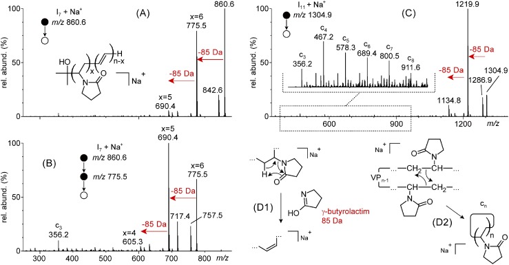
As stated in the introduction, tandem mass spectrometry and multi-stage mass spectrometry more generally speaking have been advantageously used to unambiguously characterize complex copolymers and degradation products formed upon solvolysis.27,28) It requires nevertheless the fragmentation routes of standards of the same polymer class to be carefully investigated. Considering the scarce literature about the tandem mass spectrometry of PVP, this section is thus devoted to the thorough exploration of the fragmentation pathways of a pristine PVP prior to applying these dissociation rules to the structural analysis of aged PVP. The fragmentation routes of PVP were primarily explored for the sodium adducts as they would be the main species found in the mass spectra of PVP degraded by sodium hypochlorite (NaOCl) without any addition of salt (vide infra). The ageing procedure has been voluntarily conducted with concentrated bleach for sake of kinetics, which means sodium cations are in large excess as compared to the polymer sample regardless of the dilution factor. The ESI-MS spectrum of the pristine PVP solubilized in MeOH (Fig. S1 in the Supporting Information) readily displays the sodiated oligomers of the distributions I and II over the same mass range as the MALDI mass spectrum (Fig. 1A) and with the same number average molecular weight Mn (calculated value from ESI-MS: 760 g mol−1). The isolation and consecutive activation of the sodiated 7-mer from the distribution I (noted [I7+Na]+) leads to the ESI-MS/MS spectrum depicted in Fig. 3A. Two main fragmentation routes are observed from the precursor ion consisting of 1) the elimination of a 18.0 Da neutral to produce a product ion at m/z 842.6 and 2) the elimination of one and two 85.1 Da neutrals yielding two product ions at m/z 775.5 and m/z 690.4 (red arrows in Fig. 3A). The first 18.0 Da loss is most likely a dehydration (release of water) implicating the hydroxyl group of the 2-hydroxyisopropyl termination and any hydrogen atom from the chain to produce a cyclized PVP product ion still interacting with Na+. The 85.1 Da loss has been assigned to a C4H7NO neutral, i.e., the pendant group of the vinyl pyrrolidone repeating unit. Isolated and activated in a MS3 step, the product ion at m/z 775.5 is similarly releasing one and two 85.1 Da neutrals to yield the product ions at m/z 690.4 and m/z 605.4 (Fig. 3B, red arrows). The ESI-MSn spectra with n=4–7 (isolation and fragmentation of m/z 690.4, m/z 605.4, m/z 520.3 and m/z 435.3) are depicted in Fig. S2 in the Supporting Information and all display the 85.1 Da neutral releases as the main dissociation process. A mechanism to account for this neutral expulsion is proposed in Fig. 3D1 with a charge-remote 1,5-H rearrangement26) in any vinyl pyrrolidone repeating unit leading to the expulsion of the pyrrolidone pendant group in its tautomeric γ-butyrolactim form (i.e., a 5-membered cyclic carboximidic acid). The product ions formed upon the iterative γ-butyrolactim losses are best rationalized as copolymeric backbones with vinyl pyrrolidone (VP) and acetylene (CH=CH) as the interdependent co-monomeric units (noted C3H7O–(VP)x–(CH=CH)n−x–H in inset of Fig. 3A), a new CH=CH unit being produced at each γ-butyrolactim expulsion (e.g., x=6 for m/z 775.5, x=5 for m/z 690.4). The other neutral losses observed at every MSn step (Fig. 3A & B and Fig. S2 in the Supporting Information) are −18.0 Da (H2O) and −58.0 Da (proposed to be C3H6O), both in accordance with the 2-hydroxyisopropyl termination proposed from the MALDI-MS data. The ESI-MS/MS spectrum of a larger [I11+Na]+ at m/z 1304.9 is depicted in Fig. 3C and displays the same 85.1 Da expulsions in the high mass range of the spectrum (two losses with appreciable yields leading to the product ions at m/z 1219.9 and m/z 1134.8). Contrary to the case of the 7-mer whose ESI-MS/MS spectrum is rather devoid of product ions in the low mass range, a secondary product ion series is slightly detected at m/z 356.2 and larger congeners spaced by 111.1 Da (the cutoff of the ion trap prevents from exploring the lower mass range). Based on the m/z ratios and considering the fact this series is appearing for larger precursor ions, its congeners have been described as cyclic product ions carrying no end-groups and still interacting with Na+, i.e., a cn ion series according to the nomenclature proposed by Wesdemiotis et al.26) Their formation is readily accounted for by a charge remote intramolecular C–C bond translocation in the sodium adduct of PVP (Fig. 3D2).
Fig. 3. (A) ESI-MS/MS of a sodiated 7-mer of the distribution I at m/z 860.6 and (B) ESI-MS3 spectrum of m/z 775.5 formed upon the release of γ-butyrolactim (85.1 Da) from m/z 860.6. (C) ESI-MS/MS spectrum of a sodiated 11-mer of the distribution I at m/z 1304.9. (D) Proposed charge remote mechanisms accounting for 1) the release of γ-butyrolactim through a 1,5-H rearrangement and 2) the formation of cyclic cn product ions upon intramolecular bond translocation.
In order to provide a complete description of the fragmentation routes of PVP even at the risk of being of limited use for the aged PVP samples but of definite interest for the characterization of PVP samples generally speaking, a brief description of the behaviors of multiply charged adducts (ESI notoriously promotes the formation of multiply charged polymer ions40,41)—the sample being preliminary SEC-fractionated to avoid any signal suppression effect, Fig. S3) as well as PVP carrying alternative terminations is reported in the Supporting Information. The main dissociation process of the sodiated chains with charge states of +4, +3 and +2 (Fig. S4) simply consists in the release of a naked Na+ cation to produce a [M+(n−1)Na]n−1+ precursor ion from the [M+nNa]n+ precursor ion. With the CID of the singly charged sodium adducts of PVP as a background knowledge, and relying on the MSn capability of an ion trap, it means long PVP chains could also be investigated by isolating and activating the [M+nNa]n+ adducts until the singly charged [M+Na]+ is formed and detected (in the limit of the ion trap capability, up to m/z 4,400 in a so-called “extended mass range” in our case) and ultimately activated to yield the information-rich product ions. The degradation of PVP by sodium hypochlorite could also induce chain scissions, which means the formation of new end-groups.4) In order to check at their influence, oligomers from the distribution II (inset of Fig. 1B) as well as the cn product ions formed upon the activation of the sodiated large In oligomers (Fig. 3C) have been subjected to ESI-MSn (Fig. S5). Beyond the secondary fragment ions (two water losses for [II7+Na]+, Fig. S6) or the absence of fragment ions (for c4) which highlight the differing nature of the terminations (two hydroxyisopropyl groups or no end-groups, respectively), the main dissociation pathway still consists in the release of the pyrrolidone pendant group (85.1 Da) regardless of the end-groups. It is also the most informative ion series from the analytical point of view. On one hand, counting the number of eliminations of the pendant groups has indeed been proposed as a reliable tool to confirm the degree of polymerization of the precursor ion.42,43) On the other hand and regarding the forthcoming characterization of the degraded PVP chains, it would be an elegant way to highlight the occurrence of succinimides along the polymeric backbone. The composition of the repeating unit is changing upon its oxidation (one additional C=O group in lieu of CH2, 111.1 Da→125.0 Da) and so would be the mass of the neutral expelled from the aged PVP chains under CID (expected to be a 99.0 Da neutral in lieu of the 85.1 Da neutral for the pristine PVP, +14.0 Da), validating at the molecular level the degradation processes proposed in the literature.
ESI-MSn of protonated pristine PVP
To complete the overview of the fragmentation routes of PVP and in spite of the expected dominion of the sodium adduction to the degraded PVP, the influence of the adducted cation is also worth studying. It has been reported that the use of protonation or ammonium adduction (indirect protonation, the protonated oligomer is formed upon the release of ammonia in MS/MS) opens up new fragmentation routes and/or allows a dramatic decrease of the activation energy (i.e., higher signal to noise ratio).44–46) The ESI-MS spectrum of the pristine PVP diluted in a methanolic solution of NH4Cl is depicted in Fig. S7 (Supporting Information). The ammonium adducts of PVP are of very low abundance and their activation produces the protonated PVP chains (data not shown). On the contrary, an intense signal is observed for the protonated congeners of the distribution I (2-hydroxyisopropyl-/H-terminated, noted [In+H]+, Fig. S7A), while the oligomers of the secondary distribution II with two 2-hydroxyisopropyl terminations [IIn+H]+ as well as the doubly charged [In+2H]+ adducts are seen in the background (Fig. S7B). The CID of the protonated PVP is exemplified with [I7+H]+ at m/z 838.6 and depicted in Fig. 4A. Upon activation, the protonated 7-mer is releasing water (18 Da) to yield a first [I7+H−H2O]+ product ion at m/z 820.5 from which two 85.1 Da losses are readily detected to form two product ions detected at m/z 735.5 and m/z 650.4, undoubtedly associated to the pyrrolidone neutral previously described for the sodiated oligomers. Upon activation of the dehydrated [I7+H−H2O]+ (Fig. 4B, MS3), the complete series of product ions formed upon the release of pyrrolidone is observed with appreciable intensity (from one 85.1 Da loss to form m/z 735.5 up to seven losses to form m/z 225.1) with no need for additional MSn steps. The fragmentation route of the protonated PVP thus strongly differs from its sodiated counterpart since the mechanism is highly suspected to be charge-driven, requiring a preliminary loss of water to trigger the iterative elimination of all the pyrrolidone groups in one step only. The complete MSn experiments (n=3–10, i.e., isolation and activation of the [I7+H−H2O−x*85]+ product ions formed upon the release of 85.1 Da, x=0–7) are reported in the Supporting Information (Fig. S8). A mechanism accounting for this fragmentation route is depicted in Fig. 4C. Following the release of water from the precursor ion which produces a positively charged carbon atom (noted C+) located at the chain end, a 1,5-H transfer implicating the oxygen atom of the first VP unit and a H atom in alpha of C+ produces a C=C double bond and a new positively charged carbon atom within the VP unit. It then triggers the cleavage of the C–N bond and leads to the release of the γ-butyrolactim neutral (tautomeric form of the pyrrolidone group) and the creation of a new positively charged carbon atom along the chain. This mechanism could be repeated as many times as VP units are available and leads to the production of a conjugated polyunsaturated chain (at m/z 225.1 for the last product ion in the present example, carrying seven C=C double bonds).
Fig. 4. (A) ESI-MS/MS of the protonated [I7+H]+ at m/z 838.6 and (B) ESI-MS3 of [I7+H−H2O]+ at m/z 820.5 formed upon the release of water from the precursor ion which readily eliminates the pyrrolidone pendant groups. (C) Mechanism proposed to account for the charge driven loss of pyrrolidone.
The ESI-MSn spectra (n=2–4) of the protonated 5-mer from the distribution II [II5+H]+ at m/z 674.5 are depicted in Fig. S9 (Supporting Information). In accordance with the proposed presence of two 2-hydroxyisopropyl end-groups (i.e., two hydroxyl groups), two water losses are required to trigger the formation of the complete −x*85.1 Da product ion series. The CID of the doubly protonated 8-mer of the distribution I ([I8+2H]2+, Fig. S10) does not depart from the main fragmentation routes above-described with the detection of a water loss (−9 Da for a doubly charged dissociating species). The activation of the dehydrated oligomer in MS3 then produces a couple of product ions formed upon the release of pyrrolidone (x*42.5 Da). As for the case of the sodiated oligomers, the CID of protonated PVP would be of interest for the characterization of aged samples since it also consists in the release of the pendant groups which contain the information of the aging route (e.g. oxidation in succinimide during the exposure to bleach). The advantage of the protonated species resides in the need for MS3 only to display the whole informative product ion series (in lieu of MSn with n up to 10 for the sodium adduct). In other words, if the aged polymeric sample could be precipitated or separated from the bleach solution to avoid the dominion of sodium cations, the analysis of the degradation products of PVP when exposed to NaOCl should be favorably conducted on the protonated oligomers.
ESI-MSn of sodiated and protonated aged PVP
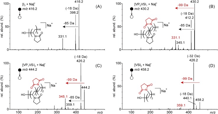
With the fragmentation pathways of the pristine PVP as a background knowledge, the aged PVP sample has then been submitted to CID to validate the copolymeric structure proposed from the MALDI high resolution MS analysis, itself based on the literature. As expected, the hypothesized P(VP-co-VSI) copolymer is readily seen in the ESI-MS spectrum of the aged sample in the form of sodium adducts owing to the use of an excess of NaOCl as oxidizing agent (Fig. S11 in the Supporting Information). Surprisingly, only the shortest chains have been detected with satisfactory intensity (up to n=6) while pristine PVP is found in the higher mass range. This variation between MALDI and ESI remains unexplained but is of no consequence on the results: since the CID of PVP mainly concerns the pendant groups of the repeating units, the length of the dissociating species is of limited impact (Fig. 2). The ESI-MS/MS spectra of the sodium adducts of PVP, P(VP-co-VSI) and PVSI 3-mers at m/z 416.3, m/z 430.2, m/z 444.2 and m/z 458.2 are depicted in Fig. 5A–D, respectively.
Fig. 5. ESI-MS/MS spectra of the sodiated PVP, P(VP-co-VSI) and PVSI 3-mers at m/z 416.3 (VP3), m/z 430.2 (VP2VSI1), m/z 444.2 (VP1VSI2) and m/z 458.1 (VSI3). The generic structure of each precursor ion is depicted in inset of the associated spectrum.
Following the behavior of its larger homologues, the sodiated PVP 3-mer at m/z 416.3 (Fig. 5A) readily releases water (−18.0 Da) and one pyrrolidone (−85.1 Da). The relatively high abundance of [VP3+Na−H2O]+ is accounted for by considering the short chain length—only three units. Indeed, the end-groups exert usually a stronger influence on the CID pattern for the shortest chains while their impact gradually decreases with the increase of the polymerization degree.47) The ESI-MS/MS spectrum of m/z 430.2—spaced by +14.0 Da from the untouched PVP oligomer and assigned to a [VP2VSI1+Na]+ co-oligomeric sodium adduct—mainly displays the same two 18.0 Da and 85.1 Da losses in addition to a new release of a 99.0 Da neutral from the precursor ion yielding a [VP2VSI1−99 Da+Na]+ product ion at m/z 331.1 (Fig. 5B). Such neutral loss is in accordance with the oxidation of one pyrrolidone pendant group into succinimide (C6H9NO, 111.1 Da→C6H7NO2, 125.0 Da). The neutral expelled by the precursor ion could then be either a γ-butyrolactim from a pristine vinyl pyrrolidone unit or a succinimide from the newly formed vinyl succinimide (more precisely its stable tautomer carrying a C=N double bond,48) C4H5NO2, 99.0 Da). The ESI-MS/MS spectrum of m/z 444.2 assigned to a VP1VSI2 sodium adduct displays the same pattern with the detection of the −18.0 Da/−85.1 Da/−99.0 Da neutral losses triplet (Fig. 5C). Since only one −85.1/−99.1 Da loss is observed from the precursor ion in MS/MS, it is not possible to discriminate VP2VSI1 from VP1VSI2. In both cases [VPxVSIy+Na−99 Da]+ is far more intense than [VPxVSIy+Na−85 Da]+ and prevents from an empirical correlation between the peak intensities and the composition of the precursor ion. The presence of an additional C=O electrophilic group within the pendant group increases the statistics of the charge-remote 1,5-H transfer to occur as well as weakens the C–N bond between the backbone and the so-formed succinimide. The fully oxidized PSI 3-mer at m/z 458.1 does not eliminate the pyrrolidone neutral any longer accordingly to its proposed composition but a new −32.0 Da neutral which has been hypothetically assigned to O2 (Fig. 5D). The mechanism of this elimination remains unclear. A last step remains to fully characterize the co-oligomeric degradation products and consists in the proper distinction of the VPxVSIy species using an additional MS3 step. The ESI-MS3 spectra of the two product ions formed upon a first release of succinimide (99 Da) from the two VPxVSIy 3-mers are depicted in Fig. 6A ([VP2VSI1+Na−99 Da]+ at m/z 331.1) and 6B ([VP1VSI2+Na−99 Da]+ at m/z 345.1). In the first case, a unique 85.1 Da loss is observed from the precursor ion (producing a [VP1VSI2+Na−99 Da−85 Da]+ fragment ion at m/z 345.1), in accordance with the presence of (two) VP repeating units only and validating the proposed structure of the precursor ion depicted in the inset of Fig. 6A. On the contrary, a dual 85.1 Da/99.0 Da neutral expulsion is detected in the ESI-MS3 spectrum of m/z 345.1 (producing two [VP1VSI2+Na−99 Da−85 Da]+ at m/z 260.1 and [VP1VSI2+Na−2*99 Da]+ at m/z 246.1, respectively) thus highlighting the presence of both VP and VSI within the backbone (inset of Fig. 6B). It is worth mentioning the composition of the two VPxVSIy degradation products can be differentiated at the MS3 step thanks to their short chain length (3-mers) but additional MSn steps (n>3) would be required to gain insights into the VP/VSI composition of larger co-oligomeric chains, making the whole procedure time-consuming. This pitfall has been overcome with the protonation of the PVP oligomers which drastically simplifies the analysis by strictly limiting the multi stage MSn to n=3 to observe the complete [M+H−H2O−x*85 Da]+ product ions series (Fig. 4). Protonated chains were thus tentatively formed by diluting the bleach solution in methanolic NH4Cl. However only the sodium adducts were detected regardless of the dilution factor. To circumvent this issue, the PVP oligomers and degradation products were partly recovered from the aqueous bleach solution by a liquid/liquid extraction using CHCl3 as the organic phase, further diluted in methanolic NH4Cl. The protonated PVP and degraded PVP chains were eventually seen with low abundance but intense enough for the MS3 spectra to be recorded. The ESI-MS3 of the dehydrated PVP and P(VP-co-VSI) 3-mers (noted [M+H−H2O]+, isolated from the ESI-MS/MS spectra of the associated [M+H]+) are depicted in Fig. 7 (the fully oxidized PSI 3-mer was not detected with enough intensity to record its spectra).
Fig. 6. ESI-MS3 of the [M+Na−99 Da]+ product ions formed upon the release of succinimide from A) VP2SI1 at m/z 430.2 yielding m/z 331.1 and B) VP1SI2 at m/z 444.2 yielding m/z 345.1. The proposed structures of the dissociating species are depicted in insets.
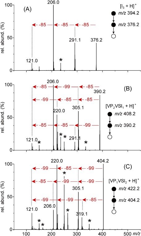
Fig. 7. ESI-MS3 spectra of dehydrated PVP and P(VP-co-VSI) 3-mers at m/z 376.2 (VP3), m/z 390.2 (VP2VSI1) and m/z 404.2 (VP1VSI2) formed upon the release of water from the protonated (co-)oligomers. Asterisks designate the product ions formed upon the loss of 58.0 Da neutral (proposed to be C3H6O) from the 2-hydroxyisopropyl termination.
As for the larger congeners of the pristine PVP (Fig. 4), [PVP3+H−H2O]+ at m/z 376.2 iteratively eliminates its three pyrrolidone pendant groups (85.1 Da loss) in a single scan yielding the tri-unsaturated [PVP3+H−H2O−3*85 Da]+ at m/z 121.0 (Fig. 7A). Following the same trend, [P(VP2-co-VSI1)+H−H2O]+ at m/z 390.2 now expels both 85.1 Da and 99.1 Da neutrals in accordance to its proposed composition. Based on the m/z ratios, considering one 99.0 Da and two 85.1 Da losses with their three possible iterative occurrence allows the main peaks to be assigned (−85.1→−85.1→−99.0 or −85.1→−99.0→−85.1 or −99.0→−85.1→−85.1, Fig. 7B). Similarly, the product ions detected in the ESI-MS3 spectrum of [P(VP1-co-VSI2)+H−H2O]+ are readily assigned by considering the combination of two 99.0 Da and one 85.1 Da losses (−85.1→−99.0→−99.0 or −99.0→−99.0→−85.1 or −99.0→−85.1→−99.0, Fig. 7C). Thanks to the production of the complete series of −85.1/−99.0 Da product ions, the two co-oligomers could thus be differentiated based on their CID fingerprint without the need for supernumerary MSn steps. It should also be noted that contrary to the case of the charge remote eliminations of 85.1/99.0 Da for the sodium adducts, the intensity of the product ions formed upon the first charge driven 85.1/99.0 Da loss from the dehydrated co-oligomers reflects their composition (−1*85.1 Da is three times more abundant than −1*99.0 Da for VP2-co-SI1, and vice-versa for VP1-co-SI2). Despite the protonation of PVP chains is not straightforward for the aged samples owing to the dominion of the sodium cations, their production should be favored whenever it is possible with regard to the advantages offered (MS3 only, lower activation energy leading to higher signal-to-noise ratios and shorter accumulation times).
CONCLUSION
The degradation of a PVP chain exposed to bleach into its vinyl succinimide copolymer has been unambiguously highlighted using high resolution MALDI spiralTOF-MS and multi-stage ESI-MSn, in great accordance with the literature but operated at a molecular level when the previously published studies were relying on chemical analyses. In particular, the fragmentation pathways of PVP oligomers adducted with sodium and proton have been thoroughly investigated in order to get a sufficient knowledge for the exploration of aged PVP samples. For both sodium and proton adductions, the main dissociation route consists in the release of the pendant pyrrolidone group of the repeating units in the form of γ-butyrolactim, upon a charge remote mechanism for the sodium adducts which requires MSn steps while a MS3 step is sufficient in the case of protonated oligomers to detect the whole associated product ion series in a charge driven process. In the case of the aged PVP, an alternative succinimide neutral loss has then been detected with similar features (charge remote in MSn for the sodium adduction, charge driven in MS3 for the protonation), validating the vinyl pyrrolidone/vinyl succinimide copolymeric structure hypothesized from the single stage mass analysis. It constitutes an example of direct extrapolation of the fragmentation routes found for a standard and used for the characterization of an unknown sample, and exemplifies the interest of the protonation (in lieu of alkali adduction) for the tandem mass spectrometry of polymer ions.
Acknowledgments
T. Fouquet and H. Sato gratefully acknowledge the ongoing financial support by the Japan Society for the Promotion of Science (JSPS) under the postdoctoral fellowship for overseas researchers program (FY2015) and a Grant-in-Aid “JSPS KAKENHI” (Grant number: JP 15F15344).
Mass Spectrom (Tokyo) 2016; 5(1): A0050
References
- 1) X. Liu, Y. Xu, Z. Wu, H. Chen. Poly(N-vinylpyrrolidone)-modified surfaces for biomedical applications. Macromol. Biosci. 13: 147–154, 2013. [DOI] [PubMed] [Google Scholar]
- 2) F. Haaf, A. Sanner, F. Straub. Polymers of N-vinylpyrrolidone: Synthesis, characterization and uses. Polym. J. 17: 143–152, 1985. [Google Scholar]
- 3) K. M. Koczkur, S. Mourdikoudis, L. Polavarapu, S. E. Skrabalak. Polyvinylpyrrolidone (PVP) in nanoparticle synthesis. Dalton Trans. 44: 17883–17905, 2015. [DOI] [PubMed] [Google Scholar]
- 4) I. M. Wienk, E. E. B. Meuleman, Z. Borneman, T. Van Den Boomcaard, C. A. Smolders. Chemical treatment of membranes of a polymer blend: Mechanism of the reaction of hypochlorite with poly(vinyl pyrrolidone). J. Polym. Sci. A Polym. Chem. 33: 49–54, 1995. [Google Scholar]
- 5) Y. Hanafi, A. Szymczyk, M. Rabiller-Baudry, K. Baddari. Degradation of poly(ether sulfone)/polyvinylpyrrolidone membranes by sodium hypochlorite: Insight from advanced electrokinetic characterizations. Environ. Sci. Technol. 48: 13419–13426, 2014. [DOI] [PubMed] [Google Scholar]
- 6) Y. Hanafi, P. Loulergue, S. Ababou-Girard, C. Meriadec, M. Rabiller-Baudry, K. Baddar, A. Szymczy. Electrokinetic analysis of PES/PVP membranes aged by sodium hypochlorite solutions at different pH. J. Membr. Sci. 501: 24–32, 2016. [Google Scholar]
- 7) B. Pellegrin, R. Prulho, A. Rivaton, S. Thérias, J.-L. Gardette, E. Gaudichet-Maurin, C. Causserand. Multi-scale analysis of hypochlorite induced PES/PVP ultrafiltration membranes degradation. J. Membr. Sci. 447: 287–296, 2013. [Google Scholar]
- 8) R. Prulho, S. Thérias, A. Rivaton, J.-L. Gardette. Ageing of polyethersulfone/polyvinylpyrrolidone blends in contact with bleach water. Polym. Degrad. Stabil. 98: 1164–1172, 2013. [Google Scholar]
- 9) L. Charles. MALDI of synthetic polymers with labile end-groups. Mass Spectrom. Rev. 33: 523–543, 2014. [DOI] [PubMed] [Google Scholar]
- 10) G. Montaudo, R. P. Lattimer. Mass spectrometry of polymers, CRC Press, Taylor and Francis Group, Oxford, 2001.
- 11) T. Gruendling, S. Weidner, J. Falkenhagen, C. Barner-Kowollik. Mass spectrometry in polymer chemistry: A state-of-the-art up-date. Polym. Chem. 1: 599–617, 2010. [Google Scholar]
- 12) M. Hakkarainen. Mass spectrometry of polymers—New techniques. Springer-Verlag, Berlin-Heidelberg-New York, 2012.
- 13) S. D. Hanton. Mass spectrometry of polymers and polymer surfaces. Chem. Rev. 101: 527–569, 2001. [DOI] [PubMed] [Google Scholar]
- 14) C. A. Jackson, W. J. Simonsick Jr. Application of mass spectrometry to the characterization of polymers. Curr. Opin. Solid State Mater. Sci. 2: 661–667, 1997. [Google Scholar]
- 15) H. Pasch, W. Schrepp. MALDI-TOF mass spectrometry of synthetic polymers, Springer-Verlag, Berlin–Heidelberg–New York, 2003.
- 16) N. Fandrich, J. Falkenhagen, S. M. Weidner, B. Staal, A. F. Thunemann, A. Laschewsky. Characterization of new amphiphilic block copolymers of N-vinylpyrrolidone and vinyl acetate, 2—Chromatographic separation and analysis by MALDI-TOF and FT-IR coupling. Macromol. Chem. Phys. 211: 1678–1688, 2010. [Google Scholar]
- 17) S. Trimpin, P. Eichhorn, H. J. Rader, K. Mullen, T. P. Knepper. Recalcitrance of poly(vinylpyrrolidone): Evidence through matrix-assisted laser desorption-ionization time-of-flight mass spectrometry. J. Chromatogr. A 938: 67–77, 2001. [DOI] [PubMed] [Google Scholar]
- 18) K. Krueger, C. Terne, C. Werner, U. Freudenberg, V. Jankowski, W. Zidek, J. Jankowski. Characterization of polymer membranes by MALDI mass-spectrometric imaging techniques. Anal. Chem. 85: 4998–5004, 2013. [DOI] [PubMed] [Google Scholar]
- 19) P. Malvagna, G. Impallomeni, R. Cozzolino, E. Spina, D. Garozzo. New results on matrix-assisted laser desorption/ionization mass spectrometry of widely polydisperse hydrosoluble polymers. Rapid Commun. Mass Spectrum. 16: 1599–1603, 2002.
- 20) S. Trimpin, S. Keune, H. J. Räder, K. Müllen. Solvent-free MALDI-MS: Developmental improvements in the reliability and the potential of MALDI in the analysis of synthetic polymers and giant organic molecules. J. Am. Soc. Mass Spectrom. 17: 661–671, 2006. [DOI] [PubMed] [Google Scholar]
- 21) R. Pfukwa. Synthesis and characterization of telechelic hydroxyl functional PVP. Dissertation (MSc), Stellenbosch University, 2008.
- 22) E. Ranucci, P. Ferruti, R. Annunziata, I. Gerges, G. Spinelli. NMR spectroscopy and MALDI-TOF MS characterisation of end-functionalised PVP oligomers prepared with different esters as chain transfer agents. Macromol. Biosci. 6: 216–227, 2006. [DOI] [PubMed] [Google Scholar]
- 23) T. Fouquet, S. Humbel, L. Charles. Tandem mass spectrometry of trimethylsilyl-terminated poly(dimethylsiloxane) ammonium adducts generated by electrospray ionization. J. Am. Soc. Mass Spectrom. 22: 649–658, 2011. [DOI] [PubMed] [Google Scholar]
- 24) T. Fouquet, J. Petersen, J. A. S. Bomfim, J. Bour, F. Ziarelli, D. Ruch, L. Charles. Electrospray tandem mass spectrometry combined with authentic compound synthesis for structural characterization of an octamethylcyclotetrasiloxane plasma polymer. Int. J. Mass Spectrom. 313: 58–67, 2012. [Google Scholar]
- 25) A. M. Yol, D. E. Dabney, S.-F. Wang, B. Laurent, M. D. Foster, R. P. Quirk, S. M. Grayson, C. Wesdemiotis. Differentiation of linear and cyclic polymer architectures by MALDI tandem mass spectrometry (MALDI-MS2). J. Am. Soc. Mass Spectrom. 24: 74–82, 2013. [DOI] [PubMed] [Google Scholar]
- 26) C. Wesdemiotis, N. Solak, M. J. Polce, D. E. Dabney, K. Chaicharoen, B. C. Katzenmeyer. Fragmentation pathways of polymer ions. Mass Spectrom. Rev. 30: 523–559, 2011. [DOI] [PubMed] [Google Scholar]
- 27) T. Fouquet, C. Chendo, V. Toniazzo, D. Ruch, L. Charles. Collision-induced dissociation of synthetic polymers containing hydride groups: The case of poly(methylhydrosiloxane) homopolymers and poly(methylhydrosiloxane)-co-(dimethylsiloxane) copolymers. Rapid Commun. Mass Spectrom. 27: 88–96, 2013. [DOI] [PubMed] [Google Scholar]
- 28) T. Fouquet, J. Bour, V. Toniazzo, D. Ruch, L. Charles. Characterization of ethanolysis products of poly(dimethylsiloxane) species by electrospray ionization tandem mass spectrometry. Rapid Commun. Mass Spectrom. 26: 2057–2067, 2012. [DOI] [PubMed] [Google Scholar]
- 29) A. P. Gies, W. K. Nonidez, M. Anthamatten, R. C. Cook, J. W. Mays. Characterization of an insoluble polyimide oligomer by matrix-assisted laser desorption/ionization time-of-flight mass spectrometry. Rapid Commun. Mass Spectrom. 16: 1903–1910, 2002. [DOI] [PubMed] [Google Scholar]
- 30) T. Satoh, H. Tsuno, M. Iwanaga, Y. Kammei. The design and characteristics features of a new time-of-flight mass spectrometer with a spiral ion trajectory. J. Am. Soc. Mass Spectrom. 16: 1969–1975, 2005. [DOI] [PubMed] [Google Scholar]
- 31) M. Strohalm, D. Kavan, P. Novák, M. Volný, V. Havlícek. mMass 3: A cross-platform software environment for precise analysis of mass spectrometric data. Anal. Chem. 82: 4648–4651, 2010. [DOI] [PubMed] [Google Scholar]
- 32) H. Sato, S. Nakamura, K. Teramoto, T. Sato. Structural characterization of polymers by MALDI Spiral-TOF mass spectrometry combined with Kendrick mass defect analysis. J. Am. Soc. Mass Spectrom. 25: 1346–1355, 2014. [DOI] [PMC free article] [PubMed] [Google Scholar]
- 33) T. Fouquet, S. Nakamura, H. Sato. MALDI SpiralTOF high-resolution mass spectrometry and Kendrick mass defect analysis applied to the characterization of poly(ethylene-co-vinyl acetate) copolymers. Rapid Commun. Mass Spectrom. 30: 973–981, 2016. [DOI] [PMC free article] [PubMed] [Google Scholar]
- 34) T. Fouquet, H. Aizawa, H. Sato. Taking MALDI SpiralTOF high-resolution mass spectrometry and mass defect analysis to the next level with ethylene vinyl acetate vinyl alcohol terpolymers. Rapid Commun. Mass Spectrom. 30: 1818–1822, 2016. [DOI] [PubMed] [Google Scholar]
- 35) L. Luo, M. Ranger, D. G. Lessard, D. Le Garrec, S. Gori, J.-C. Leroux, S. Rimmer, D. Smith. Novel amphiphilic diblock copolymer of low molecular weight poly(N-vinylpyrrolidone)-block-poly(d,l-lactide): Synthesis, characterization, and micellization. Macromolecules 37: 4008–4013, 2004. [Google Scholar]
- 36) M. Tomida, T. Nakato, S. Matsunami, T. Kakuchi. Convenient synthesis of high molecular weight poly(succinimide) by acid-catalysed polycondensation of L-aspartic acid. Polymer (Guildf.) 38: 4733–4736, 1997. [Google Scholar]
- 37) K. Matsubara, T. Nakato, M. Tomida. 1H and 13C NMR characterization of poly(succinimide) prepared by thermal polycondensation of L-aspartic acid. Macromolecules 30: 2305–2312, 1997. [Google Scholar]
- 38) J. Furukawa, T. Tsuruta, H. Fukutani, N. Yamamoto. Copolymerization of N-vinyl succinimide. J. Soc. Chem. Indust. Jpn. 60: 1085–1086, 1957. [Google Scholar]
- 39) E. Sivtsov, E. Chernikova, A. Gostev, E. Garina. Controlled free-radical copolymerization of N-vinyl succinimide and n-butyl acrylate via a reversible addition-fragmentation chain transfer (RAFT) technique. Macromol. Symp. 296: 112–120, 2010. [Google Scholar]
- 40) C. N. McEwen, W. J. Simonsick Jr., B. S. Larsen, K. Ute, K. Hatada. The fundamentals of applying electrospray ionization mass spectrometry to low mass poly(methyl methacrylate) polymers. J. Am. Soc. Mass Spectrom. 6: 906–911, 1995. [DOI] [PubMed] [Google Scholar]
- 41) G. Montaudo, R. P. Lattimer. Mass spectrometry of polymers, CRC Press, Taylor and Francis Group, Boca Raton, FL, 2002
- 42) R. Giordanengo, S. Viel, B. Allard-Breton, A. Thévand, L. Charles. Positive mode electrospray tandem mass spectrometry of poly(methacrylic acid) oligomers. Rapid Commun. Mass Spectrom. 23: 1557–1562, 2009. [DOI] [PubMed] [Google Scholar]
- 43) T. Fouquet, F. Ziarelli, H. Sato, L. Charles. Structural characterization of polymethoxymethylsiloxanes by electrospray ionization tandem mass spectrometry. Int. J. Mass Spectrom. 402: 42–48, 2016. [Google Scholar]
- 44) T. Fouquet. Mass spectrometry of synthetic polysiloxanes: From linear models to plasma polymer networks. ChemistryOpen 3: 269–273, 2014. [DOI] [PMC free article] [PubMed] [Google Scholar]
- 45) A. M. Alhazmi, P. M. Mayer. Protonating polymer oligomers in the gas phase to change fragmentation pathways. J. Am. Soc. Mass Spectrom. 20: 60–66, 2009. [DOI] [PubMed] [Google Scholar]
- 46) J. Renaud, A. M. Alhazmi, P. M. Mayer. Comparing the fragmentation chemistry of gas-phase adducts of poly(dimethylsiloxane) oligomers with metal and organic ions. Can. J. Chem. 87: 453–459, 2009. [Google Scholar]
- 47) T. Fouquet, S. Humbel, L. Charles. Dissociation characteristics of α,ω-dihydride PDMS ammonium adducts generated by ESI. Int. J. Mass Spectrom. 306: 70–76, 2011. [Google Scholar]
- 48) Y. Valadbeigi, H. Farrokhpour. Simple and water-assisted tautomerism in succinimide. Struct. Chem. 26: 539–545, 2015. [Google Scholar]


![Fig. 4. (A) ESI-MS/MS of the protonated [I7+H]+ at m/z 838.6 and (B) ESI-MS3 of [I7+H−H2O]+ at m/z 820.5 formed upon the release of water from the precursor ion which readily eliminates the pyrrolidone pendant groups. (C) Mechanism proposed to account for the charge driven loss of pyrrolidone.](https://cdn.ncbi.nlm.nih.gov/pmc/blobs/8f8b/5086575/3095cbb202b0/massspectrometry-5-1-A0050-figure004.jpg)
![Fig. 6. ESI-MS3 of the [M+Na−99 Da]+ product ions formed upon the release of succinimide from A) VP2SI1 at m/z 430.2 yielding m/z 331.1 and B) VP1SI2 at m/z 444.2 yielding m/z 345.1. The proposed structures of the dissociating species are depicted in insets.](https://cdn.ncbi.nlm.nih.gov/pmc/blobs/8f8b/5086575/0c99131bb330/massspectrometry-5-1-A0050-figure006.jpg)
