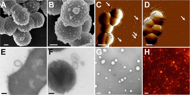
FIG 1 .
GAS cells exhibit MV protrusions during in vitro growth. Cellular morphologies of ISS3348 (A, C, and E) and SF370 (B, D, and F) were examined by SEM (A and B), AFM (C and D), and TEM (E and F). Isolated MVs from ISS3348 were visualized using negative-stain TEM (G) and staining for M1 protein and STED microscopy (H). White arrowheads indicate vesicle-like structures. Scale bars are drawn to 200 nm (A and B and E to G) or 500 nm (C, D, and H).

