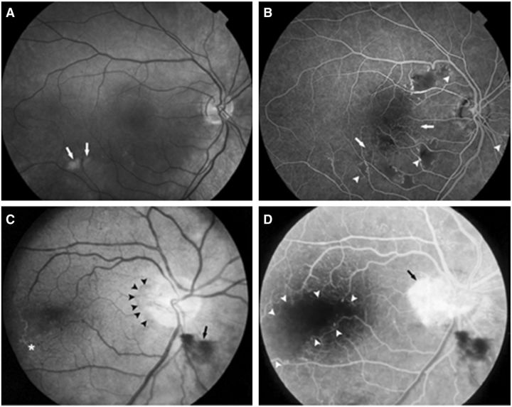
Figure 2.
Fundoscopic (A and C) and fluorescein angiogram (B and D) images of vascular retinopathy. (A and B) Right eye of a 33-year-old male with cotton-wool spots (arrows, A), extensive areas of capillary obliteration with non-perfusion (arrows, B), and intraretinal microvascular abnormalities (arrow heads, B). (C and D) Right eye of a 48-year-old female with a neovascular membrane (arrowheads, C) and preretinal haemorrhage (arrow, C). Temporal to the macula, vascular sheathing and occlusion is present (asterisk, C). The same eye shows profuse leakage from the membrane on the disc (arrow, D) and a large avascular region involving the fovea (arrow heads, D).

