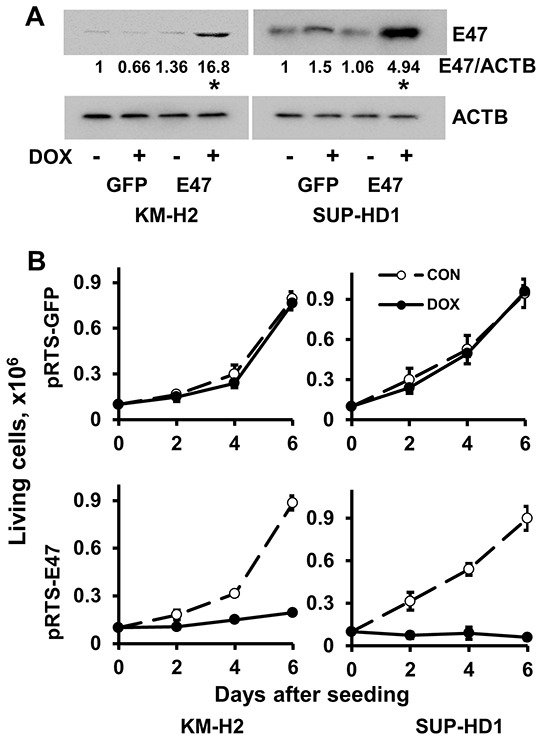
Figure 1. E47 induces cell death in cHL cell lines.

A. KM-H2 and SUP-HD1 cell lines conditionally expressing E47 were established using the pRTS vector system. After transfection, cells were selected with hygromycin for two weeks (KM-H2) and one week (SUP-HD1), more than 95% cells were GFP positive upon DOX treatment. Expression of E47 protein was measured by immunobloting using anti-TCF3 antibody. Anti-ACTB antibody was used as a loading control. * p<0.05 (expression levels upon DOX treatment compared with that of samples without DOX treatment) as it was assessed by double sided T-test. B. Cells transfected with empty pRTS vector (pRTS-GFP) or pRTS-E47 were seeded at low density (0.1 × 10⁶ cells per well in 3 ml of complete medium). DOX was added on day 0 and day 3. On day 2, day 4 and day 6, the cells were counted by flow cytometer and viable cells were discriminated using side/forward scatter parameters. C. The nuclear fragmentation was measured by Nicoletti method after 4 days of incubation with DOX. * p<0.05 as it was assessed by double sided T-test. D. The influence of E47 on cell cycle progression. Cells were seeded at density of 0.5×10⁶ per 10 ml of complete medium and treated with 0.5 μg/ml DOX. After 2 days, cell cycle distribution was measured by PI staining as described in the Materials and Methods section. Experiments were repeated three times. Results are presented as mean ± SD. * p<0.05.
