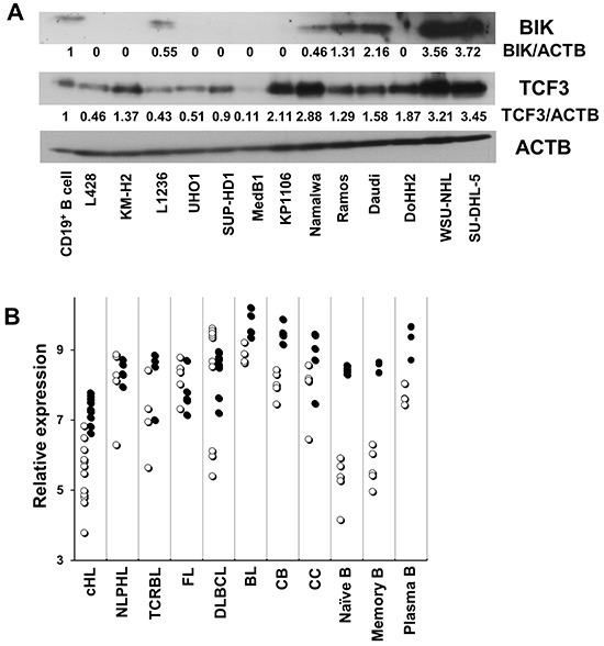
Figure 4. The expression of BIK correlates with that of TCF3/E2A.

A. The expression of BIK and TCF3 in various B cell lymphoma cell lines and CD19+ B cells was measured by immunobloting. Anti-ACTB antibody was used as a loading control. The immunoblots in (A) were quantified using ImageJ software. B. Published gene expression data of microdissected tumor cells including 12 cHL cases, 5 nodular lymphocyte predominant Hodgkin's lymphoma (NLPHL) cases, 4 T cell - rich B cell lymphoma (TCRBL) cases, 5 follicular lymphoma (FL) cases, 11 diffuse large B cell lymphoma (DLBCL) cases, 5 BL cases, and normal B cell subtypes (5 samples each) including centroblast (CB), centrocyte (CC), Naïve B cell (N), memory B cell (M), plasma cell (PC), were re-analyzed with help of the Genesifter software (Perkin Elmer, Seattle, WA). The data are shown as log2 of fluorescence intensity. The hollow circle (○) stands for BIK, the solid circle (•) stands for TCF3.
