Abstract Abstract
Rhodnius marabaensis sp. n. was collected on 12 May 2014 in the Murumurú Environmental Reserve in the city of Marabá, Pará State, Brazil. This study was based on previous consultation of morphological descriptions of 19 Rhodnius species and compared to the identification key for the genus Rhodnius. The examination included specimens from 18 Rhodnius species held in the Brazilian National and International Triatomine Taxonomy Reference Laboratory in the Oswaldo Cruz Institute in Rio de Janeiro, Brazil. The morphological characteristics of the head, thorax, abdomen, genitalia, and eggs have been determined. Rhodnius prolixus and Rhodnius robustus were examined in more detail because the BLAST analysis of a cyt-b sequence shows they are closely related to the new species, which also occurs in the northern region of Brazil. The most notable morphological features that distinguish Rhodnius marabaensis sp. n. are the keel-shaped apex of the head, the length of the second segment of the antennae, the shapes of the prosternum, mesosternum and metasternum, the set of spots on the abdomen, the male genitalia, the posterior and ventral surfaces of the external female genitalia, and the morphological characteristics of the eggs. Rhodnius jacundaensis Serra, Serra & Von Atzingen (1980) nomen nudum specimens deposited at the MCCF were examined and considered as a synonym of Rhodnius marabaensis sp. n.
Keywords: Triatominae, Rhodnius marabaensis sp. n., new species, Amazon
Introduction
Vectors of the protozoan Trypanosoma cruzi, the etiological agent of Chagas disease, include 151 species distributed into 18 genera belonging to the subfamily Triatominae (Galvão 2014, Mendonça et al. 2016). The genus Rhodnius includes 19 species (Alevi et al. 2015), of which six were described after the publication of the Lent and Wygodzinsky review (1979): Rhodnius stali Lent, Jurberg & Galvão, 1993; Rhodnius colombiensis Mejia, Galvão & Jurberg, 1999; Rhodnius milesi (Carcavallo, Rocha, Galvão & Jurberg, 2001); Rhodnius zeledoni Jurberg, Rocha & Galvão, 2009; Rhodnius montenegrensis Rosa et al., 2012, and Rhodnius barretti Abad-Franch, Palomeque & Monteiro, 2013. Rhodnius amazonicus Almeida, Santos & Sposina, 1973 was synonymized with Rhodnius pictipes by Lent and Wygodzinsky (1979) according a photograph of the holotype, but subsequently it was validated by Bérenger and Pluot-Sigwalt (2002) by morphological study of 19 characters with of the two species.
Among the Rhodnius species, only nine are found in the northern region of Brazil: Rhodnius amazonicus, Rhodnius brethesi, Rhodnius milesi, Rhodnius montenegrensis, Rhodnius neglectus, Rhodnius paraensis, Rhodnius pictipes, Rhodnius robustus, and Rhodnius stali (Galvão 2014, Meneguetti et al. 2015).
In May 2014, two Rhodnius spp. specimens were collected in Marabá, Pará, Brazil, and compared to the key described by Lent and Wygodzinsky (1979), as well as to previously described Rhodnius species, without success. These samples were compared and identified as the same species as Rhodnius jacundaensis Serra, Serra & Von Atzingen, 1980 which had been deposited at the Marabá Cultural Center in Pará. Rhodnius jacundaensis was mentioned in an abstract presented at the Fourth Annual Brazilian Parasitology Conference in Rio de Janeiro in 1980. According to Article 9 of the International Code of Zoological Nomenclature, however, this new species was not confirmed. As a result, Carcavallo et al. (1998) and Galvão et al. (2003) considered it to be Rhodnius jacundaensis Serra, Serra & Von Atzingen, 1980) nomen nudum. This article describes Rhodnius marabaensis sp. n. as the tenth species found in northern Brazil and the twentieth member of this genus (Abad Franch et al. 2013, Galvão 2014, Meneguetti et al. 2014, Alevi et al. 2015).
Materials and methods
Morphological identification and description
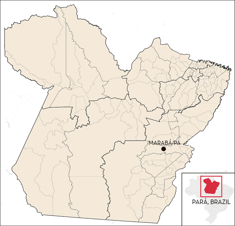
The specific description was based on the observation of two adult specimens (one female and one male) collected in a residence in the Murumuru Environmental Reserve in the city of Marabá, Pará, Brazil (coordinates: 10°10'05.1"S and 63°24'09.1"W) (Fig. 1). The description included 14 males and 14 females deposited at the MCCF and previously characterized as Rhodnius jacundaensis [nomen nudum].
Figure 1.
Localization of Marabá- PA where Rhodnius marabaensis sp. n. specimens were collected (05°21'54"S, 49°07'24"W).
The identification of samples was performed using the dichotomous key by Lent and Wygodzinsky (1979). The study also considered descriptions of Rhodnius amazonicus, Rhodnius stali, Rhodnius colombiensis, Rhodnius milesi, Rhodnius zeledoni, Rhodnius montenegrensis, and Rhodnius barretti (Almeida et al. 1973, Lent et al. 1999, Mejia et al. 1999, Valente et al. 2001, Jurberg et al. 2009, Rosa et al. 2012, Abad-Franch et al. 2013). Rhodnius marabaensis was also compared to specimens of 18 Rhodnius species held at the Brazilian National and International Triatomine Taxonomy Reference Laboratory at the Oswaldo Cruz Institute in Rio de Janeiro, Brazil. The only species that was compared only by description was Rhodnius amazonicus, which is held at the (INPA).
Genetic identification
After the identification, the mitochondrial gene fragment (cyt-b) was amplified by using suggested primers by Monteiro et al. (2003). The amplified fragments were purified and sequenced in duplicate (forward and reverse). The same haplotype was shown with 693 (bp). This sequence was evaluated by BLAST (http://www.blast.ncbi.nlm.nih.gov/Blast.cgi) to diagnose the homologous sequences in GenBank. In view of this and of the fact that all three species occur in Northern Brazil (Galvão et al. 2003, Galvão 2014), Rhodnius robustus and Rhodnius prolixus were morphologically examined and compared in more detail.
Morphological study
For the comparative morphological study, Rhodnius prolixus specimens from (CTA) 074 were used, as were Rhodnius robustus specimens from CTA085. The specimens were kept in these colonies at the Triatominae Insectarium at the (UNESP), Araraquara, São Paulo, Brazil. The Rhodnius prolixus colony had been originally collected in a sylvatic environment in Venezuela on 23 May 1983. The Rhodnius robustus colony has been maintained since February 1972 using specimens from Peru (Fig. 2).
Figure 2.
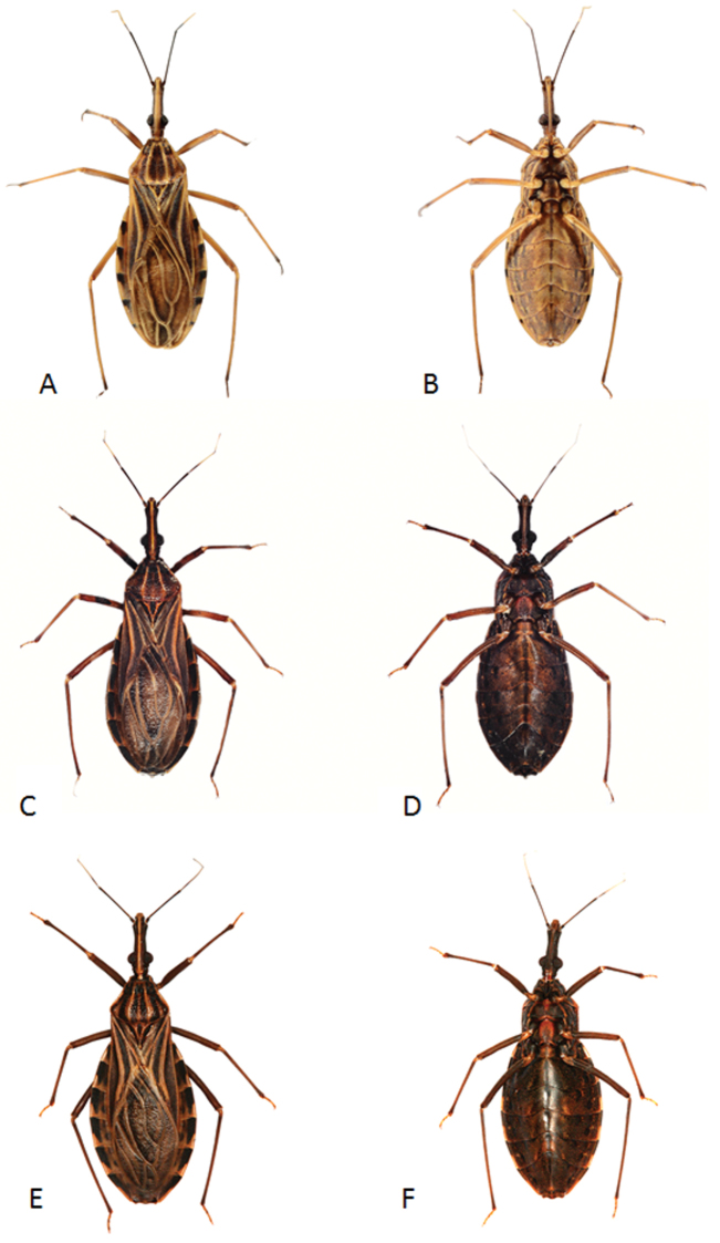
Rhodnius marabaensis sp. n. female (A dorsal side B ventral side); Rhodnius prolixus female (C dorsal side D ventral side); Rhodnius robustus female (E dorsal side F ventral side).
Optical microscopy and (SEM) were used to compare the morphology of Rhodnius marabaensis sp. n., Rhodnius prolixus, and Rhodnius robustus. The head, the ventral portion of the thorax, the scutellum, and the pygophore were studied using SEM (Figs 3–6, 8). A female of Rhodnius marabaensis sp. n. that had been collected in May 2014 was used to study the female genitalia, and 13 eggs obtained from its uterus were also analyzed by SEM (Figs 9–13).
Figure 3.
Head of Rhodnius marabaensis sp. n. (A), Rhodnius prolixus (B), Rhodnius robustus (C). V; C; AC.
Figure 6.
Thorax ventral of Rhodnius marabaensis sp. n. (A), Rhodnius prolixus (B), Rhodnius robustus (C). ms; mt.
Figure 8.
Median process of the pygophore of Rhodnius marabaensis sp. n. (A), Rhodnius prolixus (B), Rhodnius robustus (C). gp: nb; sp; wb.
Figure 9.
Female external genitalia, dorsal side of Rhodnius marabaensis sp. n. (A), Rhodnius prolixus (B), Rhodnius robustus (C). VI, VII, VIII, IX, X, tergites.
Figure 13.
Egg exochorium detail of Rhodnius marabaensis sp. n. (A), Rhodnius prolixus (B), Rhodnius robustus (C). ec; ft; ll.
Morphometric study
A Leica MZ APO stereoscope and the Motic Images Advanced System version 3.2 were used for the measurements, as well as for the study of Rhodnius marabaensis sp. n. male genitalia. For the comparative study of the male genitalia of Rhodnius marabaensis sp. n. and Rhodnius prolixus and Rhodnius robustus, the descriptions by Lent and Jurberg (1969) and by Rosa et al. (2012) were used.
The following parameters of both females (15) and males (15) were measured: (TL); (HL); the length of the four antennal segments (A1, A2, A3, and A4); the three segments of the proboscis (R1, R2, and R3); the (IE); the (OE); the (DE); the (MWA); the (MWT); and the length, width, and diameter of the opercular opening of the eggs (Lent and Wygodzinsky 1979, Dujardin et al. 1999, Rosa et al. 2000).
Taxonomy
Family Reduviidae Latreille, 1807: Subfamily Triatominae Jeannel, 1919: Genus Rhodnius Stål, 1859
Rhodnius marabaensis sp. n.
http://zoobank.org/883B9B62-9E78-4AFF-9518-021593A308A4
Holotype
♂. BRAZIL: Pará: Marabá: Reserva Ambiental Murumurú, 10°10'05.1"S, 63°24'09.1"W, 12 May 2014, N. C. B. Von Atzingen, M. B. Furtado, UNESP.
Paratypes.
1 ♀: same data as holotype (UNESP). 14 ♀, 14 ♂ BRAZIL: Pará: Jacundá/Jatobal/Marabá,N.C.B. Von Atzingen, Maraba Cultural Center Foundation – MCCF.
Synonym.
Rhodnius jacundaensis Serra, Serra and Von Atzingen (1980) [nomen nudum].
Etymology.
The name Rhodnius marabaensis was chosen because this species was found in the city of Marabá, Pará, Brazil.
Diagnosis.
The most notable morphological features that distinguish Rhodnius marabaensis sp. n. are the keel-shaped apex of the head, this feature is not accentuated in Rhodnius prolixus or Rhodnius robustus ; the second antennal segment of Rhodnius marabaensis sp. n. is 10.3 times larger than the first; in Rhodnius prolixus, it is 6.2 times larger, and in Rhodnius robustus it is 8.3 times larger. The prosternum has a longer and more clearly shaped stridulatory sulcus relative to those of Rhodnius prolixus and Rhodnius robustus. In Rhodnius marabaensis sp. n. the transverse carinae that border the mesosternum and the metasternum are elevated and prominent, and possess convex curvature in the central portion. In Rhodnius prolixus, they are less elevated and prominent, and in Rhodnius robustus they are interrupted in the central portion. The set of dark brown spots presents in the ventral abdomen of Rhodnius marabaensis sp. n. does not appear in Rhodnius prolixus or Rhodnius robustus. The ventral connective is also distinct among the three species: the black spots are smaller and, on the sixth segment, much smaller in Rhodnius marabaensis sp. n. The apex of the endosoma of male genitalia of Rhodnius marabaensis sp. n. was found to be long and straight, in Rhodnius prolixus, the apex is long and convex, and in Rhodnius robustus it is shorter, wide, and convex. The posterior surface and the ventral surface of the ninth and tenth segments of external female genitalia are distinct in the three species. Rhodnius marabaensis sp. n. eggs possess chorion rims, whereas those of Rhodnius prolixus and Rhodnius robustus do not (Figs 1, 3, 5, 6, 7, 10, 11, 12, 13).
Figure 5.
Thorax ventral of Rhodnius marabaensis sp. n. (A), Rhodnius prolixus (B), Rhodnius robustus (C). SS.
Figure 7.
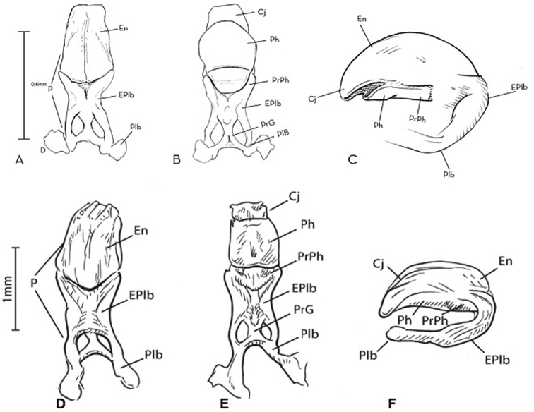
Phallus of Rhodnius marabaensis sp. n. (A dorsal view B ventral view C lateral view) and Rhodnius robustus (D dorsal view E ventral view F lateral view). Cj; En; EPlb; P; Plb; PrG; PrPh; Ph.
Figure 10.
Female external genitalia, posterior side of Rhodnius marabaensis sp. n. (A), Rhodnius prolixus (B), Rhodnius robustus (C). Ap; Gc 8; Gp 8; VII, VIII, IX: tergites; X: segment.
Figure 11.
Female external genitalia, ventral side of Rhodnius marabaensis sp. n. (A), Rhodnius prolixus (B), Rhodnius robustus (C). Gc 8; Gc 9; Gp 8; VII, IX: esternites; X: segment.
Figure 12.
Eggs general vision of Rhodnius marabaensis sp. n. (A), Rhodnius prolixus (B), Rhodnius robustus (C). cl; cr; ex; nk; op.
Description.
Measurements of 15 females and 15 males of Rhodnius marabaensis sp. n., Rhodnius prolixus, and Rhodnius robustus are detailed in the Table 1.
Table 1.
Mean of measurement (mm) of 15 females and males of Rhodnius marabaensis sp. n., Rhodnius prolixus, and Rhodnius robustus.
| Male | Female | |||||
|---|---|---|---|---|---|---|
| Rhodnius marabaensis | Rhodnius prolixus | Rhodnius robustus | Rhodnius marabaensis | Rhodnius prolixus | Rhodnius robustus | |
| HL | 4.90 a | 3.87 b | 3.82 c | 5.32 a | 3.90 b | 4.06 c |
| IE | 0.62 a | 0.53 b | 0.64 a | 0.59 a | 0.56 b | 0.67 c |
| AO | 2.21 a | 2.05 b | 2.23 c | 3.04 a | 2.26 b | 2.38 c |
| PO | 0.98 a | 0.92 b | 0.72 c | 1.06 a | 0.77 b | 0.78 c |
| DE | 1.72 a | 1.94 b | 1.00 c | 1.91 a | 1.68 b | 1.64 c |
| R1 | 0.97 a | 0.55 b | 0.91 c | 0.93 a | 0.57 b | 0.97 c |
| R2 | 3.87 a | 3.25 b | 3.02 c | 3.77 a | 3.32 b | 3.30 c |
| R3 | 0.87 a | 0.33 b | 0.92 c | 0.77 a | 0.39 b | 0.96 c |
| TL | 20.41 a | 19.98 b | 20.20 c | 22.35 a | 20.98 b | 21.28 c |
| MWT | 4.25 a | 4.62 b | 4.06 c | 4.88 a | 4.82 b | 4.12 c |
| MWA | 6.22 a | 5.93 b | 6.03 c | 6.92 a | 6.75 b | 6.56 c |
| A1 | 0.48 a | 0.38 b | 0.37 c | 0.45 a | 0.37 b | 0.38 c |
| A2 | 4.72 a | 3.04 b | 3.28 c | 4.47 a | 2.88 b | 3.18 c |
| A3 | 2.68 a | 2.25 b | 2.32 c | 3.05 a | 1.94 b | 2.41 c |
| A4 | 1.05 a | 0.98 b | 1.54 c | 1.15 a | 0.94 b | 1.64 c |
HL; IE; AO; PO (excluding neck); DE; R1, R2 and R3: lengths of first, second and third rostral; TL; MWT; MWA; A1, A2, A3 and A4: 1st, 2nd, 3rd, and 4th antennal segments, respectively; a,b,c: Lower case letters indicate significant differences between specimens with Tukey's test: p < 0,05. Values in bold indicate the main findings.
Head with apex (central longitudinal dorsal portion), which is elevated, straw yellow, and keel shaped. This keel-shaped section presents the same shape from the beginning of the clypeus to the posterior portion of the ocelli; thus, the border of the clypeus is visible around/from the gena and the jugum (1+1), which are located laterally. However, the gena begin before the beginning of the clypeus. Thus, the gena go toward the anteclypeus which are rounded. The species presents crystalline ocelli and eyes with black and yellow ommatidia. The first and second segments of the antennae are yellow, whereas the posterior two thirds of the third segment are white, and the fourth segment is completely white. The species presents a second antennal segment that is significantly larger than the others (10.3 times larger than the first; 1.65 times larger than the third, and 4.3 times larger than the fourth) (Table 1).
At the juncture between the neck and the thorax, there is a ring that is anteriorly black and posteriorly yellow; the anterolateral angles (1+1) are yellow. The dorsal thorax (pronotum) is shaped like a trapezoid and surrounded by a yellow carina. There are two yellow submedian carinae running lengthwise around the pronotum, from the anterior portion to the posterior one. The submedian carina border three anterior lobes, each of which has a set of black spots, and three posterior lobes with two parallel black stripes on each that are connected to the set of black spots on the anterior lobes. The triangular scutellum is marked laterally and is very clear because of its black color. The internal portion is also triangular and yellow, and it is bordered by thick and obvious carina. When the wings are removed, the posterior portion (tip) of the scutellum covers 2/3 of the I urotergite process (Figs 4, 5).
Figure 4.
Escutellum and process of I urotergit of Rhodnius marabaensis sp. n. (A), Rhodnius prolixus (B), Rhodnius robustus (C). pr; sc; sb; sg; cd; le; ap; pu; tg; fr.
From the ventral surface of the thorax, a prosternum with deep, well-defined stridulatory black sulcus is visible; in the posterior portion, it takes on a funnel shape and ends as a tip between the anterior pair of legs (Fig. 5). The mesosternum has two elevated black areas that are separated by a yellow depression. The border between the mesosternum and the metasternum is formed by a set of three elevated and prominent carinae. The two lateral carinae are black, and the central carina is yellow. These three carinae are curved backward. The central carina, which is elevated and prominent, possesses a semicircular depression in the central portion at the border with the metasternum. The metasternum is slightly rectangular in shape. The central portion is black and outlined by two yellow carinae (Fig. 6).
The legs have an overall yellowish tone. The coxae have yellow and black spots; the trochanters are yellow and do not have spots; the femurs are yellow with black spots running lengthwise; the tibias are yellow except for the posterior sixth segment, which continues the black pattern of the tarsi (Fig. 2).
The ventral surface of the abdomen is predominantly yellow, with three sets of black stripes: one on the central longitudinal portion and (2+2) on the side portions above the connectives (Fig. 2). The first abdominal segment has a longitudinal black spot between the two larger yellow spots. The second, third, fourth, and fifth ventral abdominal segments possess (1+1) curved sets of dark brown spots. These spots begin at the anterior dividing line and extend diagonally along the central portion of the segments (Fig. 2). The dorsal connective includes yellow and black spots that cover half of each segment. They are wide in the anterior portion and become thinner in the inner posterior portion. The black spot of the connective of the sixth segment is smaller than those of the fifth, fourth, third, and second segments (Fig. 2). The first tergite, which is visible when the wings are removed, is essentially formed by two parts. The anterior portion has a striated cuticula that contrasts with the surrounding smooth cuticula and which is triangular in shape on its upper level. It possesses a clearly defined transverse sulcus. The second portion is posterior to the first and is at a lower level. It consists of a set of transverse and straight fringes (Fig. 5).
When the male genitalia is seen from the dorsal surface, it is clear that the (P) is formed by an (En), by the (EPlb), and by the (Plb). When seen from the ventral surface, the (P) is formed by the (Cj), the (ph), the (PrPh), the (EPlb), the (PrG), and the (Plb). When seen from the lateral surface, the phallus is formed by the (Cj), the (En), the (EPlb), the (Plb), the (Ph), and the (PrPh) (Fig. 8).
The dorsal surface of the external female genitalia was examined using (SEM), which showed that the seventh segment is separated from the eighth segment by a slightly irregular line and forms (1+1) triangular tips at the border between the connective and the eighth segment. The eighth segment is trapezoid shaped. The ninth segment appears as a protrusion. The tenth segment appears as a small curve in the central portion where it delimits with the eighth segment (Fig. 9)
From the posterior surface, (1+1) appendages can be seen on the border between the eighth and ninth segments. The tenth segment is semicircular in shape with a pronounced central slit in the shape of an upside-down V and with (1+1) protrusions at the posterior edge of the gonocoxite 8. Display is also a (1+1) gonocoxite 8 and a (1+1) gonapophysis 8 (Fig. 10).
From the ventral surface, the lateral portions of the line that divides the seventh segment and the gonocoxites 8 and the gonapophysis 8 are curved, and the line then forms small (1+1) ascending curves and a slight depression in the central portion. The ninth segment forms (1+1) lateral flaps at the border with the tenth segment and presents transverse slits at the sub-intermediate position (1+1). The transverse slits then form into two triangles, whose tips are separated in the central portion. The tenth segment is the outer edge of the external female genitalia and is presented as a narrow curved and convex band (Fig. 11).
The egg shells measure 1.59 mm in length and 0.71 mm in width. They present prominent collar and chorial rim (Fig. 12 and Table 2). The exochorion cells are clearly demarcated, with internal granulations organized into a circle. The follicular tubes of each exochorion cell do not differ in diameter (Fig. 13).
Table 2.
Mean of measurements (mm) of 13 eggs of Rhodnius marabaensis sp. n., Rhodnius prolixus, and Rhodnius robustus.
| Measurement | Rhodnius marabaensis | Rhodnius prolixus | Rhodnius robustus |
|---|---|---|---|
| L (mm) | 1.54 ± 0.04 a | 1.73 ± 0.02 b | 1.61 ± 0.04 c |
| W (mm) | 0.87 ± 0.01 a | 0.71 ± 0.06 b | 0.93 ± 0.01 c |
| Oo (mm) | 0.49 ± 0.01 a | 0.67 ± 0.01 b | 0.73 ± 0.01 c |
L (40×); W (40×); Oo (80×); a,b,c: Lower case letters indicate significant differences between specimens with Tukey's test: p < 0,05.
The molecular study shown the same haplotype for the find sequences of the cyt-b (693 bp) and the evaluation by BLAST have shown that Rhodnius marabaensis sp. n. is closely related to Rhodnius robustus and Rhodnius prolixus (until 99% and 94% of identity, respectively).
Discussion
In epidemiological terms (i.e., considering the role of the species as vectors of Trypanosoma cruzi), the three main Triatominae genera are Panstrongylus, Rhodnius, and Triatoma. Distinguishing among these three genera is not difficult because they can be characterized macroscopically based on the format of the head and the position of the antenniferous tubercle, as described by Pinto (1931). However, distinguishing among Rhodnius species requires a more detailed examination through optical microscopy. The difficulty in identifying Rhodnius species was first noted by Neiva and Pinto (1923). At the time, there were five known species: Rhodnius nasutus, Rhodnius prolixus, Rhodnius pictipes, Rhodnius brethesi, and Rhodnius domesticus (Lent and Wygodzinsky 1979). Including this description of Rhodnius marabaensis sp. n., there are currently 20 species (Rosa et al 2012, Abad Franch 2013); therefore, specific distinction is even more difficult and requires more characteristics to be considered. As a result, the characterization of Rhodnius marabaensis sp. n. is discussed comparatively using Rhodnius prolixus and Rhodnius robustus. Rhodnius marabaensis is closely related to Rhodnius robustus and Rhodnius prolixus (until 99% and 94% of identity, respectively). In view of this and of the fact that all three species occur in Northern Brazil (Galvão 2014), Rhodnius robustus and Rhodnius prolixus were morphologically examined and compared in more detail (Table 3).
Table 3.
Triatominae species found in the Amazon region.
| Species | Descriptors | Distribution | References |
|---|---|---|---|
| Rhodnius amazonicus | Almeida, Santos & Sposina, 1973 | Brazil (Amazonas), French Guiana (Cacao, Saul) | Galvão 2014 |
| Rhodnius barretti | Abad-Franch, Palomeque & Monteiro, 2013 | Colombia (Puerto de Assís), Ecuador (Lagro Ágrio) | Abad-Franch et al. 2013 |
| Rhodnius brethesi | Matta, 1919 | Brazil (Amazonas), Venezuela (Aragua) | Galvão 2014 |
| Rhodnius colombiensis | Mejia, Galvão, Jurberg, 2009 | Colombia (Tolima) | Guhl et al. 2007 |
| Rhodnius dalessandroi | Carcavallo & Barreto, 1976 | Colombia (Meta) | Guhl et al. 2007 |
| Rhodnius ecuadoriensis | Lent & Leon, 1958 | Ecuador (Manabi, Eujias, Loja), Peru (Amazonas, Tumbis, Piura, Cojamarca, La libertad, Lambayeque e San Martin) | Galvão et al. 2003 |
| Rhodnius neivai | Lent, 1953 | Colombia (Cesar), Venezuela (Lara, Falcón, Zulia) | Galvão et al. 2003 |
| Rhodnius milesi | Carcavallo et al., 2001 | Brazil (Pará) | Galvão 2014 |
| Rhodnius montenegrensis | Rosa et al., 2012 | Brazil (Rondônia, Acre) | Galvão 2014 |
| Rhodnius pallescens | Barber, 1932 | Colombia (Bolívar, Sucre) | Arboleda et al. 2009 |
| Rhodnius paraensis | Sherlock, Guitton & Miles, 1977 | Brazil (Amazonas,Pará), French Guiana | Galvão 2014 |
| Rhodnius pictipes | Stal, 1872 | Brazil, French Guiana, Colombia, Peru, Suriname, Venezuela | Galvão 2014 |
| Rhodnius prolixus | Stal, 1859 | Brazil, Bolivia, Colombia, Guatemala | Galvão et al. 2003 |
| Rhodnius robustus | Larousse, 1927 | Brazil, Bolivia, Colombia, Venezuela, French Guiana | Galvão 2014 |
| Rhodnius stali | Lent, Jurberg & Galvão, 1993 | Brazil (Mato Grosso do Sul, Acre), Bolivia (Santa Cruz, La Paz) | Matias et al. 2003 |
Source: Galvão 2014; Galvão et al. 2003; Matias et al. 2003.
Rhodnius marabaensis sp. n. is predominantly yellow, whereas Rhodnius prolixus is black and Rhodnius robustus is brown. The legs of the three species are also consistent in these color schemes (Fig. 2). Another very clear characteristic of Rhodnius marabaensis sp. n. is that it is larger than Rhodnius prolixus and Rhodnius robustus (Figs 3–5 and Table 1).
The head of Rhodnius marabaensis sp. n. has four very clear characteristics that help to distinguish it from the others:
It possesses a keel-shaped longitudinal dorsal portion (apex) running from the clypeus to the ocelli. This feature is not accentuated in Rhodnius prolixus or Rhodnius robustus (Fig. 3);
It possesses a rounded anteclypeus. In Rhodnius prolixus and Rhodnius robustus, the anteclypeus is flat (Fig. 3);
It possesses an indistinguishable clypeus. In Rhodnius prolixus, it is narrow in the anterior portion and wide in the posterior one. In Rhodnius robustus it is wide in the anterior portion, narrow in the medial portion, and wide in the posterior one (Fig. 3);
The second segment of the antenna is significantly larger than the other three, with the following length ratio among the four antennal segments: 2nd > 3rd > 4th > 1st. This relative length pattern of the four antennal segments is the same as the pattern observed by Rosa et al. (2010) in Rhodnius neglectus and Rhodnius prolixus adults; however, the differences in size between the largest and the smallest segments are distinct: the second antennal segment of Rhodnius marabaensis sp. n. is 10.3 times larger than the first; in Rhodnius prolixus, it is 6.2 times larger, and in Rhodnius robustus it is 8.3 times larger (Table 1).
For the description of Rhodnius marabaensis sp. n. five features of the thorax allow for its distinction:
The scutellum is larger and includes two prominent internal lateral carinae. It is therefore distinct from Rhodnius prolixus and Rhodnius robustus, which present smaller scutellum and whose carinae are not pronounced (Fig. 4);
The first urotergite has a pronounced transverse groove and inferior fringe consisting of long and straight filaments. In Rhodnius prolixus, the transverse groove does not appear and the fringe possesses short and irregular filaments. On the other hand, in Rhodnius robustus the transverse groove is not accentuated, and the fringe possesses short and straight filaments, as shown in (Fig. 5).
It possesses a longer and more clearly shaped stridulatory sulcus relative to those of Rhodnius prolixus and Rhodnius robustus (Fig. 5). This specific differentiation among these three Rhodnius species adds to the observations by Lent and Wygodzinsky (1979), who verified that the stridulatory sulcus in six species from different genera presented characteristics that allowed for species identification;
The transverse carinae that border the mesosternum and the metasternum differ among the three species. In Rhodnius marabaensis sp. n., they are elevated and prominent, and possess convex curvature in the central portion. In Rhodnius prolixus, they are less elevated and prominent, and in Rhodnius robustus they are interrupted in the central portion (Fig. 6);
The metasternum is slightly rectangular in shape in Rhodnius marabaensis sp. n., whereas in Rhodnius prolixus and Rhodnius robustus they are slightly triangular (Fig. 6).
From the view of the ventral surface, the abdomen of Rhodnius marabaensis sp. n. is distinct because of its yellow colouring; this area is brown in Rhodnius prolixus and black in Rhodnius robustus (Fig. 11). The set of dark brown spots does not appear in Rhodnius prolixus or Rhodnius robustus, and the ventral connective is also distinct among the three species: the black spots are smaller and, on the sixth segment, much smaller in Rhodnius marabaensis sp. n. (Fig. 2).
When the male genitalia was examined from the dorsal surface, the apex of the endosoma of Rhodnius marabaensis sp. n. was found to be long and straight; in Rhodnius prolixus, the apex is long and convex, and in Rhodnius robustus it is shorter, wide, and convex (Lent and Jurberg 1969) (Fig. 7). The analysis also revealed that the two final portions of the basal piece are turned to the side in Rhodnius marabaensis sp. n., whereas in Rhodnius robustus they face the posterior region. From the ventral surface, the phallosoma of Rhodnius marabaensis sp. n. is rounded, whereas in Rhodnius prolixus the anterior portion is convex and in Rhodnius robustus it is square (Fig. 7). The pygophore of Rhodnius marabaensis sp. n., Rhodnius prolixus, and Rhodnius robustus are all in the shape of an isosceles triangle. However, the sides of the pygophores are straight in Rhodnius robustus and curved in Rhodnius prolixus and Rhodnius marabaensis sp. n. The pygophore of Rhodnius marabaensis sp. n. is also larger at the tip, thicker, and circular (Fig. 8).
The external female genitalia was also considered. From the dorsal surface, Rhodnius marabaensis sp. n. and Rhodnius robustus show no differences; however, the format of the ninth segment of the dorsal surface in Rhodnius prolixus presents different characteristics (Fig. 9). The posterior surface and the ventral surface of the ninth and tenth segments are distinct in the three species (Fig. 10). These characteristics are also distinct from 12 other Rhodnius species, as described by Rosa et al. (2014).
Rhodnius marabaensis sp. n. eggs possess chorion rims, whereas those of Rhodnius prolixus and Rhodnius robustus do not. The diameter of the follicular tubes of the exochorion cells in Rhodnius prolixus are larger than those of Rhodnius marabaensis sp. n. and Rhodnius robustus; however, they are regular in Rhodnius marabaensis sp. n. and varied in Rhodnius robustus (Figs 12, 13 and Table 2).
The data obtained corroborate the status of Rhodnius marabaensis sp. n. as a new species and indicates that the use of morphology for the description of Triatominae species offers phenotypic information (morphological and morphometric) to define the status of species. It is also important to note that molecular analysis generates data that can help phylogenetic relationships and taxonomic studies of Triatominae (Abad-Franch and Monteiro 2005, Justi et al. 2014). BLAST analysis of cyt-b sequence shows Rhodnius marabaensis sp. n. as closely related to Rhodnius robustus and Rhodnius prolixus, so this new species must be included in the Rhodnius prolixus complex (Carcavallo et al. 2000). Complementary approaches using molecular data must be encouraged to establish the phylogenetic placement of this new species based on the evaluation of other gene fragments and a more robust assessment.
Supplementary Material
Acknowledgents
We want to thank Dr. Oswaldo Pinto Serra and Raquel Guglielmetti Serra (in memoriam), who briefly characterized Rhodnius jacundaensis [nomen nudum] in 1980. We also want to thank Nelson Papavero from the Zoological Museum of USP and Cleber Galvão from the Oswaldo Cruz Institute for suggesting the scientific name of the new species. We are grateful to José Jurberg, who allowed us to observe the Herman Lent and Rodolfo Carcavallo collection at the Oswaldo Cruz Institute. We thank Marcelo Ornaghi Orlandi, Mario Cilense, Juan Carlos Guerrero Barreto, and Sebastião Dameto from the Institute of Chemistry at São Paulo State University, Araraquara, for their help with the electronic microscope, as well as Danila Blanco de Carvalho, Aline Ribeiro Rimoldi, and Felipe Fatobene from the School of Pharmaceutical Sciences at São Paulo State University, Araraquara, for their collaboration. Special thanks are given to Marcos Takashi Obara from the University of Brasília (UnB) and to Mara Cristina Pinto from the School of Pharmaceutical Sciences at São Paulo State University, Araraquara, for their suggestions, and finally, to Davi Antonio da Rosa for the photos and drawings. Financial Support: São Paulo Research Foundation (FAPESP) grant process number 2010/15386-3; Brazilian Federal Agency for the Support and Evaluation of Graduate Education (CAPES), grant process number 23038-005285/2011-2012, Program for the Improvement and Scientific Development of the Pharmaceutical Sciences, School of Pharmaceutical Sciences at São Paulo State University, Araraquara - PADC/FCF/UNESP; Brazilian National Council for Scientific Technological Development (CNPq).
Citation
Souza ES, Von Atzingen NCB, Furtado MB, Oliveira J, Nascimento JD, Vendrami DP, Gardim S, Rosa JA (2016) Description of Rhodnius marabaensis sp. n. (Hemiptera, Reduviidae, Triatominae) from Pará State, Brazil. ZooKeys 621: 45–62. doi: 10.3897/zookeys.621.9662
References
- Abad-Franch F, Monteiro FA. (2005) Molecular research and the control of Chagas disease vectors. Annals of the Brazilian Academy of Sciences 77: 437–454. doi: 10.1590/S0001-37652005000300007 [DOI] [PubMed] [Google Scholar]
- Abad-Franch F, Pavan MG, Jaramillo O, Palomeque FS, Dale C, Chaverra D, Monteiro FA. (2013) Rhodnius barretti, a new species of Triatominae (Hemiptera: Reduviidae) from western Amazonia. Memórias Instituto Oswaldo Cruz 108: 92–99. doi: 10.1590/0074-0276130434 [DOI] [PMC free article] [PubMed] [Google Scholar]
- Alevi KCC, Ravazi A, Mendonça VJ, Rosa JA, Azeredo-Oliveira MTV. (2015) Karyotype of Rhodnius montenegrensis (Hemiptera, Triatominae). Genetics and Molecular Research 12: 222–226. doi: 10.4238/2015.January.16.5 [DOI] [PubMed] [Google Scholar]
- Almeida FB, Santos EI, Sposina G. (1973) Triatomíneos da Amazonia III. Acta Amazonica 3: 43–66. [Google Scholar]
- Bérenger JM, Pluot-Sigwalt D. (2002) Rhodnius amazonicus Almeida, Santos & Sposina, 1973, bona species, close to R. pictipes Stål, 1872 (Heteroptera: Reduviidae: Triatominae). Memórias do Instituto Oswaldo Cruz 97: 73–77. doi: 10.1590/S0074-02762002000100011 [DOI] [PubMed] [Google Scholar]
- Carcavallo RU, Galvão C, Lent H. (1998) Triatoma jurbergi sp.n. do Norte do Estado do Mato Grosso, Brasil (Hemiptera, Reduviidae, Triatominae) com uma Atualização das Sinonímias e outros Táxons. Memórias do Instituto Oswaldo Cruz 93: 459–464. doi: 10.1590/S0074-02761998000400007 [DOI] [PubMed] [Google Scholar]
- Carcavallo RU, Jurberg J, Lent H, Noireau F, Galvão C. (2000) Phylogeny of the Triatominae (Hemiptera: Reduviidae). Proposals for taxonomic arrangements. Entomología y Vectores 7: 1–99. [Google Scholar]
- Dujardin J, Steinden M, Chavez T, Machane M, Schofield C. (1999) Changes in the Sexual Dimorphism of Triatominae in the Transition From Natural to Artificial Habitats. Memórias Instituto Oswaldo Cruz 94: 565–569. doi: 10.1590/S0074-02761999000400024 [DOI] [PubMed] [Google Scholar]
- Galvão C, Carcavallo R, Rocha DS, Juberg JA. (2003) Checklist of the current valid species of the subfamily Triatominae Jeannel, 1919 (Hemiptera, Reduviidae) and their geographical distribuition, with nomenclatural and taxonomic note. Zootaxa 202: 1–36. doi: 10.11646/zootaxa.202.1.1 [Google Scholar]
- Galvão C. (2015) Vetores da doença de Chagas no Brasil. Sociedade Brasileira de Zoologia, Curitiba, 289 pp. [Google Scholar]
- Jurberg J, Rocha DS, Galvão C. (2009) Rhodnius zeledoni sp. nov. afim de Rhodnius paraensis Sherlock, Guitton e Milles, 1977 (Hemíptera, Reduviidae, Triatominae). Biota Neotropica 9: 123–128. doi: 10.1590/S1676-06032009000100014 [Google Scholar]
- Justi AS, Russo CAM, Mallet JRS, Obara MT, Galvão C. (2014) Molecular phylogeny of Triatomini (Hemiptera: Reduviidae: Triatominae). Parasites & Vectors 7: . doi: 10.1186/1756-3305-7-149 [DOI] [PMC free article] [PubMed] [Google Scholar]
- Lent H, Juberg J. (1969) O gênero Rhodnius Stål, 1859 com um estudo sobre a genitália das espécies (Hemiptera, Reduviidae, Triatominae). Brazilian Journal of Biology 29: 487–560. [Google Scholar]
- Lent H, Wygodzysnky P. (1979) Revision of the Triatominae (Hemiptera - Reduviidae) and their significance as vectors of Chagas’ disease. Bulletin of the American Museum of Natural History 163: 125–520. [Google Scholar]
- Lent H, Juberg J, Galvão C. (1993) Rhodnius stali n. sp., Afim de Rhodnius pictipes, 1872 (Hemíptera, reduviidae, triatominae). Memórias do Instituto Oswaldo Cruz 88: 605–614. doi: 10.1590/S0074-02761993000400019 [Google Scholar]
- Meija JM, Galvão C, Juberg J. (1999) Rhodnius colombiensis sp. n. da Colômbia, com quadros comparativos entre estruturas fálicas do gênero Rhodnius Stål, 1859 (Hemíptera, Reduviidae, Triatominae). Entomología y Vectores 6: 601–617. [Google Scholar]
- Mendonça VJ, Chaboli AKC, Pinotti H, Gurgel-Gonçalves R, Pita S, Guera AL, Panzera F, Araújo RF, Vilela de Azeredo-Oliveira MT, Rosa JA. (2016) Revalidation of Triatoma bahiensis Sherlock & Serafim, 1967 (Hemiptera: Reduviidae) and phylogeny of the T. brasiliensis species complex. Zootaxa 4107: 239–254. doi: 10.11646/zootaxa.4107.2.6 [DOI] [PubMed] [Google Scholar]
- Meneguetti DUO, Castro GVS, Castro MALR, Souza JLS, Oliveira J, Rosa JA, Aranha Camargo LMA. (2016) First report of Rhodnius stali (Hemiptera, Reduviidae, Triatominae) in the State of Acre and in the Brazilian Amazon. Journal of the Brazilian Society of Tropical Medicine 49: 365–368. doi: 10.1590/0037-8682-0066-2016 [DOI] [PubMed] [Google Scholar]
- Monteiro FA, Barrett TV, Fitzpatrick S, Cordon-Rosales C, Feliciangeli D, Beard CB. (2003) Molecular Phylogeography of the Amazonian Chagas disease vectors Rhodnius prolixus and Rhodnius robustus. Molecular Ecology 12: 997–1006. doi: 10.1046/j.1365-294X.2003.01802.x [DOI] [PubMed] [Google Scholar]
- Neiva A, Pinto C. (1923) Estado actual dos conhecimentos sobre o gênero Rhodnius Stål, com a descrição de uma nova espécie. Brasil-Médico 37: 20–24. [Google Scholar]
- Pinto C. (1931) Valor do rostro e antenas na caracterização dos gêneros de triatomideos. (Hemiptera, Reduviidae). Boletim biológico de São Paulo 19: 45–136. [Google Scholar]
- Rosa JA, Freitas SC, Malara FF, Rocha CS. (2010) Morphometry and morphology of the antennae of Panstrongylus megistus Burmeister, Rhodnius neglectus Lent, Rhodnius prolixus Stål and Triatoma vitticeps Stål (Hemiptera: Reduviidae). Neotropical Entomology 39: 214–220. doi: 10.1590/S1519-566X2010000200011 [DOI] [PubMed] [Google Scholar]
- Rosa JA, Barata JMS, Santos JLF, Cilense M. (2000) Morfologia de ovos de Triatoma circummaculata e Triatoma rubrovaria (Hemiptera, Reduviidae) Egg morphology of Triatoma circummaculata and Triatoma rubrovaria (Hemiptera, Reduviidae). Journal of Public Health 34: 538–542. [DOI] [PubMed] [Google Scholar]
- Rosa JA, Mendonça VJ, Rocha CS, Gardim S, Cilense M. (2010) Characterization of the external female genitalia of six species of Triatominae (Hemiptera: Reduviidae) by scanning electron microscopy. Memórias do Instituto Oswaldo Cruz 105: 286–292. doi: 10.1590/S0074-02762010000300007 [DOI] [PubMed] [Google Scholar]
- Rosa JA, Rocha CS, Gardim S, Pinto MC, Mendonça VJ, Filho JCRF, Carvalho EOC, Camargo LMA, Oliveira JO, Nascimento JD, Cilense M, Almeida CD. (2012) Description of Rhodnius montenegresis n. sp. (Hemiptera: reduviidae: Triatominae) from the state of Rondônia, Brazil. Zootaxa 3478: 62–76. [Google Scholar]
- Rosa JA, Mendonça VJ, Gardim S, Blanco DC, Oliveira J, Nascimento JD, Pinotti H, Pinto MC, Cilense M, Galvão C, Barata JMS. (2014) Study of the external female genitalia of 14 Rhodnius species (Hemiptera, Reduviidae, Triatominae) using scanning electron microscopy. Parasite & Vectors 7: . doi: 10.1186/1756-3305-7-17 [DOI] [PMC free article] [PubMed] [Google Scholar]
- Serra OP, Serra RG, Atzigen NCBV. (1980) Nova espécie do gênero Rhodnius da Amazônia, Estado do Pará, Brasil. Anais do V Congresso Brasileiro de Parasitologia, Rio de Janeiro, 120 pp. [Google Scholar]
- Valente VC, Valente S, Carcavallo RU, Rocha DS, Galvão C, Juberg J. (2001) Considerações sobre uma nova espécie do gênero Rhodnius Stål, do estado do Pará, Brasil (Hemíptera, Reduviidae, Triatominae). Entomología y Vectores 8: 65–80. [Google Scholar]
Associated Data
This section collects any data citations, data availability statements, or supplementary materials included in this article.


