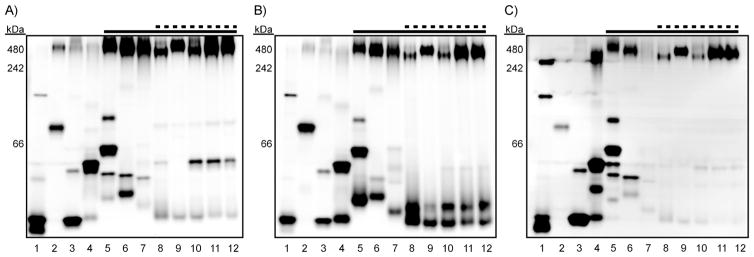
Figure 3.
Fluorogenic esterase probes reveal distinct mycobacterial esterase activities. Mycobacterial lysates (1–8 μg of total protein per lane) were resolved by using native PAGE (10–20 % Tris·HCl gradient gel). NativeMark molecular weight ladder was run on each gel (not shown), and the apparent molecular weights are provided. The dashed line indicates species that cause pulmonary disease, and the solid line indicates members of the MTBC. Each gel was incubated for 5 min in 10 mM HEPES (pH 7.3) with A) 1 μM DDAO-AME 1, B) 1 μM DDAO-AME 2, or C) 5 μM Res-AME. The gels were imaged to reveal fluorescent bands corresponding to hydrolyzed probe. Lane assignments: 1) M. smegmatis; 2) M. marinum; 3) M. flavescens; 4) M. nonchromogenicum; 5) M. kansasii; 6) M. avium; 7) M. intracellulare; 8) M. bovis (BCG); 9) M. africanum; 10) M. tuberculosis Erdman; 11) M. tuberculosis H37Rv; 12) M. tuberculosis CDC1551.

