Abstract
Unilateral phrenic nerve palsy as initial presentation of the retrosternal goitre is extremely rare event. This is a case report of a 57-year-old woman with history of cough and breathlessness of 3 months duration, unaware of the thyroid mass. She had large cervico-mediastinal goiter and chest radiograph revealed raised left sided hemidiaphragm. Chest CT scan did not reveal any lung parenchymal or mediastinal pathology. The patient underwent a total thyroidectomy through a cervical approach. The final pathology was in favor of multinodular goitre. Even after 1 year of follow up, phrenic nerve palsy did not improve indicating permanent damage. Phrenic nerve palsy as initial presentation of the retrosternal goitre is unusual event. This case is reported not only because of the rare nature of presentation, but also to make clinicians aware of the entity so that early intervention may prevent attendant morbidity.
Keywords: Phrenic nerve palsy, Raised hemi diaphragm, Retrosternal goitre, Thyroidectomy, Complications
Introduction
Although the phrenic nerves cross the thyroid gland at the thoracic inlet, palsy of this nerve is a rare complication of retro-sternal extension of the multinodular goitre [1]. Large goitres, especially with significant retro-sternal extension usually present as compressive symptoms like dysphagia or dyspnea due to oesophageal or tracheal compression respectively. The phrenic nerve may be compressed by a large goitre along its course in the neck, but the more likely site of injury is the point at which it enters the thoracic cavity adjacent to the first rib. [1] Unilateral diaphragmatic paralysis is usually asymptomatic and may only be accidentally detected on a chest radiograph. As compared to this bilateral phrenic nerve palsy secondary to a benign substernal goitre may present as acute respiratory failure requiring intubation or tracheostomy [1]. Unilateral phrenic nerve paralysis following surgery for cervico-mediastinal goitre although rare, has been reported well in the literature [2]. We report a case of 57 year old female where left sided phrenic nerve palsy was seen at initial presentation, which is very rare event to occur. This case is also reported to make clinicians aware of the presentation so that early thyroidectomy is done to prevent permanent damage to the nerve.
Case Report
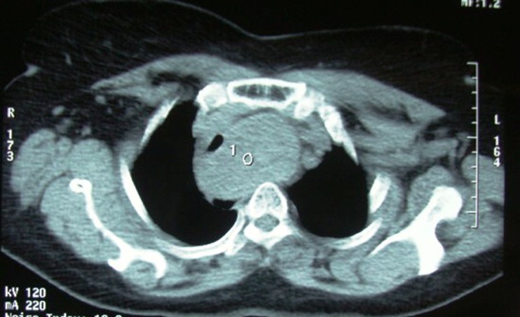
A 57 year old female was referred to our head and neck surgery unit from the department of pulmonology with 3 months history of recurrent cough and breathlessness on exertion. Chest radiograph revealed left side elevated hemi- diaphragm and significant compression of the trachea to the right side due to large retro-sternal thyroid mass enlargement (Fig. 1). On neck palpation there was large swelling in the lower neck, the lower end of which could not be palpated. Her thyroid function tests were normal. Fine needle aspiration cytology was in favor of benign thyroid disease. Computer tomography of the neck and thorax revealed multinodular goiter of thyroid gland with large retro-sternal component causing significant deviation and compression of the trachea to the right side (Fig. 2). Sagittal sections of computer tomography of neck and thorax showing 128.4 × 45.2 mm multinodular goiter of thyroid gland with significant superior mediastinal involvement (Fig. 3). Chest CT scan did not reveal any primary lung or mediastinal pathology except significant retro-sternal extension of the multinodular goitre. She underwent total thyroidectomy with excision of retrosternal component through cervical incision without sternotomy. Left phrenic nerve was examined and found intact grossly. Her post operative period was uneventful. Histopathology was in favor of multinodular goiter without evidence of any malignant change. Even after 1 year of follow up, the phrenic nerve palsy did not improve indicating permanent damage to the nerve (Fig. 4).
Fig. 1.
Chest radiograph showing large thyroid mass with retro-sternal extension with deviation of the trachea to the right (thin arrows). Raised left sided hemidiaphragm can also be noted (thick arrows)
Fig. 2.
Axial section of computer tomography of the neck and thorax revealed multinodular goiter of thyroid gland with large retro-sternal component causing significant deviation and compression of the trachea to the right side
Fig. 3.
Sagittal sections of computer tomography of neck and thorax showing 128.4 × 45.2 mm multinodular goiter of thyroid gland with significant superior mediastinal involvement
Fig. 4.
Chest radiograph after 1 year after total thyroidectomy showing persistent elevation of left hemidiaphragm. Tracheal shadow is now in midline
Discussion
Retrsternal goiters usually present with symptoms related to compression of adjacent vital structures like trachea, esophagus, nerves and the blood vessels [3]. Our case presented with signs and symptoms of phrenic nerve palsy as the patient was unaware of the large retrosternal goiter. Compression of the phrenic nerve appears to be unusual manifestation of an retrosternal goiter and thus far has been rarely reported [1, 4]. On the other hand iatrogenic phrenic nerve paralysis although not common, is well documented in the literature [2, 3, 5–8]. Manning et al. was first to report the bilateral phrenic nerve palsy associated with the benign thyroid goiter [1]. Van Doorn et al. also reported a case where partial unilateral phrenic nerve palsy was caused due to large retrosternal goiter [4]. L. Rosato on the other hand reported the phrenic nerve paralysis following total thyroidectomy for large retrosternal goiter. He attributed particular anatomic conditions like adhesion which may be found at the time of surgery rather than the operative technique for the iatrogenic paralysis [2].
The natural history of the retrosternal goiter is of a slow progressive increase in size, often detected incidentally on chest radiograph in 5th or 6th decade. Phrenic nerve palsy because of retrosternal extension of multinodular goitre can be explained on the anatomical basis. The phrenic nerve arises in the neck from the 3rd, 4th, and 5th nerves of the cervical plexus. It runs vertically downward across the front of the scalenus anterior muscle and enters the thorax by passing in front of the subclavian artery. It enters the chest and settles on the pleuric cupule descending into the anterior mediastinum. On the right it follows the side of the superior vein cava, runs along the pericardium and finally reaches the diaphragm. On the left side, the nerve descends laterally to the aortic arch on the side wall of the pericardium and reaches the diaphragm just behind the top of the heart. The phrenic nerve is the only motor nerve supply to the diaphragm. As the thyroid increases in size and weight, it slowly descends into the mediastinum. Factors which favour its descent are short neck, greater width of thoracic inlet, increase of the size of the lower part of the thyroid lobes, elasticity of the upper vascular pedicles. The negative thoracic pressure and the movement of the prethyroid muscles promote further descent of goiter into the mediastinum. Phrenic nerve paralysis occurs due to compression especially where the nerve enters the mediastinum, through the thoracic inlet, behind the first rib. In fact it is here, between the capsule of the intrathoracic goiter and the nerve; significant adhesions may form more often [4].
Iatrogenic injury to the phrenic nerve occurs relative more frequently than compression due to large retrosternal goiter. It mainly occurs in the cardiovascular thoracic surgery procedures and oncologic procedures of the cervico-mediastinal region [3, 5, 6]. If adhesions occur between the thyroid gland and the phrenic nerve in case of retrosternal extension of goiter, immediately beyond the upper thoracic outlet, it is possible to damage the nerve during surgical procedures [2]. The phrenic nerve in that region is neither under direct vision, nor is there a way of identifying and preserving the nerve [2]. Surgical technique may prevent unnecessary injury to the phrenic nerve during removal of the large retrosternal goiter. Most of the retosternal goiters can be removed via cervical approach. It is preferable to perform lobectomy with isthmusectomy on the contralateral side first. After that superior vascular pedicle on the ispilateral side is identified and ligated and slowly mediastinal portion should be freed with gentle traction upwards avoiding sharp dissection [2]. This will help in preserving vital structures like recurrent laryngeal and phrenic nerves.
Phrenic nerve palsy although very rare, should be suspected in goitre with significant retro-sternal extension and associated breathlessness. This condition, caused by the rise of the diaphragm, may determine a reduction in the respiratory space due to compression of the pulmonary paraenchyma and a restrictive syndrome with a slight hypoxia. Early recognition of the complication and timely intervention may prevent permanent damage and consequent morbidity.
Acknowledgments
Authors’ Contributions
AHH wrote the draft of the article. AHH, IHH and FJW helped in the final writing of the paper and gave final approval of the article. AHH, IHH and FJW participated in the article revision.
All authors read and approved the final manuscript.
Compliance with Ethical Standards
Competing interests
The authors declare that they have no competing interests.
Financial Support
None.
Consent
The informed consent was obtained from the patient for the publication of this report and any accompanying images. A copy of the written consent is available for review by the Editor-in-Chief of this journal.
References
- 1.Manning PB, Thompson NW. Bilateral phrenic nerve palsy associated with benign thyroid goiter. Acta Chir Scand. 1989;155:429–30. [PubMed] [Google Scholar]
- 2.Rosato L, Nasi PG, Porcellana V, Varvello G, Mondini G, Bertone P. Unilateral phrenic nerve paralysis: a rare complication after total thyroidectomy for a large cervico-mediastinal goitre. G Chir. 2007;28(4):149–52. [PubMed] [Google Scholar]
- 3.Anders HJ. Compression in syndromes caused by substernal goitres. Postgrad Med J. 1998;74:701–2. doi: 10.1136/pgmj.74.872.327. [DOI] [PMC free article] [PubMed] [Google Scholar]
- 4.Van Doorn LG, Kranendonk SE. Partial unilateral phrenic nerve paralysis caused by allarge intrathoracic goitre. Neth J Med. 1996;48:216–9. doi: 10.1016/0300-2977(95)00083-6. [DOI] [PubMed] [Google Scholar]
- 5.Salame N, Winiszewski P, Rossier S, Picard A. Goitre endothoracique rétrotrachéal: exérèse par voie cervicale. J Chir. 1997;134(7–8):311–13. [PubMed] [Google Scholar]
- 6.De Jong AA, Manni JJ. Phrenic nerve paralysis following neck dissection. Eur Arch Otorhinolaryngol. 1991;248:132–4. doi: 10.1007/BF00178921. [DOI] [PubMed] [Google Scholar]
- 7.Goffart Y, Moreau P, Biquet JF, Melon J. Phrenic nerve paralysis complicating cervicofacial surgery. Acta Otorhinolaryngol Belg. 1988;42:564–70. [PubMed] [Google Scholar]
- 8.Mc Caul JA, Hislop WS. Transient hemi-diaphragmatic paralysis following neck surgery: report of a case and review of the literature. J R Coll Surg Edinb. 2001;46:186–8. [PubMed] [Google Scholar]


