Abstract
Extraosseous osteogenic sarcoma is a very rare malignant neoplasm. The most common sites are the extremities, thorax, and the abdomen. Retroperitoneal osteosarcomas are rare and very few cases have been reported. They are similar in their biology to high-grade soft tissue sarcomas. R0 resection appears to be the best possible treatment for these tumors but there are no published cases on how to manage them when it involves posterior and intra-spinal regions. We report a 62-year-old male who presented with a backache, and investigations revealed a large retroperitoneal fibrosarcoma invading into the lumbar spine, but was found to be an extra osseous osteosarcoma on final histopathological examination. It is important to emphasize that due to the rarity of soft tissue sarcomas as well as the uniqueness of the multimodal treatment plan for each subtype, soft tissue sarcomas involving the spine are best managed by a multi disciplinary team. Overall, patients with soft tissue sarcomas involving the spine usually present a poor long-term prognosis. Therefore, whenever feasible, “en bloc” resection of such lesions has been shown to play a crucial role in improving the overall and recurrence-free survival rates.
Keywords: Extraosseous, Osteosarcoma, Retroperitoneum, Soft tissue sarcoma
Introduction
Extraosseous osteosarcoma (EOO) or soft tissue osteosarcoma (STO) is a malignant mesenchymal neoplasm located in the soft tissues without direct attachment to the skeletal system, and produces osteoid, bone, or chondroid material. The first case of EOO was reported by Wilson in 1941 [1]. Extra osseous osteosarcoma most commonly occur in the extremities, followed by the thorax, and rarely in the retroperitoneum, and only few cases have been reported [2]. Consequently, their biological behavior and response to treatment are yet to be fully understood.
Case Report
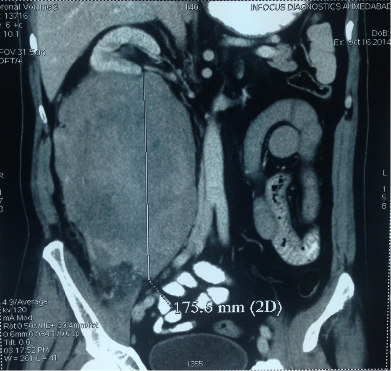
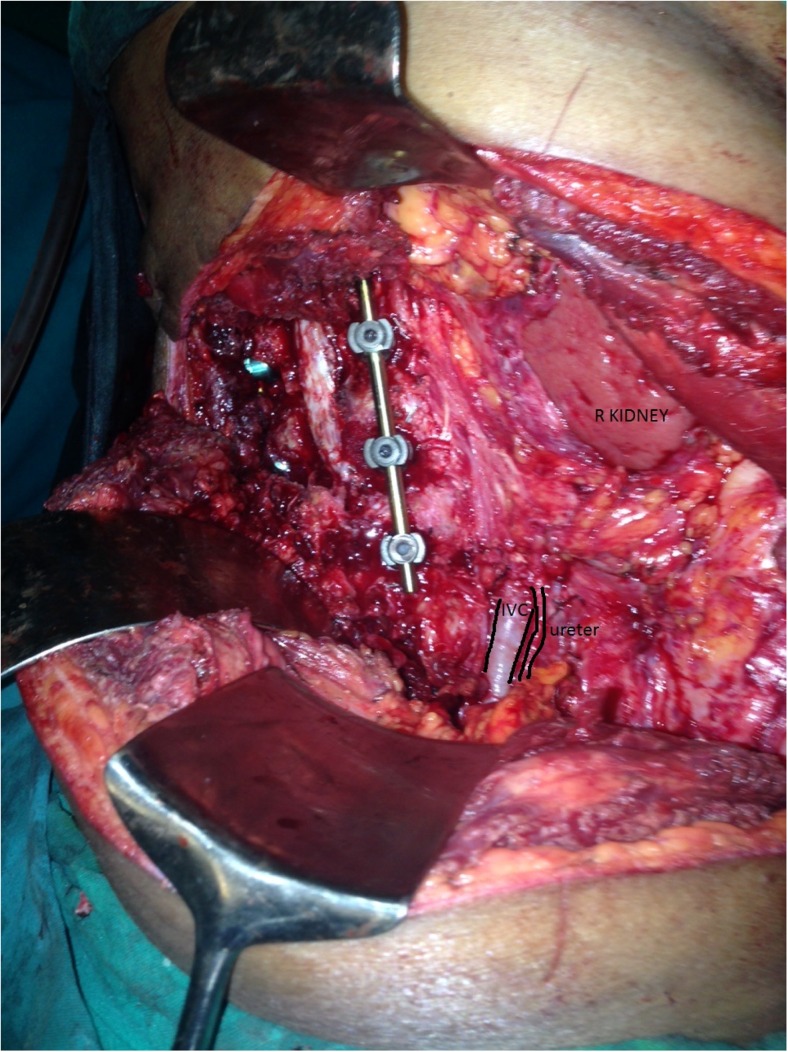
A 62-year-old male was investigated for chronic backache of duration 3–4 months and magnetic resonance imaging (MRI) of the spine revealed erosion of L3 and L4 vertebrae with a mass lesion in the retroperitoneum. Contrast enhanced computed tomography (CECT) of the abdomen showed right-sided large 20 cm × 15 cm heterogeneous retroperitoneal mass with contrast enhancement, which did not envelope any major organ or vessel, and calcification but was invading into the spine [Fig. 1]. Core needle biopsy was suggestive of epitheloid fibrosarcoma and immunohistochemistry showed positive SMA marker (myofibroblastic marker) and focal positive desmin muscle marker. Due to the large size and invasion into the spine, pre-operative radiotherapy was given. Patient underwent surgery with right extended lumbar incision with an en bloc resection of the retroperitoneal mass with partial vertebrectomy and spine fixation [Fig. 2]. The tumor was not involving the peritoneum and other great vessels but a major portion of psoas major and two spinal nerve roots had to be sacrificed. Pathologic examination revealed spindle cell sarcoma which shows fasicles of pleomorphic spindle cells amidst collagenised stroma at places resembling osteoid with interspersed foci which showed multinucleated osteoclast-like giant cells and few hemosiderin-laden macrophages. The final histopathogical diagnosis was extraosseous osteogenic sarcoma.
Fig. 1.
Computerized tomography image of the lesion
Fig. 2.
Intra-operative post resection—dura “uninvolved”
Currently 16 months after diagnosis and treatment, patient has no recurrence.
Discussion
Sarcomas are rare malignant tumors that arise from mesenchymal tissue at any site in the body. Ten to twenty percent of STS occur in the retroperitoneum [3]. Leiomyosarcoma, liposarcoma, and fibrosarcoma are the most common histologic types of retroperitoneal sarcomas [3]. Extraosseous osteogenic sarcoma arising from the soft tissue constitutes 1.2 % of all soft tissue sarcomas and the most common primary sites are the thighs and buttocks [4].
In our case, the tumor invaded the spine but the neural elements were not involved. So an en bloc resection was planned preoperatively. To maximize possibility of R0 resection preoperative radiotherapy with “tumor painting” was employed. The spinal surgeon started the surgery with a plan to remove the spinal attachment with the main tumor en bloc. The bone elements of the spinal column (vertebral body, pedicles, and lamina) were broken in a wide enough manner to allow dissection around the spinal cord. Partial vertebrectomy was done and the spine was stabilized with screw fixation. Later, the main tumor was approached and en bloc resection was done avoiding opening the peritoneum. The biopsy showed R0 resection with diagnosis of extra osseous osteosarcoma. The patient recovered well postoperatively and is tumor free post 6 months of surgery with no neurological problem.
The retroperitoneum provides a widely expansible anatomic location for tumors arising there, and these tumors often become very large before symptoms manifest. The median size at diagnosis is approximately 15 cm [5]. Complete tumor resection is the treatment of choice but is rarely feasible due to invasion of adjacent structures by tumors, and is perhaps the most important factor in survival outcome. An R0 resection, if possible, is the best initial modality of treatment. Radical surgery is the most effective treatment available till date. The results of surgery are quite inferior to that of extremity sarcomas because of the large size at presentation and difficulty in resecting the tumor because of its anatomical location.
Adjuvant therapy with radiation is commonly recommended for patient with high-grade resected tumors. The benefits of adjuvant chemotherapy remain controversial. These tumors have been found to be chemoresistant. The 5-year survival has ranged from 12 % to about 25 % [6]. Treatment of this rare neoplasm is complicated by the large size of the tumor at diagnosis and frequent presence of metastases; therefore, prognosis is poor.
Conclusion
Patients with soft tissue sarcomas involving the spine usually present a poor long-term prognosis. Therefore, whenever feasible, en bloc resection with microscopic negative margins of such lesions has been shown to play a crucial role in improving the overall and recurrence-free survival rates. Local recurrence remains a difficult problem, with increased associated morbidity and psychological stress for affected patients. These tumors are best managed by multidisciplinary evaluation and treatment in a tertiary referral center.
Contributor Information
Anish P. Nagpal, Email: dranagpal@gmail.com
Somesh Chandra, Email: someshchandra@hotmail.com.
References
- 1.Wilson H. Extra skeletal ossifying tumors. Ann Surg. 1941;113:95–112. doi: 10.1097/00000658-194101000-00013. [DOI] [PMC free article] [PubMed] [Google Scholar]
- 2.Lorentzon R, Larsson SE, Boquist L. Extra osseous osteosarcoma—a clinical and histopathological study of four cases. J Bone Joint Surg (Br) 1971;61:205–8. doi: 10.1302/0301-620X.61B2.285933. [DOI] [PubMed] [Google Scholar]
- 3.Catton CN, O’Sullivan B, Kotwall C, Cummings B, Hao Y, Fornasier V. Outcome and prognosis in retroperitoneal soft tissue sarcoma. Int J Radiat Oncol Biol Phys. 1994;29:1005–10. doi: 10.1016/0360-3016(94)90395-6. [DOI] [PubMed] [Google Scholar]
- 4.Sordillo PP, Hajdu SI, Magill GB, Golbey RB. Extraosseous osteogenic sarcoma: a review of 48 patients. Cancer. 1982;51:727–34. doi: 10.1002/1097-0142(19830215)51:4<727::AID-CNCR2820510429>3.0.CO;2-I. [DOI] [PubMed] [Google Scholar]
- 5.Stoeckle E, Coindre JM, Bonvalot S, Kantor G, Terrier P, Bonichon F, Nguyen BB. Prognostic factors in retroperitoneal sarcoma: a multivariate analysis of a series of 165 patients of the French Cancer Center Federation Sarcoma Group. Cancer. 2001;92:359–68. doi: 10.1002/1097-0142(20010715)92:2<359::AID-CNCR1331>3.0.CO;2-Y. [DOI] [PubMed] [Google Scholar]
- 6.Mattei TA, Teles AR, Mendel E. Modern surgical techniques for management of soft tissue sarcomas involving the spine: outcomes and complications. J Surg Oncol. 2014 doi: 10.1002/jso.23805. [DOI] [PubMed] [Google Scholar]


