Abstract
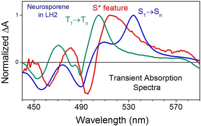
Carotenoids are a class of natural pigments present in all phototrophic organisms, mainly in their light-harvesting proteins in which they play roles of accessory light absorbers and photoprotectors. Extensive time-resolved spectroscopic studies of these pigments have revealed unexpectedly complex photophysical properties, particularly for carotenoids in light-harvesting LH2 complexes from purple bacteria. An ambiguous, optically forbidden electronic excited state designated as S* has been postulated to be involved in carotenoid excitation relaxation and in an alternative carotenoid-to-bacteriochlorophyll energy transfer pathway, as well as being a precursor of the carotenoid triplet state. However, no definitive and satisfactory origin of the carotenoid S* state in these complexes has been established, despite a wide-ranging series of studies. Here, we resolve the ambiguous origin of the carotenoid S* state in LH2 complex from Rba. sphaeroides by showing that the S* feature can be seen as a combination of ground state absorption bleaching of the carotenoid pool converted to cations and the Stark spectrum of neighbor neutral carotenoids, induced by temporal electric field brought by the carotenoid cation–bacteriochlorophyll anion pair. These findings remove the need to assign an S* state, and thereby significantly simplify the photochemistry of carotenoids in these photosynthetic antenna complexes.
Introduction
Carotenoids are present in all phototrophic organisms and play important roles in various light-harvesting complexes as accessory light absorbers and photoprotectors of numerous types of (bacterio)chlorophylls.1−4 In light-harvesting (LH) complexes from purple bacteria, carotenoids play a structural role5 and serve as auxiliary light absorbers, harvesting light at wavelengths not covered by bacteriochlorophyll a (BChl a). As the lowest lying excited state S1 is optically forbidden,6 absorption of carotenoids, responsible for a bright yellow or orange coloration of the pigments, is associated with the strongly allowed S0 → S2 electronic transition. In the LH2 complex, this harvested energy is transferred to both the B800 and B850 spectral forms of BChls (Figure 1). Finally, carotenoids are also excellent photoprotectors, efficiently quenching BChl a triplet states, harmful reactive singlet oxygen,7,8 and in some cases also BChl a excited singlet state.9
Figure 1.
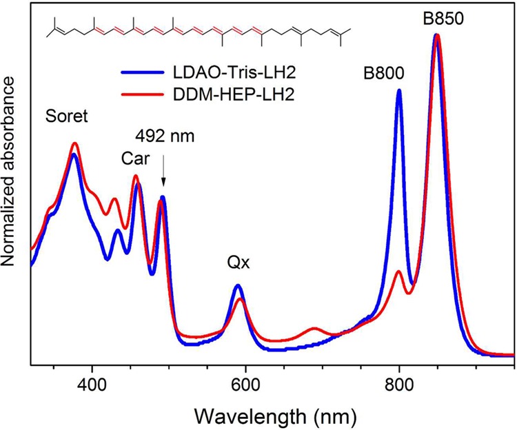
Steady-state absorption spectra of two batches of LH2 complex from the ΔcrtC mutant of Rba. sphaeroides along with the chemical structure of carotenoid neurosporene. The major absorption bands are marked. The excitation wavelength used for time-resolved studies is indicated by an arrow.
The standard three-state S0–S1–S2 model satisfactorily accounts for the typical spectroscopic properties of carotenoids in solvents and in protein environments. However, experimental results from femtosecond time-resolved absorption spectroscopy, first applied to photophysical studies of carotenoids in the 1990s,10,11 demonstrated that additional optically silent electronic states may lie in the vicinity of the S1 and S2 states.12−15 The most controversial state, S*, was first introduced to explain ambiguous spectral features in transient absorption (TA) spectra of the open-chain carotenoid spirilloxanthin,14 although similar spectral features had been observed previously for long-chain homologues of β-carotene.16 In the case of spirilloxanthin, the S* feature was observed in organic solvents as well as in the LH1 protein complex, and it was natural to use the same term in both cases.14 Two hypotheses were introduced to explain the nature S* state of carotenoids dissolved in solvents. One assumed that the S* is an electronic excited state, resembling the S1 state of a carotenoid with a twisted geometry,17,18 whereas the second argued that it is a highly vibrationally excited (“hot”) ground state, S0, populated via impulsive stimulated Raman scattering19 or intramolecular vibrational redistribution.20 Most recent investigations suggest that the S* feature can be associated with vibronic transition from either S1 or from vibrationally excited levels of S0 state, depending on the carotenoid conjugation.21
With respect to protein-bound carotenoids the S* feature TA band was initially observed in the LH1 complex from R. rubrum containing spirilloxanthin,14,22 a carotenoid with a long conjugated carbon–carbon double bond system (N = 13). Subsequently, TA signals were assigned as an ambiguous S* state in numerous LH2 antenna complexes with carotenoids such as neurosporene (N = 9), spheroidene (N = 10), and rhodopin glucoside (N = 11).22−24 Some studies on LH2 suggested that S*, although very minor, is an energy donor to BChl a being consistent with S* as an excited state.23,24 It was also argued that the “S* state” is a precursor in the ultrafast formation of a carotenoid triplet, further supporting the excited state hypothesis.14,22,25
However, the assumption that the S* feature in LH2 is associated with a distinct excited state of a carotenoid has a major drawback. Comparison of lifetimes of the S* feature, obtained for various carotenoids in several LH complexes, did not reveal the expected dependence on the carotenoid conjugation length, N,14,22−24 in apparent contradiction of the energy gap law.26 This problem could be disregarded for solvents in which the S*-to-S0 internal conversion depends on the rate of preceding conformational change of molecule; however, this argument does not hold within a protein environment in which the carotenoid geometry is much more rigid due to pigment–protein interactions and steric considerations.
Interestingly, explanations of the origin of the carotenoid S* feature in LH2 complexes have not taken into account the possibility that it may be somehow associated with the short-lived carotenoid radical cation–B800 anion pair (Car+•B800–) that is formed after direct excitation of a carotenoid, during excitation relaxation from its S2 state. Ultrafast formation of the Car+•B800– pair in LH2 complexes was not known at the time when S* was introduced,27 and the potential relation between the Car+•B800– pair and the S* feature in LH2 complexes has not been further evaluated.
As formation of a Car+•B800– pair reduces the initial pool of carotenoids in the excited state, spectral features of Car+•, visible as transient absorption band in the NIR spectral range, should also be accompanied by bleaching of a ground state absorption of a carotenoid’s neutral form in the vis range. This fact has not been taken under consideration in fitting models of transient absorption data of carotenoids in LH2 complexes. The TA data show that an S* feature always comprises negative bands that more or less correspond to carotenoid ground state bleaching. In addition, an S* feature also consists of an adjacent positive band that has been interpreted as a transient absorption band of the ambiguous S* state.
However, the S* feature in the LH2 complexes may have an alternative origin. It seems apparent that the short-lived Car+•B800– pair will temporarily form quite a strong electric dipole that will provide additional polarization of the nearest environment. It is not difficult to imagine that this temporal polarization brought by the local electrostatic field of a Car+•B800– pair will lead to an electrochromic response of neighboring pigments (like carotenoid). In such a scenario, TA spectra of the LH2 complex (upon carotenoid excitation) recorded in the range of the carotenoid band should also consist of a spectral feature that will be a mixture of two elements: ground state bleaching (associated with portion of carotenoids converted to Car+•) and electrochromic spectrum of a neutral carotenoid nearby to a Car+•B800– pair. The temporal characteristics of this spectral feature should be completely independent from the conjugation of an involved carotenoid; its decay will be coupled to recombination of Car+• back to a carotenoid neutral form. As shown below, this hypothetical temporal spectral feature fits quite well to the spectral and temporal properties of the S* feature in LH2 complexes.
To demonstrate that the S* feature is indeed associated with the Car+•B800– pair, as suggested above, we have studied two neurosporene-containing LH2 complexes from a Rba. sphaeroides mutant strain,28 either with a native or a severely attenuated B800 BChl a absorption band. These preparations will be referred to as LDAO-Tris-LH2 and DDM-HEP-LH2 where LDAO and DDM are abbreviations for the detergents used during purification processes: N,N-dimethyldodecylamine N-oxide and β-dodecyl maltoside, respectively.
Time-resolved absorption spectroscopy studies combined with simple modeling of changes in a local electric field brought by the Car+•B800– pair show that the transient spectral feature previously assigned to an S* state can be very adequately modeled as combination of a bleaching of ground state absorption of the fraction of the excited carotenoids that had been converted to radical cations and electrochromic response of the neighbor neutral carotenoid. In order to experimentally test this hypothesis, we have also studied LH2 with a depleted B800 BChl a band. It is known that in this type of LH2 a Car+•B800– pair cannot be formed;29 consequently, an S* feature should also be absent in TA spectra.
Materials and Methods
Bacterial Growth, Isolation, and Purification of LH2 Complexes
Cells of a Rba. sphaeroides ΔcrtC, neurosporene-containing mutant were grown according to the protocol described previously.28 The LH2 complexes were prepared using two different protocols, yielding preparations with a well-developed or severely depleted B800 BChl a absorption band. The LH2 complexes with an intense B800 absorption band were prepared using a protocol published previously.30 The LH2 preparation with a reduced B800 band was obtained according to the protocol published in ref (28). The final sample buffer (pH = 7.5) consisted of 20 mM HEPES and 0.03% β-dodecyl maltoside (β-DDM). As shown in ref (30), this protocol and buffer are perfect for simultaneous preparation of LH2 and LH1 complexes, although prolonged storage of LH2 complex leads to a slow oxidation of B800 BChl a to 3-acetyl-Chl a, probably due to the oxidative properties of HEPES.
Steady-State, Femtosecond, and Nanosecond Time-Resolved Absorption Spectroscopy
Transient absorption experiments were carried out using a Helios femtosecond time-resolved and EOS, nanosecond time-resolved pump–probe absorption spectrometers (Ultrafast Systems LCC, Sarasota, FL) coupled to a Spectra-Physics femtosecond laser system described previously (Spectra-Physics, Santa Clara, CA).31 The excitation wavelength was preferentially set to excite the first vibronic band of the S0 → S2 carotenoid absorption at 490 nm. The energy of the excitation beam was kept between 200 and 400 nJ, corresponding to an intensity of ∼0.5–1 × 10¹⁴ photons/cm2 per pulse, low enough to prevent singlet–singlet annihilation within the B850 BChl a array. Steady-state absorption measurements were performed using a Shimadzu UV-1800 spectrophotometer.
Data Processing and Fitting
Group velocity dispersion in TA data sets was corrected using Surface Xplorer software provided by Ultrafast Systems by building a dispersion correction curve from a set of initial times of transient signals obtained from single wavelength fits of representative kinetics. Target analysis, a directed kinetic modeling of TA results, was performed using CarpetView, data viewing and analysis software for ultrafast spectroscopy measurements (Light Conversion Ltd., Vilnius, Lithuania). The fitting employed the kinetic models with anticipated realistic branches mimicking the true decay pathways following excitation of the carotenoid. If the underlying assumptions are correct, targeted kinetic analysis separates spectral components such as excited state absorption (ESA) of the specific excited state of molecule, etc. The results are commonly abbreviated as SADS or SAS, species associated (decay) spectra.32 The instrument temporal response function was assumed to have a Gaussian-like shape with the full width at half-maximum (fwhm) of ∼200 fs and was used as a fixed parameter in the fitting procedure.
Results
Steady-State Absorption
Steady-state absorption spectra of the LDAO-Tris-LH2 and DDM-HEP-LH2 complexes containing neurosporene are given in Figure 1.
The spectra consist of features associated with electronic transitions of BChl a: Soret bands with maxima at ∼370 nm, Qx bands at 590 nm and two Qy bands, B800 (∼800 nm) and B850 (∼850 nm) associated with monomeric and excitonically coupled forms of BChl a, respectively. The vibronic peaks of the neurosporene S0 → S2 band appear between 400 and 500 nm. The absorption spectrum of the DDM-HEP-LH2 shows a substantial reduction in intensity of the B800 peak and the presence of a new weak band at ∼690 nm, indicating that the B800 BChls a are not released from the protein but were oxidized to 3-acetyl-chlorophyll a (ac-Chl a). This structural modification of the pigments in the B800 ring only marginally affects the spectroscopic properties of the carotenoid, which are manifested as a small, ∼2 nm, hypsochromic shift of the absorption band. A similar but bathochromic shift of the B850 band in respect to their counterpart from the LDAO-Tris-LH2 is also observed. The overall integrity of the DDM-HEP-LH2 complex is preserved, including carotenoid-B850 BChl a pigment interactions, as shown in previous studies.28 Because there is a mismatch in the excited state energies between neurosporene and ac-Chl a, the energetic coupling between them will be substantially weakened.
Excited-State Dynamics of LH2-Bound Neurosporene
Transient absorption results taken in the vis and NIR spectral ranges of the LDAO-Tris-LH2 excited at the neurosporene absorption band are given in Figure 2. Figure 2A,B shows 3D-pseudocolor contour maps of TA within 8 ps after excitation in vis and NIR spectral ranges, respectively. Figure 2C,D highlights exemplary TA spectra extracted at various time delays.
Figure 2.
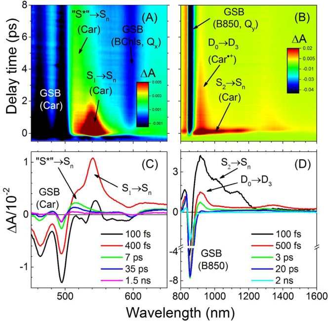
Transient absorption data of the LDAO-Tris-LH2 complex recorded after excitation into the (0–0) vibronic band of carotenoid neurosporene; (A, B) TA contour maps highlighting ESA bands and bleaching of ground state absorption (GSB) of either carotenoid (Car) or BChls present within 8 ps after excitation. The states from which the transition occurs are indicated. (C, D) Exemplary transient absorption spectra taken at various delay times after excitation. The main transient ESA bands are indicated.
The main ESA bands associated with the carotenoid neurosporene are indicated along with spectral features corresponding to bleaching of ground state absorption (GSB) of either carotenoid or BChls. In the LDAO-Tris-LH2 sample, the S1 → Sn ESA band with maximum at 540 nm is instantaneously populated and then fades away within 3 ps. The remaining spectrally broad signal with a maximum at ∼520 nm represents the ESA associated with an ambiguous “S* state”. Another noticeable band present in the NIR range appearing only within a few hundred femtoseconds after excitation (Figure 2B, deep-red color; Figure 2D, black line) is associated with the short-lived neurosporene S2 state. Previous experimental and theoretical evaluations of that band demonstrated that it is associated with the S2 → Sn transition.33 Simultaneously with the S* feature, another spectral band is clearly present in the NIR range between 920 and 1000 nm. This band is associated with D0 → D3 absorption of carotenoid radical cation23,27,29,34 that is formed shortly after excitation via interaction with B800 BChl a that leads to ultrafast formation of the temporarily existing Neu•+B800– pair. Simultaneous appearance and decay of both transient bands (D0 → D3 and S*) suggests that those may have common temporal characteristics.
Transient absorption results taken in vis and NIR spectral ranges of the DDM-HEP-LH2 excited in the neurosporene absorption band are given in Figure 3.
Figure 3.
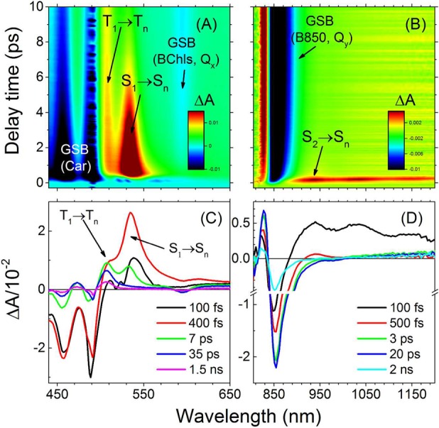
Transient absorption data of the DDM-HEP-LH2 complex recorded after excitation into the (0–0) vibronic band of carotenoid neurosporene. (A, B) TA contour maps highlighting ESA bands and bleaching of ground state absorption (GSB) of either the carotenoid (Car) or BChls present within 10 ps after excitation. The states from which the transition occurs are indicated. (C, D) Exemplary transient absorption spectra taken at various delay times after excitation. Note that the D0 → D3 absorption band of Neu•+ has disappeared and the S* spectral feature is replaced by a noticeably sharper and blue-shifted transient absorption band.
Contour maps of TA shown within the first 10 ps after excitation (Figure 3A,B) demonstrate few significant changes in respect to LH2 with a fully developed B800 absorption band. The neurosporene S1 → Sn ESA band appears at 535 nm and persists for a much longer time indicating weakened energetic coupling with (B)Chls. At the very early delay times, the S1 → Sn ESA band is also red-shifted, representing vibrational equilibration of the S1 state. Most importantly, the broad spectral feature characteristic of “S*” has substantially changed. It has been replaced by a sharp and slightly blue-shifted positive transient band with a maximum at 507 nm. In the NIR spectral range the band associated with absorption of Neu•+ completely disappeared, demonstrating that neurosporene is not able to form the cation–anion pair with ac-Chl a “B800”.34 Further spectroscopic evaluation by measurements in the submicrosecond time regime demonstrated that this transient band is identical to triplet–triplet (T1 → Tn) ESA of LH2-bound neurosporene, as demonstrated in Figure 4. It shows the TA spectrum of the LDAO-Tris-LH2 taken at 7 ps, corresponding to so-called S* spectral feature overlaid with equivalent TA spectrum of the DDM-HEP-LH2 (because neurosporene decays longer in this LH2 batch it was taken at 30 ps).
Figure 4.
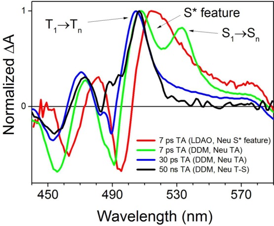
Comparison of transient absorption spectra taken for both LH2 batches (LDAO and DDM preps) at time delays at which the main S1 → Sn ESA of neurosporene decayed. For the LDAO-Tris-LH2, the TA spectrum taken at 7 ps corresponds to S* spectral feature. For comparison, also TA spectrum of the DDM-HEP-LH2 at the same delay time is shown. The 30 ps spectrum (free of S1 → Sn ESA band) is identical to the triplet-minus-singlet (T – S) spectrum of neurosporene taken at 50 ns after excitation. All spectra were normalized to unity at their maxima.
The fourth spectrum corresponds to TA of the DDM-HEP-LH2 taken at 50 ns, and due to the long time delay, it can be only associated with the so-called triplet-minus-singlet (T–S) spectrum of neurosporene. It is apparent that both 30 ps and 50 ns TA spectra are essentially identical but are very different from the spectral envelope of the S* feature. It is worth noting that, thus, the triplet T1 → Tn band is already clearly resolved in the TA spectrum at 1 ps (Figure 3C).
Discussion
Electric Field Established by Neu•+B800– Pair
Experimental results shown above clearly suggest a connection between the Neu•+B800– pair and the S* transient spectral feature. As we have hypothesized that the S* band can be associated with an electrochromic response of neutral carotenoid to the electric field temporarily induced by the Car•+B800– pair, it would be particularly interesting to know what level of electric field the neighboring neutral carotenoid may sense from the cation–anion pair. In order to calculate this, pigment coordinates from the Rps. acidophila LH2 crystal structure have been used.35 As crystal structure of Rba. sphaeroides LH2 is not known, the results should be interpreted rather as “qualitative” evidence used to support the concept. Because the pigments are not infinitesimally small, in order to make this simple modeling feasible it was necessary to approximate them by points that lie on the surface that is designated by the plane of the B800 macrocycle. With these assumptions, the arrangement of the pigments of interest can be represented as in Figure 5. The point symbolizing B800 BChl a has been placed in the center of the pigment’s macrocycle. To simplify calculations, the frame of reference was placed in the midpoint between B800– and Car+.
Figure 5.
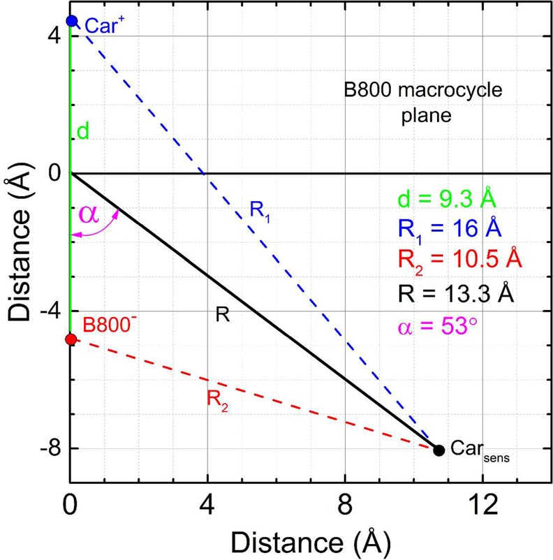
Simplified arrangement of the Car•+B800– pair and the nearest neutral carotenoid (Carsens) based on pigment coordinates from Rps. acidophila LH2.35 The axis surface represents the B800 BChl a macrocycle plane. Point symbolizing B800 BChl a is placed in the center of the pigment’s macrocycle.
The electric field that the nearest neutral carotenoid senses from the electric dipole formed by the Car•+B800– pair can be described by following equation:
| 1 |
The parameters d, R, and α are provided in Figure 5; q is the electron charge, and εp is the relative dielectric constant of the surrounding protein medium. Its value has been estimated for various LH2 complexes in the 1.2–1.3 range,36 and a value of 1.2 has been used in this model. Calculations show that the nearest neutral carotenoid (Carsens) senses an electric field of 4.4 MV/cm.
Previous studies of electrochromic responses of carotenoids bound to LH2 suggested that the strong bathochromic shift of carotenoid absorption observed in the LH2 protein, with respect to low polarizable solvents like n-hexane, is caused by a local electric field of 4 MV/cm that is most likely brought by protein residues like βArg-10.37,38 If those two above-mentioned factors, a static local electric field from protein residue (Eloc) and a temporal electric field brought by Car•+B800– pair (ET), will simultaneously perturb the carotenoid S2 state, the overall temporal electrochromic shift of the carotenoid absorption spectrum (Δυ̃, in wavenumbers) can be calculated on the basis of the following equation for a Stark shift:39
| 2 |
Here, Eloc and ET are electric field vectors; Δμ and Δα are changes in the vector of dipole moment and polarizability tensor, respectively, of the carotenoid between its S0 and S2 states. For simplicity, Δα is assumed to be a scalar with value of Δα = 1350 Å3 that was previously calculated for structurally similar open-chain carotenoid lycopene40 (1 Å3 = 1.113 × 10–40 C m2 V–1). The values of electric vectors are Eloc = 4 MV/cm,38ET = 4.4 MV/cm, and 1/hc = 0.5 × 1025 C J–1 m–1. It was also assumed that the first term in eq 2 is negligible. According to the simplistic model in Figure 5, ET and Δμ vectors would be orthogonal to each other (Δμ will be essentially in line with carotenoid conjugation that crosses the graph surface while ET will lie on the graph surface). Then, if ET and Eloc are mutually oriented such that the dot product is maximized and positive (similar orientation, parallel), the second term will give an electrochromic shift of 1330 cm–1 while the third one will be 730 cm–1 and an overall red-shift of 2060 cm–1 in respect to absorption in a low polarizable medium like n-hexane. As in n-hexane, the (0–0) vibronic band of neurosporene appears at 467 nm (21 410 cm–1),41 so that the shifted spectrum should appear at 19 350 cm–1, which corresponds to 517 nm.
Reconstructing the S* Transient Spectral Feature
We have shown that this carotenoid may temporarily experience additional a 26 nm red-shift (∼1000 cm–1) of its steady-state absorption in LH2 (normally the (0–0) band is at 491 nm). As demonstrated in Figure 6, this ultrafast Stark shift is strong enough to very adequately reconstruct the S* transient spectral feature. Figure 6A shows the expected transient Stark spectrum of the neurosporene, induced by the electric field of the Neu•+B800– pair (red line). The spectrum is a spectral difference of shifted (dashed black line) and nonshifted (blue solid line) absorption spectra of neurosporene with spectral shift of 26 nm. The pure transient Stark spectrum is similar to the S* transient feature; however, it does not mimic it correctly, as demonstrated in Figure 6B. This suggests that other factor(s) could influence its spectral shape. In order to be sure that raw S* feature spectrum is not significantly distorted by coexisting transient bands of BChls, those bands have been removed. It was done by subtracting a corresponding transient absorption spectrum of the carotenoidless LH2 from Rba. sphaeroides R.26.1 (Figure 6B, red line). The “pure” S* feature spectrum (blue line) still substantially deviates from the Stark spectrum, mainly in the range of bleaching of the carotenoid absorption band. However, it should be emphasized that, in addition to the transient Stark spectrum, an additional transient band should be also observed. This band will correspond to bleaching of a ground-state absorption (GSB) of the carotenoid pool fraction that is involved in ultrafast formation of the Neu•+B800– pair. Our model of pigment interactions assumed that a single molecule of Neu•+ affects a single molecule of the neutral carotenoid, and such stoichiometry should be also reflected in the spectroscopic measurements. Therefore, the amplitude of GSB should be equal with the amplitudes of shifted and nonshifted absorption spectra as shown in Figure 6A (green line). Because both spectral features, GSB and Stark spectrum, have identical temporal characteristics, both should appear as one spectro-temporal feature that spectrally will resemble the sum of both features. This spectral-temporal feature is given in Figure 6C (red line), along with the “pure” transient S* spectrum (blue line). Importantly, now both spectral profiles match each other very well.
Figure 6.
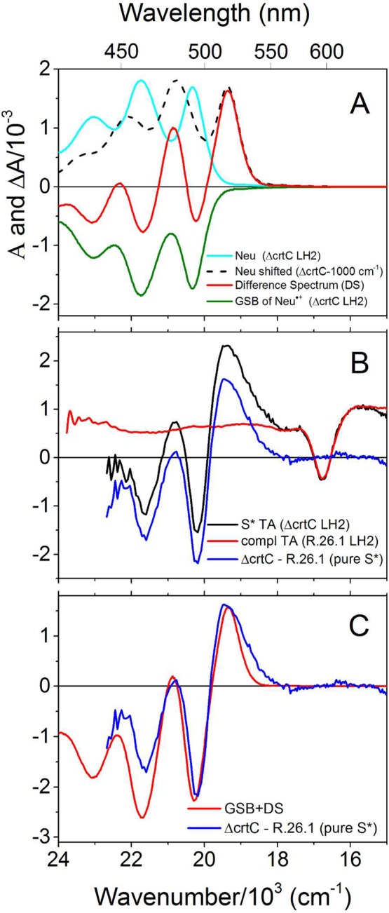
Reconstruction of the S* transient spectral feature. (A) Spectral components involved in reconstructing the S* transient feature: cyan line, ground-state absorption spectrum of neurosporene in the LH2; dashed black line, ground-state absorption spectrum of neurosporene affected by the electric field induced by Neu•+B800– pair; red line, difference spectrum (Stark spectrum) of the previous two spectra; green line, ground-state absorption bleaching of neurosporene involved in formation of Neu•+B800– pair. (B) The S* spectral feature cleaned from contribution of transient absorption of BChls by subtracting complementary spectrum of carotenoidless LH2. (C) Pure S* feature overlaid with its anticipated spectrum (for details refer to the main text): TA, transient absorption, GSB, ground-state absorption bleaching.
Formation of Carotenoid Triplet State
Another interesting aspect revealed by this study is the ultrafast formation of the carotenoid triplet state in situations when the Car•+B800– pair cannot be formed due to an absence of B800 BChl a. Triplet formation within 1 ps after excitation strongly suggests that the carotenoid triplet pool is not populated in the typical way via sensitization by BChls triplets, and therefore, carotenoid singlet fission has to be involved. This process in carotenoids was first suggested after observation of a high quantum yield of carotenoid triplet formation in whole cells of the purple bacterium R. rubrum.42 It has been suggested that S1 and 11Bu–, a state without a precise Sx designation (as it may lie below or above S2 depending on length of carotenoid conjugation), could be viewed as a singlet-coupled combination of two triplets localized in the different parts of a carotenoid chromophore: 11Bu– = T2⊗T1 and S1 = T1⊗T1.43 Singlet fission, in which T⊗T decouples into physically independent triplet states, was reported to have very low yields in solvents,44−46 but it is high (up to 30%) in proteins,25,43,47 suggesting that the decoupling process could be facilitated by a conformational distortion of a carotenoid structure provided by the protein environment. The TA data from Figure 3 show that T1 → Tn and S1→ Sn ESA bands appear simultaneously, suggesting that the S1 state cannot be a state in which singlet fission takes place. The second candidate, an optically forbidden 11Bu– state, has been proposed to lie just below the S2 state for carotenoids with N ≥ 946 (and that includes neurosporene), and its lifetime could be too short to be temporally resolved from the initially excited S2 state.48 Therefore, the detailed mechanism of carotenoid singlet fission was not further investigated in this study.
Simplified Carotenoid Excitation Decay Pathway in LH2
Target model analysis (Figure 7) can be used to review the study presented here and propose a new, simplified S*-free carotenoid excitation decay pathway in the LH2 complex from purple bacteria.
Figure 7.
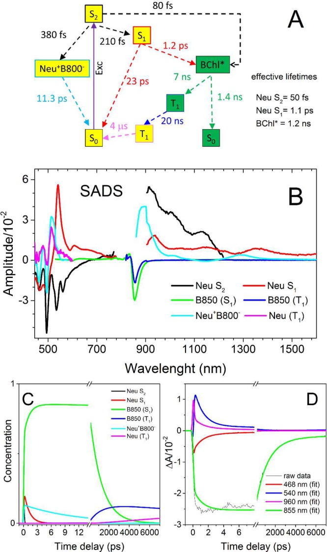
Target analysis of the vis–NIR combined TA data set of the LDAO-Tris-LH2 complex. (A) Kinetic model employed to simulate the TA data set. (B) SADS amplitudes resulting from the kinetic analysis. The yellow compartments in the kinetic models symbolize carotenoid neurosporene (with exception of the Neu•+B800– pair) while green compartments correspond to BChl a. Note that the SADS amplitudes do not correspond to actual contribution of the component in the raw TA data and should be weighted by their time-dependent concentrations; Exc = excitation.
The model should be seen as data simulation that incorporates numerous known parameters adapted from previous studies of neurosporene in solvents and in LH2. The so-called “S* state” has been replaced by the Neu•+B800– pair, evolving from the carotenoid S2 state during its relaxation. It was assumed that the population of BChl a triplets is entirely quenched by neurosporene molecules with time constant of ∼20 ns, a value obtained from fitting the recovery of BChl a ground state absorption bleaching (Figure S1). This value is similar to triplet lifetime of BChl a in other LH2 complexes.49 Neurosporene triplet lifetime was obtained from fitting of the T1 → Tn band decay (Figure S2). The effective lifetime of the B850 BChl a of ∼1.2 ns was obtained from fitting of the kinetic trace recorded at 850 nm (Figure S3) and was decomposed to microscopic decays of 1.4 ns (S1 → S0, radiative and nonradiative) and 7 ns (intersystem crossing, ISC, S1 → T1, to be in agreement with triplet yield). Due to the temporal resolution limitation of the spectrometer, the effective lifetime of the carotenoid S2 state in LH2 complexes could not be measured and instead was set to ∼50 fs as demonstrated in previous studies.24,25 Microscopic parameters of neurosporene S2 state depopulation are described with the following details: the S2 → S1 decay was assumed to have the same characteristics as in solvent; the S2 → BChls decay was set in order to give an overall carotenoid to BChl a energy transfer efficiency of 87% that is known from previous studies of this complex.9 For simplicity both spectral forms of BChl a, B800 and B850, were combined as unity. It was assumed, in agreement with experimental data, that Neu•+ is populated with ∼13% quantum efficiency from the S2 state of the neutral form. The SADS (species associated decay spectra) are given in Figure 7B. It should be clarified that the contribution of the SADS component in the raw TA spectra should not be judged on the basis of its relative amplitude but on the basis of the product of SADS amplitude and its concentration at a specific delay time.
Conclusions
This study clarifies the origin of the S* feature in LH2 complexes from purple bacterium Rba. sphaeroides. We propose that the S* transient spectral feature corresponds to a combination of bleaching of the ground-state absorption of carotenoid involved in the Car•+B800– pair and the Stark spectrum of the nearest neutral carotenoid. The target analysis model of the TA data set also supports this view. This eliminates the so-called “S* state” from existing kinetic schemes of the carotenoid excitation decay pathway in the LH2 complex, and thereby provides a new framework for investigating the excited states of carotenoids in these photosynthetic antenna complexes.
Acknowledgments
All spectroscopic work was done in the PARC ultrafast laser facility supported as part of the Photosynthetic Antenna Research Center (PARC), an Energy Frontier Research Center funded by the U.S. Department of Energy, Office of Science, Office of Basic Energy Sciences under Award DE-SC 0001035. PARC also provided support for D.M.N. and partial support for C.N.H. C.N.H. also gratefully acknowledges financial support from the Biotechnology and Biological Sciences Research Council (BBSRC UK), award number BB/M000265/1, and an Advanced Award 338895 from the European Research Council. We would like to thank Prof. Harry Frank from the University of Connecticut for providing carotenoidless LH2 from Rba. sphaeroides strain R.26.1.
Supporting Information Available
The Supporting Information is available free of charge on the ACS Publications website at DOI: 10.1021/acs.jpcb.6b08639.
Recovery of bleaching of the B850 band in the LDAO-Tris-LH2 sample recorded in 7 and 150 ns time windows and decay of neurosporene triplet state (PDF)
The authors declare no competing financial interest.
Supplementary Material
References
- Takaichi S.Carotenoids and Carotenogenesis in Anoxygenic Photosythetic Bacteria. In Photochemistry of Carotenoids; Frank H. A., et al. , Ed.; Kluwer Academic Publishers: Dordrecht, 1999; pp 39–69. [Google Scholar]
- Demmig-Adams B.; Adams W. W. I.. The Xanthophyll Cycle. In Carotenoids in Photosynthesis; Young A.J., Britton D., Eds.; Chapman and Hall: London, 1993; pp 206–251. [Google Scholar]
- Staleva H.; Komenda J.; Shukla M. K.; Slouf V.; Kana R.; Polivka T.; Sobotka R. Mechanism of Photoprotection in the Cyanobacterial Ancestor of Plant Antenna Proteins. Nat. Chem. Biol. 2015, 11, 287–291. 10.1038/nchembio.1755. [DOI] [PubMed] [Google Scholar]
- Niedzwiedzki D. M.; Tronina T.; Liu H.; Staleva H.; Komenda J.; Sobotka R.; Blankenship R. E.; Polivka T. Carotenoid-Induced Non-Photochemical Quenching in the Cyanobacterial Chlorophyll Synthase-Hlic/D Complex. Biochim. Biophys. Acta, Bioenerg. 2016, 1857, 1430–1439. 10.1016/j.bbabio.2016.04.280. [DOI] [PubMed] [Google Scholar]
- Lang H. P.; Hunter C. N. The Relationship between Carotenoid Biosynthesis and the Assembly of the Light-Harvesting LH2 Complex in Rhodobacter Sphaeroides. Biochem. J. 1994, 298, 197–205. 10.1042/bj2980197. [DOI] [PMC free article] [PubMed] [Google Scholar]
- Schulten K.; Karplus M. On the Origin of a Low-Lying Forbidden Transition in Polyenes and Related Molecules. Chem. Phys. Lett. 1972, 14, 305–309. 10.1016/0009-2614(72)80120-9. [DOI] [Google Scholar]
- Krinsky N. I.Function. In Carotenoids; Isler O., Guttman N., Solms U., Eds.; Birghäuser Verlag: Basel, Switzerland, 1971; pp 669–716. [Google Scholar]
- Šlouf V.; Chabera P.; Olsen J. D.; Martin E. C.; Qian P.; Hunter C. N.; Polivka T. Photoprotection in a Purple Phototrophic Bacterium Mediated by Oxygen-Dependent Alteration of Carotenoid Excited-State Properties. Proc. Natl. Acad. Sci. U. S. A. 2012, 109, 8570–8575. 10.1073/pnas.1201413109. [DOI] [PMC free article] [PubMed] [Google Scholar]
- Dilbeck P. L.; Tang Q.; Mothersole D. J.; Martin E. C.; Hunter C. N.; Bocian D. F.; Holten D.; Niedzwiedzki D. M. Quenching Capabilities of Long-Chain Carotenoids in Light Harvesting-2 Complexes from Rhodobacter Sphaeroides with an Engineered Carotenoid Synthesis Pathway. J. Phys. Chem. B 2016, 120, 5429–5443. 10.1021/acs.jpcb.6b03305. [DOI] [PMC free article] [PubMed] [Google Scholar]
- Frank H. A.; Farhoosh R.; Gebhard R.; Lugtenburg J.; Gosztola D.; Wasielewski M. R. The Dynamics of the S1 Excited-States of Carotenoids. Chem. Phys. Lett. 1993, 207, 88–92. 10.1016/0009-2614(93)85016-H. [DOI] [Google Scholar]
- Frank H. A.; Cua A.; Chynwat V.; Young A.; Gosztola D.; Wasielewski M. R. Photophysics of the Carotenoids Associated with the Xanthophyll Cycle in Photosynthesis. Photosynth. Res. 1994, 41, 389–395. 10.1007/BF02183041. [DOI] [PubMed] [Google Scholar]
- Cerullo G.; Polli D.; Lanzani G.; De Silvestri S.; Hashimoto H.; Cogdell R. J. Photosynthetic Light Harvesting by Carotenoids: Detection of an Intermediate Excited State. Science 2002, 298, 2395–2398. 10.1126/science.1074685. [DOI] [PubMed] [Google Scholar]
- Larsen D. S.; Papagiannakis E.; van Stokkum I. H. M.; Vengris M.; Kennis J. T. M.; van Grondelle R. Excited State Dynamics of Beta-Carotene Explored with Dispersed Multi-Pulse Transient Absorption. Chem. Phys. Lett. 2003, 381, 733–742. 10.1016/j.cplett.2003.10.016. [DOI] [Google Scholar]
- Gradinaru C. C.; Kennis J. T.; Papagiannakis E.; van Stokkum I. H.; Cogdell R. J.; Fleming G. R.; Niederman R. A.; van Grondelle R. An Unusual Pathway of Excitation Energy Deactivation in Carotenoids: Singlet-to-Triplet Conversion on an Ultrafast Timescale in a Photosynthetic Antenna. Proc. Natl. Acad. Sci. U. S. A. 2001, 98, 2364–2369. 10.1073/pnas.051501298. [DOI] [PMC free article] [PubMed] [Google Scholar]
- Koyama Y.; Rondonuwu F. S.; Fujii R.; Watanabe Y. Light-Harvesting Function of Carotenoids in Photosynthesis: The Roles of the Newly Found 11Bu– State. Biopolymers 2004, 74, 2–18. 10.1002/bip.20034. [DOI] [PubMed] [Google Scholar]
- Andersson P. O.; Gillbro T. Photophysics and Dynamics of the Lowest Excited Singlet State in Long Substituted Polyenes with Implications to the Very Long Chain Limit. J. Chem. Phys. 1995, 103, 2509–2519. 10.1063/1.469672. [DOI] [Google Scholar]
- Niedzwiedzki D.; Koscielecki J. F.; Cong H.; Sullivan J. O.; Gibson G. N.; Birge R. R.; Frank H. A. Ultrafast Dynamics and Excited State Spectra of Open-Chain Carotenoids at Room and Low Temperatures. J. Phys. Chem. B 2007, 111, 5984–5998. 10.1021/jp070500f. [DOI] [PubMed] [Google Scholar]
- Niedzwiedzki D. M.; Sullivan J. O.; Polivka T.; Birge R. R.; Frank H. A. Femtosecond Time-Resolved Transient Absorption Spectroscopy of Xanthophylls. J. Phys. Chem. B 2006, 110, 22872–22885. 10.1021/jp0622738. [DOI] [PubMed] [Google Scholar]
- Lenzer T.; Ehlers F.; Scholz M.; Oswald R.; Oum K. Assignment of Carotene S* State Features to the Vibrationally Hot Ground Electronic State. Phys. Chem. Chem. Phys. 2010, 12, 8832–8839. 10.1039/b925071a. [DOI] [PubMed] [Google Scholar]
- Ehlers F.; Scholz M.; Schimpfhauser J.; Bienert J.; Oum K.; Lenzer T. Collisional Relaxation of Apocarotenals: Identifying the S* State with Vibrationally Excited Molecules in the Ground Electronic State S0*. Phys. Chem. Chem. Phys. 2015, 17, 10478–10488. 10.1039/C4CP05600K. [DOI] [PubMed] [Google Scholar]
- Balevicius V. Jr.; Abramavicius D.; Polivka T.; Galestian Pour A.; Hauer J. A Unified Picture of S* in Carotenoids. J. Phys. Chem. Lett. 2016, 7, 3347–3352. 10.1021/acs.jpclett.6b01455. [DOI] [PMC free article] [PubMed] [Google Scholar]
- Papagiannakis E.; Kennis J. T. M.; van Stokkum I. H. M.; Cogdell R. J.; van Grondelle R. An Alternative Carotenoid-to-Bacteriochlorophyll Energy Transfer Pathway in Photosynthetic Light Harvesting. Proc. Natl. Acad. Sci. U. S. A. 2002, 99, 6017–6022. 10.1073/pnas.092626599. [DOI] [PMC free article] [PubMed] [Google Scholar]
- Cong H.; Niedzwiedzki D. M.; Gibson G. N.; LaFountain A. M.; Kelsh R. M.; Gardiner A. T.; Cogdell R. J.; Frank H. A. Ultrafast Time-Resolved Carotenoid to-Bacteriochlorophyll Energy Transfer in LH2 Complexes from Photosynthetic Bacteria. J. Phys. Chem. B 2008, 112, 10689–10703. 10.1021/jp711946w. [DOI] [PMC free article] [PubMed] [Google Scholar]
- Papagiannakis E.; van Stokkum I. H.; Vengris M.; Cogdell R. J.; van Grondelle R.; Larsen D. S. Excited-State Dynamics of Carotenoids in Light-Harvesting Complexes. 1. Exploring the Relationship between the S1 and S* States. J. Phys. Chem. B 2006, 110, 5727–5736. 10.1021/jp054633h. [DOI] [PubMed] [Google Scholar]
- Papagiannakis E.; Das S. K.; Gall A.; van Stokkum I. H. M.; Robert B.; van Grondelle R.; Frank H. A.; Kennis J. T. M. Light Harvesting by Carotenoids Incorporated into the B850 Light-Harvesting Complex from Rhodobacter Sphaeroides R-26.1: Excited-State Relaxation, Ultrafast Triplet Formation, and Energy Transfer to Bacteriochlorophyll. J. Phys. Chem. B 2003, 107, 5642–5649. 10.1021/jp027174i. [DOI] [Google Scholar]
- Chynwat V.; Frank H. A. The Application of the Energy Gap Law to the S1 Energies and Dynamics of Carotenoids. Chem. Phys. 1995, 194, 237–244. 10.1016/0301-0104(95)00017-I. [DOI] [Google Scholar]
- Polivka T.; Zigmantas D.; Herek J. L.; He Z.; Pascher T.; Pullerits T.; Cogdell R. J.; Frank H. A.; Sundstrom V. The Carotenoid S1 State in LH2 Complexes from Purple Bacteria Rhodobacter Sphaeroides and Rhodopseudomonas Acidophila: S1 Energies, Dynamics, and Carotenoid Radical Formation. J. Phys. Chem. B 2002, 106, 11016–11025. 10.1021/jp025752p. [DOI] [Google Scholar]
- Chi S. C.; Mothersole D. J.; Dilbeck P.; Niedzwiedzki D. M.; Zhang H.; Qian P.; Vasilev C.; Grayson K. J.; Jackson P. J.; Martin E. C.; et al. Assembly of Functional Photosystem Complexes in Rhodobacter Sphaeroides Incorporating Carotenoids from the Spirilloxanthin Pathway. Biochim. Biophys. Acta, Bioenerg. 2015, 1847, 189–201. 10.1016/j.bbabio.2014.10.004. [DOI] [PMC free article] [PubMed] [Google Scholar]
- Polivka T.; Pullerits T.; Frank H. A.; Cogdell R. J.; Sundstrom V. Ultrafast Formation of a Carotenoid Radical in LH2 Antenna Complexes of Purple Bacteria. J. Phys. Chem. B 2004, 108, 15398–15407. 10.1021/jp0483019. [DOI] [Google Scholar]
- Niedzwiedzki D. M.; Dilbeck P. L.; Tang Q.; Mothersole D. J.; Martin E. C.; Bocian D. F.; Holten D.; Hunter C. N. Functional Characteristics of Spirilloxanthin and Keto-Bearing Analogues in Light-Harvesting LH2 Complexes from Rhodobacter Sphaeroides with a Genetically Modified Carotenoid Synthesis Pathway. Biochim. Biophys. Acta, Bioenerg. 2015, 1847, 640–655. 10.1016/j.bbabio.2015.04.001. [DOI] [PubMed] [Google Scholar]
- Niedzwiedzki D. M.; Bina D.; Picken N.; Honkanen S.; Blankenship R. E.; Holten D.; Cogdell R. J. Spectroscopic Studies of Two Spectral Variants of Light-Harvesting Complex 2 (LH2) from the Photosynthetic Purple Sulfur Bacterium Allochromatium Vinosum. Biochim. Biophys. Acta, Bioenerg. 2012, 1817, 1576–1587. 10.1016/j.bbabio.2012.05.009. [DOI] [PubMed] [Google Scholar]
- van Stokkum I. H. M.; Larsen D. S.; van Grondelle R. Global and Target Analysis of Time-Resolved Spectra. Biochim. Biophys. Acta, Bioenerg. 2004, 1657, 82–104. 10.1016/j.bbabio.2004.04.011. [DOI] [PubMed] [Google Scholar]
- Yoshizawa M.; Kosumi D.; Komukai M.; Hashimoto H. Ultrafast Optical Responses of Three-Level Systems in β-carotene: Resonance to a High-Lying N1ag– Excited State. Laser Phys. 2006, 16, 325–330. 10.1134/S1054660X06020204. [DOI] [Google Scholar]
- Polivka T.; Niedzwiedzki D.; Fuciman M.; Sundstrom V.; Frank H. A. Role of B800 in Carotenoid-Bacteriochlorophyll Energy and Electron Transfer in LH2 Complexes from the Purple Bacterium Rhodobacter Sphaeroides. J. Phys. Chem. B 2007, 111, 7422–7431. 10.1021/jp071395c. [DOI] [PubMed] [Google Scholar]
- McDermott G.; Prince S. M.; Freer A. A.; Hawthornthwaite-Lawless A. M.; Papiz M. Z.; Cogdell R. J.; Isaacs N. W. Crystal Structure of an Integral Membrane Light-Harvesting Complex from Photosynthetic Bacteria. Nature 1995, 374, 517–521. 10.1038/374517a0. [DOI] [Google Scholar]
- Georgakopoulou S.; Frese R. N.; Johnson E.; Koolhaas C.; Cogdell R. J.; van Grondelle R.; van der Zwan G. Absorption and Cd Spectroscopy and Modeling of Various LH2 Complexes from Purple Bacteria. Biophys. J. 2002, 82, 2184–2197. 10.1016/S0006-3495(02)75565-3. [DOI] [PMC free article] [PubMed] [Google Scholar]
- Herek J. L.; Polivka T.; Pullerits T.; Fowler G. J. S.; Hunter C. N.; Sundstrom V. Ultrafast Carotenoid Band Shifts Probe Structure and Dynamics in Photosynthetic Antenna Complexes. Biochemistry 1998, 37, 7057–7061. 10.1021/bi980118g. [DOI] [PubMed] [Google Scholar]
- Herek J. L.; Wendling M.; He Z.; Polivka T.; Garcia-Asua G.; Cogdell R. J.; Hunter C. N.; van Grondelle R.; Sundstrom V.; Pullerits T. Ultrafast Carotenoid Band Shifts: Experiment and Theory. J. Phys. Chem. B 2004, 108, 10398–10403. 10.1021/jp040094p. [DOI] [Google Scholar]
- Ratsep M.; Wu H. M.; Hayes J. M.; Blankenship R. E.; Cogdell R. J.; Small G. J. Stark Hole-Burning Studies of Three Photosynthetic Complexes. J. Phys. Chem. B 1998, 102, 4035–4044. 10.1021/jp980421r. [DOI] [Google Scholar]
- Krawczyk S.; Olszowka D. Spectral Broadening and Its Effect in Stark Spectra of Carotenoids. Chem. Phys. 2001, 265, 335–347. 10.1016/S0301-0104(01)00314-7. [DOI] [Google Scholar]
- Carotenoids Handbook; Britton G., Liaaen-Jensen S., Pfander H., Eds.; Birkhäuser Basel: Basel, 2004. [Google Scholar]
- Rademaker H.; Hoff A. J.; Vangrondelle R.; Duysens L. N. M. Carotenoid Triplet Yields in Normal and Deuterated Rhodospirillum Rubrum. Biochim. Biophys. Acta, Bioenerg. 1980, 592, 240–257. 10.1016/0005-2728(80)90185-1. [DOI] [PubMed] [Google Scholar]
- Tavan P.; Schulten K. Electronic Excitations in Finite and Infinite Polyenes. Phys. Rev. B: Condens. Matter Mater. Phys. 1987, 36, 4337–4358. 10.1103/PhysRevB.36.4337. [DOI] [PubMed] [Google Scholar]
- Niedzwiedzki D. M.; Kajikawa K.; Aoki K.; Katsumura S.; Frank H. Excited States Energies and Dynamics of Peridinin Analogues and Thenature of the Intramolecular Charge Transfer State in Carbonyl-Containing Carotenoids. J. Phys. Chem. B 2013, 117, 6874–6887. 10.1021/jp400038k. [DOI] [PubMed] [Google Scholar]
- Zhang J. P.; Inaba T.; Koyama Y. The Role of the Newly-Found 1Bu– State of Carotenoid in Mediating the 1Bu+-to-2Ag– Internal Conversion and the Excited-State Dynamics of Carotenoid and Bacteriochlorophyll in a Bacterial Antenna Complex. J. Mol. Struct. 2001, 598, 65–78. 10.1016/S0022-2860(01)00806-7. [DOI] [Google Scholar]
- Rondonuwu F. S.; Watanabe Y.; Fujii R.; Koyama Y. A First Detection of Singlet to Triplet Conversion from the 11Bu– to the 13Ag State and Triplet Internal Conversion from the 13Ag to the 13Bu State in Carotenoids: Dependence on the Conjugation Length. Chem. Phys. Lett. 2003, 376, 292–301. 10.1016/S0009-2614(03)00983-7. [DOI] [Google Scholar]
- Polivka T.; Balashov S. P.; Chabera P.; Imasheva E. S.; Yartsev A.; Sundstrom V.; Lanyi J. K. Femtosecond Carotenoid to Retinal Energy Transfer in Xanthorhodopsin. Biophys. J. 2009, 96, 2268–2277. 10.1016/j.bpj.2009.01.004. [DOI] [PMC free article] [PubMed] [Google Scholar]
- Polívka T.; Sundström V. Dark Excited States of Carotenoids: Consensus and Controversy. Chem. Phys. Lett. 2009, 477, 1–11. 10.1016/j.cplett.2009.06.011. [DOI] [Google Scholar]
- Kosumi D.; Horibe T.; Sugisaki M.; Cogdell R. J.; Hashimoto H. Photoprotection Mechanism of Light-Harvesting Antenna Complex from Purple Bacteria. J. Phys. Chem. B 2016, 120, 951–956. 10.1021/acs.jpcb.6b00121. [DOI] [PubMed] [Google Scholar]
Associated Data
This section collects any data citations, data availability statements, or supplementary materials included in this article.


