Abstract
PURPOSE
Considering the importance of retention in the success and long-term clinical service of fixed partial dentures (FPDs) as well as the existing controversy regarding the effect of GLUMA desensitizer on the retention of full metal crowns cemented with RelyX U200 self-adhesive cement, this study aimed to assess the effect of GLUMA desensitizer on the retention of full metal crowns cemented using RelyX U200.
MATERIALS AND METHODS
In this experimental study, 20 sound human premolars were prepared; a 0.5 mm chamfer finish line was prepared above the cementoenamel junction. The teeth were randomly assigned to two groups: a desensitizer group (n = 10, treated with GLUMA desensitizer) and a control (n = 10, no surface treatment). Full metal crowns were fabricated of base metal alloy and had a ring. All crowns were cemented with RelyX U200 and subjected to retention test by using a universal testing machine. The data were analyzed using SPSS version 20 and independent t-test.
RESULTS
The mean tensile bond strength was significantly higher in the GLUMA desensitizer group (230.63 ± 63.8 N) compared to the control group (164.45 ± 39.3 N) (P≤.012).
CONCLUSION
GLUMA desensitizer increases the tensile bond strength of RelyX U200 self-adhesive cement to dentin.
Keywords: Gluma desensitizer, Retention, Crowns, Self-adhesive cement
INTRODUCTION
Retention is an important factor in determining the success and clinical service of FPDs.1 The retention of crown is based on the presence of two almost parallel vertical surfaces from tooth preparation; Al-Omari et al. suggested that the mean convergence angle between 22.4 and 25.3 degrees was clinically acceptable.2 Zidan and Ferguson recommended 5 - 12° taper to be ideal.3 Optimal retention for extra-coronal restorations depends on the morphology of the prepared tooth and factors such as the degree of taper, the prepared surface area, roughness of the internal surfaces of crown, retentive grooves, texture of the treated surfaces, and the type of cement.4 Inadequate retention can lead to microleakage through the cement, development of secondary caries beneath the crown, cement washout beneath the crown, chipping and fracture of the crown, and the crown's eventual failure.2,4
Most FPD patients experience pain or discomfort in the prepared tooth during and some time after the cementation of restoration, which may be due to dentin hypersensitivity. 5 To overcome this problem, desensitizing agents have been introduced.5 However, a question raises that whether the application of desensitizer agents, such as GLUMA desensitizer, affects the retention of full-coverage crowns cemented with Rely-X U200 self-adhesive cements. Sailer et al.,6 in 2012, reported that application of GLUMA desensitizer enhanced the shear bond strength of cement. However, Jalandar et al.,5 in 2012, reported that GLUMA desensitizer had no significant effect on the retention of crowns. Considering the existing controversies, this study aimed to assess the effect of GLUMA desensitizer on the retention of full crowns cemented using RelyX U200.
MATERIALS AND METHODS
1-Study design: This was an in vitro experimental study.
2-Methodology: This study was done in the Prosthodontics Department of Azad University-dental Branch and was conducted on 20 sound premolar teeth (no caries or restoration) with approximately the same size, which had been extracted for orthodontic purposes in a dental clinic in Tehran in 2013. The teeth were immersed in 0.1% thymol solution for 48 hours for disinfection. The soft tissue residues were also removed.5 The teeth were then prepared as follows:
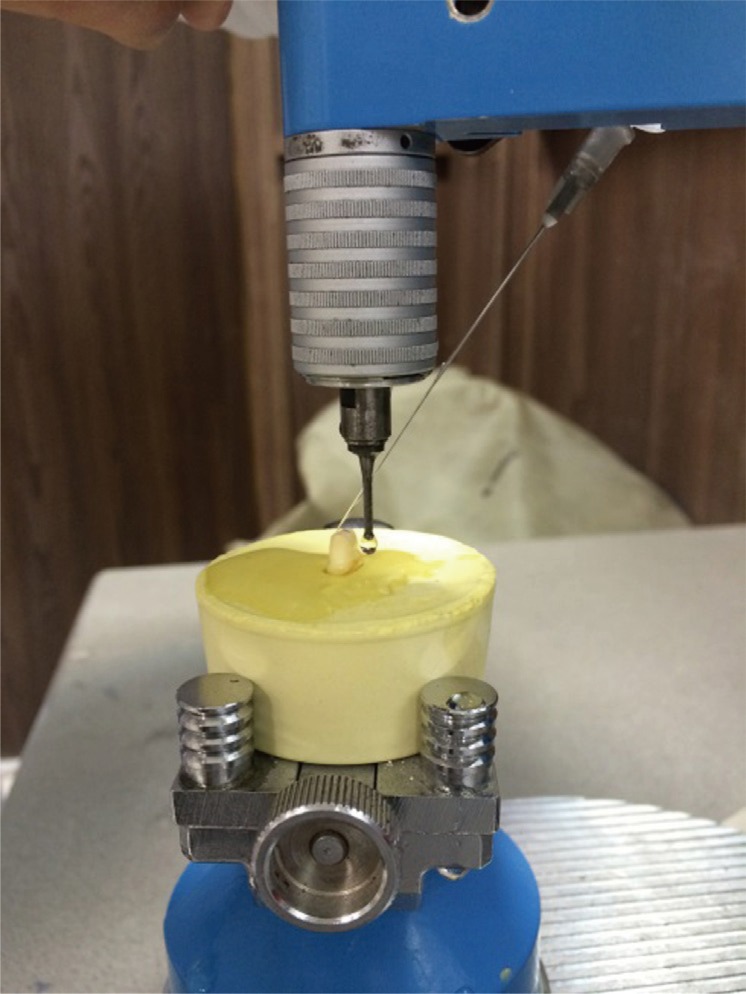
3-Preparation of samples: The teeth were prepared using a milling machine (Degussa, Germany). For this purpose, the teeth were mounted on dental stone molds (Ariadent, Tehran, Iran) and placed on the milling machine (Fig. 1). For axial and occlusal reductions, a round-end taper diamond bur (Dia-Burs, Mani Inc. Tochigi, Japan) was used.5 Standard tooth cylinders with 6° taper were obtained as such. Tooth preparation was in such a way that 4 mm5 of the tooth height remained after occlusal reduction in order to have equal surface areas in all samples. A 0.5 mm5 deep chamfer finish line was prepared above the cementoenamel junction. The teeth were then finished using a round-end taper fine-grit diamond bur (Dia-Burs, Mani Inc. Tochigi, Japan). All the line angles were rounded using abrasive strips.5
Fig. 1. Tooth preparation using a milling machine. The teeth were mounted on dental stone molds for this purpose.
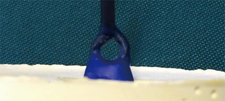
Wax patterns were then fabricated directly on the teeth using type II modeling wax (S-U-Underlay & S-U Modeling wax, Schuler, Ulm, Germany). The margins were adapted to the teeth and the excess wax was eliminated by a PKT using a magnifier. The wax pattern thickness was 0.5 mm, measured with a gauge (S-U-Iwanson-Feeler Tweezer II for metal; Schular-Dental, Ulm, Germany). A ring was then connected to the occlusal surface of wax patterns (Fig. 2). This ring, later casted on the metal crown, was used for jig attachment for retention and tensile strength testing in a universal testing machine.5
Fig. 2. Wax pattern. A ring was placed on the occlusal surface, sprued, and casted. This ring was used for attachment to the jig of the universal testing machine for tensile strength testing.
Wax patterns were then invested using high-strength phosphate investment stone (Wirovest; Bego Corp., Hanau-Wolfgang, Germany); looped full metal crowns were fabricated by investment casting.5
The cast metal crowns were then gently placed on the teeth and their marginal fit and complete seating were ensured. Defective crowns were replaced. Metal crowns were finished by metal finishing stones and burs and sandblasted using 50 µm aluminum oxide particles (AX-B5; Twin-Pen sandblaster, Titanjin Aixin medical equipment co. Ltd., Tianjin, China).5 They were then cleaned using an ultrasonic bath (Transonic 470/H, Elma, Singen, Germany) for 60 seconds.5
The teeth were then gently removed from the stone mold and mounted on metal cubes filled with auto-polymerizing acrylic resin (Ariadent, Tehran, Iran). In order to prevent dislodgement of the teeth from the acrylic resin during the tensile strength testing, some undercuts were created on the root surfaces using a #2 round bur (Dia-Burs, Mani Inc. Tochigi, Japan) and low speed handpiece prior to their placement into the acrylic resin. Care was taken not to weaken the tooth structure.
The teeth were then randomly divided into two groups of case and control (n = 10). In the case group, GLUMA desensitizer (Heraeus Kulzer, Germany) was applied to the tooth surfaces for 30 to 60 seconds as recommended by the manufacturer.5 Compressed air was sprayed on the surface until the surface was no longer shiny. The teeth were then rinsed with water.5 The remaining 10 samples (control group) received no intervention.
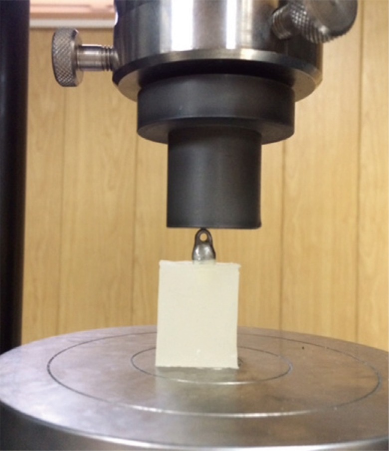
To achieve equal cement thickness in all samples, the RelyX U200 cement (3M ESPE, St. Paul, MN, USA) was mixed according to the manufacturer's instructions. Each paste was dispensed until a click was heard. The two pastes were mixed for 20 seconds.7 The crowns were filled with cement and placed on the teeth with finger fissure. The tooth and crown complex was then transferred to a universal testing machine (Zwick Z050; Roell Group, Ulm, Germany). The upper compartment of the device was attached to the ring on the crown and 5 kg axial load was constantly applied to each crown for 10 minutes (Fig. 3).8 After setting, excess cement was removed by an explorer. After cementation, all samples were incubated at 37℃ for 24 hours prior to retention testing.
Fig. 3. Constant application of 5 kg axial load to each crown for 10 minutes by the universal testing machine.
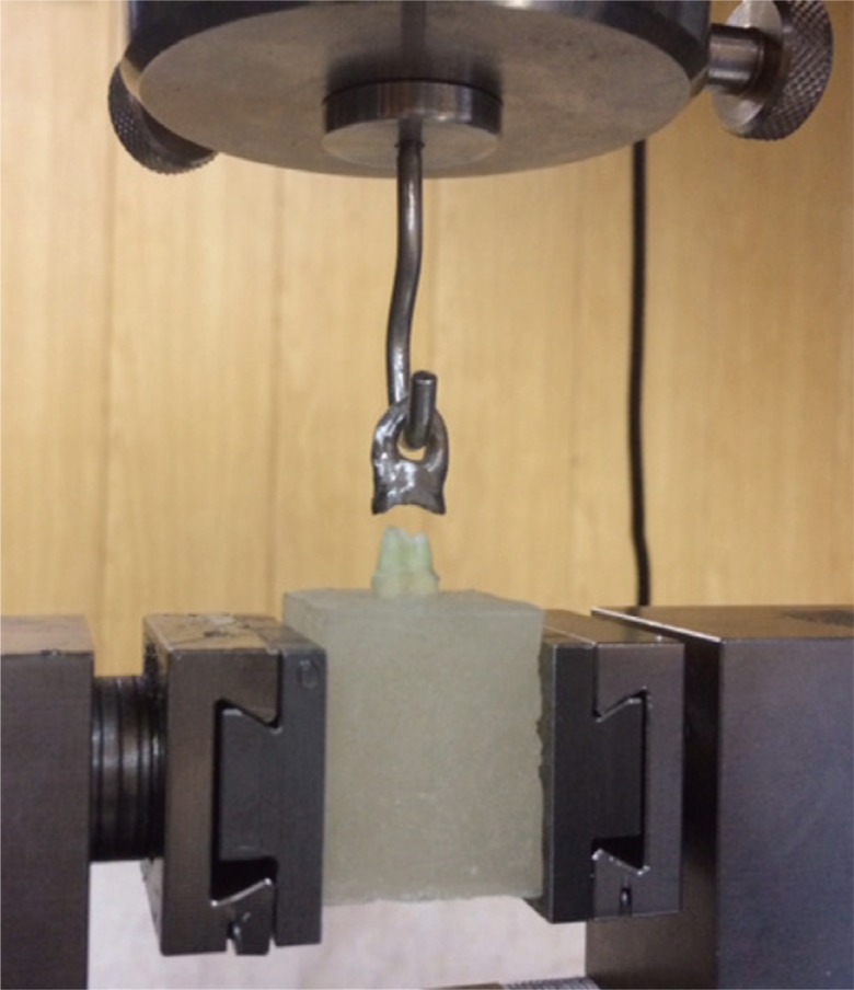
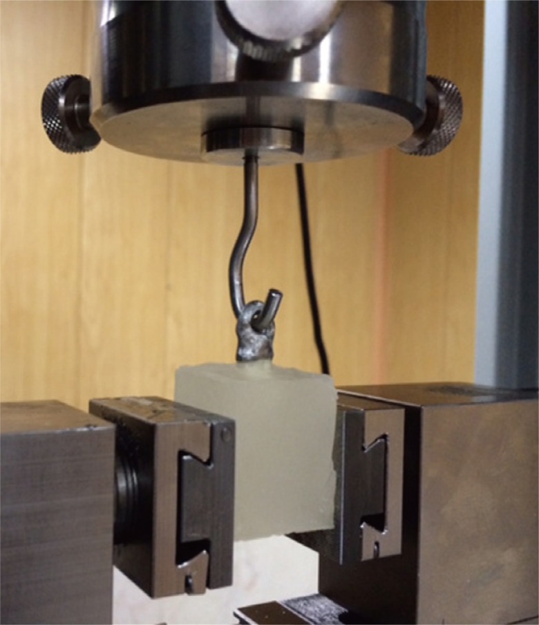
Retention test was done using a universal testing machine (Zwick Z050; Roell Group, Ulm, Germany) using a custom-made metal jig attached to the upper compartment of the device. The teeth with cemented crowns were placed on the lower compartment, and the upper vertical stylus was lowered until the pin passed through the ring (Fig. 4). By doing so, a vertical tensile load was applied to the crown. The load application was continued until the crown was detached from the tooth (Fig. 5).
Fig. 4. Load was applied until the crown was completely separated from the tooth.
Fig. 5. Load application parallel to the longitudinal axis of the tooth at a crosshead speed of 0.5 mm/min.
Load was applied at a crosshead speed of 0.5 mm/min as recommended by the ADA standards for cement testing.9
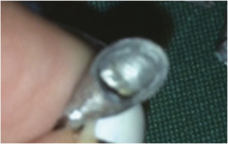
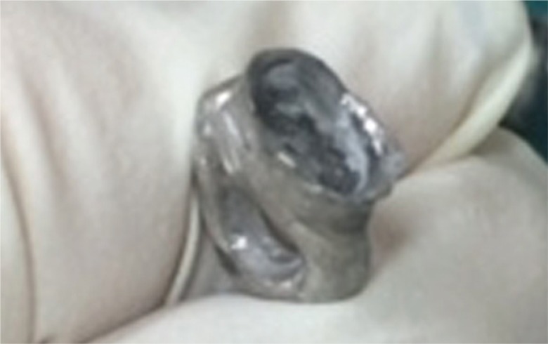
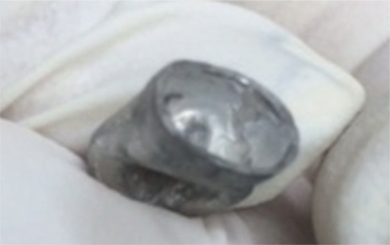
After separation of the crowns, debonded surfaces were evaluated visually under a magnifier to determine the mode of failure, which was later categorized into five groups5:
1. Cohesive failure: Cement remained mainly on the prepared tooth surfaces (Fig. 6).
2. Mixed failure: Cement remained on both the crown and tooth surfaces (Fig. 7).
3. Adhesive failure: Cement remained mainly on the crown (Fig. 8).
4. Tooth fracture or dislodgement from the acrylic mold
5. Crown fracture
Fig. 6. Cohesive failure: Cement remained mainly on the prepared tooth surfaces and hardly on the crown.
Fig. 7. Mixed failure: Cement remained on both the crown and tooth surfaces.
Fig. 8. Adhesive failure: Cement remained mainly on the crown.
The obtained retention values were compared between the two groups with the Kolmogorov-Smirnov test and independent t-test using SPSS version 20 software. P value less than .05 was considered statistically significant.
RESULTS
This study was performed on prepared human premolar teeth in two groups of with and without GLUMA desensitizer applied to crowns cemented with RelyX U200. Table 1 shows the results of Kolmogorov-Smirnov test regarding the tensile bond strength of the two groups (in N). The tensile bond strength values of the two groups had normal distributions (P > .05).
Table 1. Results of t-test for comparison of tensile bond strength between the two groups.
| Groups/Tensile bond strength | Mean ± SD | CV | P value |
|---|---|---|---|
| Without GLUMA | 164.45 ± 39.3 | 23.9 | .012 |
| With GLUMA | 230.63 ± 63.8 | 27.66 |
Table 2 shows the results of independent t-test for comparison of the bond strength values between the two groups. As seen in Table 1, the tensile bond strength was 164.45 ± 39.3 N with CV=23.9 in the control group (without GLUMA desensitizer) and 230.63 ± 63.8 N with CV = 27.66 in the case group (with GLUMA desensitizer). The tensile bond strength in the case group was significantly higher than that in the control group by 40.24% (66.18 N) (P = .012).
Table 2. Frequency of the modes of failure after retention test.
| Groups/Mode of failure | Cohesive | Adhesive | Mixed |
|---|---|---|---|
| Without GLUMA | 0 | 1 | 9 |
| With GLUMA | 2 | 0 | 8 |
In the case group, cohesive failure was noted in two samples, and the remaining failures were mixed and cement was seen mostly on the prepared tooth surfaces. In other words, mixed failures in this group were close to cohesive. In the control group, adhesive failure was noted in one sample and the failure was mixed in the remaining samples. Of the remaining samples, seven had cement remained mainly on the crown, and, in two samples, cement remained mainly on the tooth surfaces. In other words, mixed failures in this group were similar to adhesive failure (Table 2).
DISCUSSION
Most FPD patients experience pain or discomfort of the prepared teeth during and some time after the cementation of restoration, which may be due to dentin hypersensitivity. 5 To overcome this problem, desensitizer agents were introduced.5 However, considering the importance of cement-tooth bond strength in success and long-term clinical service of FPDs, a question raises that whether the application of desensitizer agents, such as GLUMA desensitizer, affects the retention of full crowns cemented with Rely-X U200 self-adhesive cement. The results of this experimental study showed that the tensile strength was significantly higher in GLUMA desensitizer group compared to the control group. Thus, within the limitations of this study, our null hypothesis regarding no effect of GLUMA desensitizer on RelyX U200 self-adhesive cement was rejected.
In the recent years, several studies have assessed the effect of desensitizers on dentin bonding to different cements, including the studies by Acar et al. in 2014,10 Aranha et al. in 2006,11 Stawarczyk et al. in 2012,12 Sailer et al. in 2012,6 Jalandar et al. in 2012,5 Stawarczyk et al. in 2011,13 Oshima et al. in 2010,14 Dündar et al. in 2010,15 Saraç et al. in 2009,16 Huh et al. in 2008,17 and Soeno et al. in 2001.18
In the current study, we used full metal crowns to better simulate the clinical setting. Irrespective of the type of cement and desensitizer agent used, only limited studies have used the same methodology as this study, including the studies by Acar et al. in 2014,10 Aranha et al. in 2006,11 Stawarczyk et al. in 2012,12 Jalandar et al. in 2012,5 and Soeno et al. in 2001.18 Thus, findings of the studies other than those mentioned above cannot be compared with this study.
Acar et al.,10 in 2014, assessed the effects of GLUMA, Aqua-Prep F, BisBlock, Cervitec Plus, Smart Protect, and Nd:YAG laser on microtensile bond strength of RelyX U200 self-adhesive cement to dentin and reported that GLUMA increased the microtensile bond strength, but not significantly; this result was somehow similar to our finding. The difference between their study and this study was in that we used premolar teeth, while they used molars. They also thermocycled the teeth, which was not performed in this study. Moreover, the number of samples per group in their study was half the amount in this study. Also, they used composite blocks instead of crown restorations. They applied 2 kg load during 60 seconds, whereas we applied 5 kg load for 10 minutes.8 Similar to our study, they tested GLUMA desensitizer and RelyX U200.
Stawarczyk et al.,12 in 2012, evaluated the effect of GLUMA desensitizer on tensile bond strength of zirconia crowns to dentin and reported that self-adhesive resin cement used with GLUMA resulted in more favorable long-term stability than Panavia 21 and which was in agreement with our results. They used full crowns and assessed the tensile strength, similar to this study. However, they used zirconia crowns, while we used metal crowns. Moreover, they performed chewing simulation, which was not performed in our study. Furthermore, in tensile strength testing, they applied load at a crosshead speed of 1 mm/min, while we used a crosshead speed of 0.5 mm/min; our protocol has been recommended in the ADA standards for cement testing.9 Despite these differences, the similarities between the two studies can explain the similar results obtained.
Aranha et al.11 evaluated the effect of several dentin desensitizers on microtensile bond strength of different adhesives. Application of GLUMA had no significant effect on microtensile bond strength, but other materials decreased the microtensile bond strength. They concluded that GLUMA effectively decreased dentin hypersensitivity. Their findings confirmed the positive effects of GLUMA, which is in line with our findings. They used composite build-ups as restorations, whereas we used metal crowns. Both studies were conducted on GLUMA.
Jalandar et al.,5 in 2012, evaluated the effect of GLUMA desensitizer and GC Tooth Mousse on retention of cast crowns cemented with zinc phosphate, glass ionomer, and resin modified glass ionomer cements. They reported that GLUMA desensitizer had no effect on retention of crowns, which is different from our finding. This difference may be due to the use of different materials and methodology. The model of the Instron machine was also different in the two studies. They used molar teeth, while we used premolars. The cements used were also different; thus, the results of the two studies cannot be well compared.
Soeno et al.,18 in 2001, evaluated the effect of three dentin desensitizers, namely GLUMA CPS, MS Coat, and Safordie, on dentin bond strength of Panavia Fluoro Cement and Super-Bond C&B. They concluded that GLUMA desensitizer had no effect on bond strength of cements to dentin. Their findings are in contrast to ours, the difference which may be due to different materials and methods of the two studies. They evaluated different cements in their study and their sample size in each group was half the size of our study groups. Also, they used steel rods as restorations.
With respect to the type of cement and desensitizing agents, only a few studies have evaluated the effect of GLUMA on bond strength of self-adhesive cements to dentin, such as studies by Stawarczyk et al.,12 in 2012, Sailer et al.,6 in 2012, Stawarczyk et al.,13 in 2011, and Sailer et al.,19 in 2010.
Sailer et al.,6 in 2012, assessed the effect of GLUMA desensitizer on RelyX Unicem self-adhesive cement, compared with other desensitizers and cements. They reported that GLUMA significantly increased the bond strength of RelyX Unicem compared to the control group; no such effect was noted for other cements evaluated in their study. This finding was in accordance with our result, which may be attributed to the fact that both studies used GLUMA and self-adhesive cements (although we used RelyX U200). However, they performed shear bond strength testing, which was different from the retention test performed in our study; they did not use crowns in their study either. Stawarczyk et al.,13 in 2011, evaluated the effect of GLUMA desensitizer on shear bond strength of RelyX Unicem self-adhesive cement after water storage and thermocycling. They reported that application of GLUMA desensitizer on dentin before cementation significantly enhanced the shear bond strength of self-adhesive cement. This finding was similar to our result. In their study, two conventional and three self-adhesive resin cements were evaluated. Also, they assessed the shear bond strength, while we performed the retention test. In their study, blocks instead of crowns were cemented to teeth. Moreover, they performed thermocycling, which was not performed in our study.
Sailer et al.,19 in 2010, evaluated the effect of GLUMA desensitizer on shear bond strength of RelyX Unicem self-adhesive resin cement and reported that GLUMA desensitizer enhanced its shear bond strength. Their findings were in accordance with this study. They used Panavia 21 cement in their control group, while, in our study, self-adhesive resin cement without GLUMA was used in the control group. Another difference between our study and theirs was that they measured the shear bond strength, while we performed the retention test. In their study, cements were used in the form of blocks on teeth and no crown was used. However, similar findings were obtained in the two studies.
In terms of mode of failure in our study, among the case group samples, 2 failures were cohesive and the rest were mixed with cement visible on the prepared tooth. In this group, failures were close to cohesive. In the control group, one adhesive failure was noted and the remaining failures were mixed; in seven of the remaining failures, cement remained on the crowns, and, in two, cement remained mainly on the tooth surface. In this group, mode of fracture was close to adhesive failure.
Only a few similar studies have assessed dentin and cement debonded surfaces, including the studies by Acar et al.10 in 2014, Sailer et al.6 in 2012, Hitz et al.20 in 2012, DiHipólito et al.21 in 2012, Stawarczyk et al.13 in 2012, and Jalandar et al.5 in 2012. Sailer et al.,6 in 2012, assessed the effect of GLUMA desensitizer on bond strength of RelyX Unicem self-adhesive cement compared with other desensitizers and cements. They reported that, in the control group, all failures were cohesive, while, in GLUMA group, failure was adhesive in one sample and cohesive in the remaining samples. Their findings regarding the control group were different from this study, while their results in GLUMA group were similar to our findings.
Hitz et al.,20 in 2012, measured the shear bond strength of six self-adhesive resin cements to dentin and glass ceramic after 24 hours and longer periods of time and compared the results with that of conventional resin cement. They reported that when self-adhesive resin cement was directly used on dentin surfaces (with no pre-treatment of dentin surface prior to cementation), only adhesive failure was seen in the samples. Their findings were similar to our results in the control group. In their study, RelyX U200 was not used. Also, they performed shear bond strength testing, while we performed the retention test. They performed aging in their study, which was not conducted in ours. Despite these differences, similar results were obtained in the two studies.
Stawarczyk et al.,13 in 2012, evaluated the effect of GLUMA desensitizer on tensile bond strength of zirconia crowns to dentin and reported that all samples underwent cohesive failure before and after aging. Their findings in GLUMA group were similar to our findings, while the results in the control groups of the two studies were different.
Acar et al.,10 in 2014, assessed the effects of GLUMA, Aqua-Prep F, BisBlock, Cervitec Plus, Smart Protect, and Nd:YAG laser on microtensile bond strength of RelyX U200 to dentin and showed that 85% of the samples in the control group experienced adhesive and 15% experienced mixed failure; this result was similar to our observations in the control group. They showed 80% adhesive and 20% mixed failure in GLUMA group,10 which was in contrast to our finding. Di Hipólito et al.,21 in 2012, measured the microtensile bond strength and assessed the mode of failure of self-adhesive cement bonded to dentin pre-treated with different concentrations of chlorhexidine. They demonstrated cohesive failure in non-thermocycled samples when self-adhesive resin cement was used on dentin with no pre-treatment. Mode of failure was adhesive in dentin samples treated with chlorhexidine. Their results in the control group were different from ours, which may be due to differences in the materials and methods of the two studies. They used RelyX U100 cement, which is different from RelyX U200 used in our study. Also, they used composite discs instead of crowns and measured the microtensile bond strength, which is different from the retention test conducted in our study. These factors may explain the differences in results.
Jalandar et al.,5 in 2012, evaluated the effect of GLUMA desensitizer and GC Tooth Mousse on retention of cast crowns using zinc phosphate, glass ionomer, and resin modified glass ionomer cements. They did not report the mode of failure in the control group. Also, accurate comparison between their study and this study was not feasible since they used different cements.
GLUMA desensitizer is composed of glutaraldehyde and hydroxyethyl methacrylate (HEMA). Glutaraldehyde results in the coagulation of amino acids and proteins in dentinal tubules and works as an effective disinfectant. HEMA can effectively seal the dentinal tubules.22 Our hypothesis regarding the effect of GLUMA desensitizer on self-adhesive cement bond to dentin and the selection of these two materials were based on the study by Munksgaard and Asmussen in 1984. Their study showed that resin cement bond strength strongly depended on the concentration of HEMA with maximum effect at 35% concentration and was almost independent of the concentration of glutaraldehyde in concentrations over 3%.23 On the other hand, Qin et al.24 stated that the glutaraldehyde present in GLUMA could not form cross-links with mineralized dentin. It has been stated that desensitizers containing glutaraldehyde and HEMA lead to formation of a collagen-glutaraldehyde layer at the interface of desensitizer-dentin. Chemical bonds are then formed between HEMA molecules in the composition of desensitizer and the collagen-glutaraldehyde complex. Eventually, copolymerization occurs between resin cement and HEMA complex, which may enhance the resin cement bond strength.23,24
Nakabayashi et al.25 stated that HEMA decreased the surface tension of water molecules and subsequently increased monomer penetration into dentin. Arrais et al.26 demonstrated a thin resinous layer penetrating into the dentinal tubules and occluding them. They did not report septum formation in the tubules in contrast to Kolker et al..27 Moreover, glutaraldehyde/HEMA contains water. Thus, it may serve as rewetting agents.10 Although data regarding the effect of glutaraldehyde/HEMA on smear layer and buffering capacity of self-adhesive resin cements are scarce, HEMA may be responsible for the increase in bond strength.10 Stawaczyk et al.,13 in 2011, discussed that the reaction between glutaraldehyde and phosphate might result in a strong and stable bond between GLUMA desensitizer and self-adhesive resin cement. Therefore, further studies are required to find out the mechanism of this increase in bond strength of self-adhesive resin cements to dentin pretreated with GLUMA desensitizer.
Adhesive cements have higher technical sensitivity than conventional cements and their clinical success may be compromised by technical errors. Self-adhesive resin cements were introduced to overcome the limitations of adhesive cements. RelyX U200, used in our study, is a recently introduced self-adhesive cement with an extra monomer and a new rheology modifier enhanced with filler particles. This new formula has improved mechanical properties.16 Use of this cement was the strength of our study; by using this cement, complications and possible confounders related to the use of multi-step conventional resin cements (etch, primer, bond) were prevented. Another strength of this study was that it was an independent study not funded by dental manufacturers.
Since our study had an in vitro design, our findings cannot be directly generalized to the clinical setting, and further studies are required to simulate the oral clinical setting by chewing simulation or thermocycling.
Previous studies have not frequently used the method of this study (tooth preparation and fabrication of full coverage crowns) due to difficulties associated with this method, which made accurate comparison of our results with those of the previous studies difficult, if not impossible. Similar studies on other cements and desensitizers are also required. In the current study, pressure of the pulp and dentinal fluid was not simulated. However, the teeth were stored in water, and, therefore, water was present in dentinal tubules.
CONCLUSION
Within the limitations of this study, the results showed that GLUMA desensitizer significantly increased the bond strength of RelyX U200 self-adhesive cement to dentin. Therefore, we recommend the application of GLUMA on prepared teeth with hypersensitivity prior to cementation of crowns with self-adhesive cements.
Conflict of interest: Authors disclose any financial and personal relationships with other people or organizations that could inappropriately influence (bias) in our work.
ACKNOWLEDGEMENTS
We are thankful to those who helped us in this article including research center of Islamic Azad university-Dental Branch.
References
- 1.Orsi IA, Varoli FK, Pieroni CH, Ferreira MC, Borie E. In vitro tensile strength of luting cements on metallic substrate. Braz Dent J. 2014;25:136–140. doi: 10.1590/0103-6440201302290. [DOI] [PubMed] [Google Scholar]
- 2.Al-Omari WM, Al-Wahadni AM. Convergence angle, occlusal reduction, and finish line depth of full-crown preparations made by dental students. Quintessence Int. 2004;35:287–293. [PubMed] [Google Scholar]
- 3.Zidan O, Ferguson GC. The retention of complete crowns prepared with three different tapers and luted with four different cements. J Prosthet Dent. 2003;89:565–571. doi: 10.1016/s0022-3913(03)00182-3. [DOI] [PubMed] [Google Scholar]
- 4.Ayad MF, Johnston WM, Rosenstiel SF. Influence of tooth preparation taper and cement type on recementation strength of complete metal crowns. J Prosthet Dent. 2009;102:354–361. doi: 10.1016/S0022-3913(09)60192-X. [DOI] [PubMed] [Google Scholar]
- 5.Jalandar SS, Pandharinath DS, Arun K, Smita V. Comparison of effect of desensitizing agents on the retention of crowns cemented with luting agents: an in vitro study. J Adv Prosthodont. 2012;4:127–133. doi: 10.4047/jap.2012.4.3.127. [DOI] [PMC free article] [PubMed] [Google Scholar]
- 6.Sailer I, Oendra AE, Stawarczyk B, Hämmerle CH. The effects of desensitizing resin, resin sealing, and provisional cement on the bond strength of dentin luted with self-adhesive and conventional resincements. J Prosthet Dent. 2012;107:252–260. doi: 10.1016/S0022-3913(12)60070-5. [DOI] [PubMed] [Google Scholar]
- 7.Chen C, Xie H, Song X, Burrow MF, Chen G, Zhang F. Evaluation of a commercial primer for bonding of zirconia to two different resin composite cements. J Adhes Dent. 2014;16:169–176. doi: 10.3290/j.jad.a31809. [DOI] [PubMed] [Google Scholar]
- 8.Yim NH, Rueggeberg FA, Caughman WF, Gardner FM, Pashley DH. Effect of dentin desensitizers and cementing agents on retention of full crowns using standardized crown preparations. J Prosthet Dent. 2000;83:459–465. doi: 10.1016/s0022-3913(00)70042-4. [DOI] [PubMed] [Google Scholar]
- 9.Johnson GH, Hazelton LR, Bales DJ, Lepe X. The effect of a resin-based sealer on crown retention for three types of cement. J Prosthet Dent. 2004;91:428–435. doi: 10.1016/S0022391304000770. [DOI] [PubMed] [Google Scholar]
- 10.Acar O, Tuncer D, Yuzugullu B, Celik C. The effect of dentin desensitizers and Nd:YAG laser pre-treatment on microtensile bond strength of self-adhesive resin cement to dentin. J Adv Prosthodont. 2014;6:88–95. doi: 10.4047/jap.2014.6.2.88. [DOI] [PMC free article] [PubMed] [Google Scholar]
- 11.Aranha AC, Siqueira Junior Ade S, Cavalcante LM, Pimenta LA, Marchi GM. Microtensile bond strengths of composite to dentin treated with desensitizer products. J Adhes Dent. 2006;8:85–90. [PubMed] [Google Scholar]
- 12.Stawarczyk B, Hartmann L, Hartmann R, Roos M, Ender A, Ozcan M, Sailer I, Hämmerle CH. Impact of Gluma Desensitizer on the tensile strength of zirconia crowns bonded to dentin: an in vitro study. Clin Oral Investig. 2012;16:201–213. doi: 10.1007/s00784-010-0502-y. [DOI] [PubMed] [Google Scholar]
- 13.Stawarczyk B, Hartmann R, Hartmann L, Roos M, Ozcan M, Sailer I, Hämmerle CH. The effect of dentin desensitizer on shear bond strength of conventional and self-adhesive resin luting cements after aging. Oper Dent. 2011;36:492–501. doi: 10.2341/10-292-L. [DOI] [PubMed] [Google Scholar]
- 14.Oshima M, Hamba H, Sadr A, Nikaido T, Tagami J. Effect of polymer-based desensitizer with sodium fluoride on prevention of root dentin demineralization. Am J Dent. 2015;28:123–127. [PubMed] [Google Scholar]
- 15.Dündar M, Cal E, Gökçe B, Türkün M, Ozcan M. Influence of fluoride- or triclosan-based desensitizing agents on adhesion of resin cements to dentin. Clin Oral Investig. 2010;14:579–586. doi: 10.1007/s00784-009-0328-7. [DOI] [PubMed] [Google Scholar]
- 16.Saraç D, Külünk S, Saraç YS, Karakas O. Effect of fluoride-containing desensitizing agents on the bond strength of resin-based cements to dentin. J Appl Oral Sci. 2009;17:495–500. doi: 10.1590/S1678-77572009000500026. [DOI] [PMC free article] [PubMed] [Google Scholar]
- 17.Huh JB, Kim JH, Chung MK, Lee HY, Choi YG, Shim JS. The effect of several dentin desensitizers on shear bond strength of adhesive resin luting cement using self-etching primer. J Dent. 2008;36:1025–1032. doi: 10.1016/j.jdent.2008.08.012. [DOI] [PubMed] [Google Scholar]
- 18.Soeno K, Taira Y, Matsumura H, Atsuta M. Effect of desensitizers on bond strength of adhesive luting agents to dentin. J Oral Rehabil. 2001;28:1122–1128. doi: 10.1046/j.1365-2842.2001.00756.x. [DOI] [PubMed] [Google Scholar]
- 19.Sailer I, Tettamanti S, Stawarczyk B, Fischer J, Hämmerle CH. In vitro study of the influence of dentin desensitizing and sealing on the shear bond strength of two universal resin cements. J Adhes Dent. 2010;12:381–392. doi: 10.3290/j.jad.a17714. [DOI] [PubMed] [Google Scholar]
- 20.Hitz T, Stawarczyk B, Fischer J, Hämmerle CH, Sailer I. Are self-adhesive resin cements a valid alternative to conventional resin cements? A laboratory study of the long-term bond strength. Dent Mater. 2012;28:1183–1190. doi: 10.1016/j.dental.2012.09.006. [DOI] [PubMed] [Google Scholar]
- 21.Di Hipólito V, Rodrigues FP, Piveta FB, Azevedo Lda C, Bruschi Alonso RC, Silikas N, Carvalho RM, De Goes MF, Perlatti D'Alpino PH. Effectiveness of self-adhesive luting cements in bonding to chlorhexidine-treated dentin. Dent Mater. 2012;28:495–501. doi: 10.1016/j.dental.2011.11.027. [DOI] [PubMed] [Google Scholar]
- 22.Joshi S, Gowda AS, Joshi C. Comparative evaluation of NovaMin desensitizer and Gluma desensitizer on dentinal tubule occlusion: a scanning electron microscopic study. J Periodontal Implant Sci. 2013;43:269–275. doi: 10.5051/jpis.2013.43.6.269. [DOI] [PMC free article] [PubMed] [Google Scholar]
- 23.Munksgaard EC, Asmussen E. Bond strength between dentin and restorative resins mediated by mixtures of HEMA and glutaraldehyde. J Dent Res. 1984;63:1087–1089. doi: 10.1177/00220345840630081701. [DOI] [PubMed] [Google Scholar]
- 24.Qin C, Xu J, Zhang Y. Spectroscopic investigation of the function of aqueous 2-hydroxyethylmethacrylate/glutaraldehyde solution as a dentin desensitizer. Eur J Oral Sci. 2006;114:354–359. doi: 10.1111/j.1600-0722.2006.00382.x. [DOI] [PubMed] [Google Scholar]
- 25.Nakabayashi N, Watanabe A, Gendusa NJ. Dentin adhesion of "modified" 4-META/MMA-TBB resin: function of HEMA. Dent Mater. 1992;8:259–264. doi: 10.1016/0109-5641(92)90096-u. [DOI] [PubMed] [Google Scholar]
- 26.Arrais CA, Chan DC, Giannini M. Effects of desensitizing agents on dentinal tubule occlusion. J Appl Oral Sci. 2004;12:144–148. doi: 10.1590/s1678-77572004000200012. [DOI] [PubMed] [Google Scholar]
- 27.Kolker JL, Vargas MA, Armstrong SR, Dawson DV. Effect of desensitizing agents on dentin permeability and dentin tubule occlusion. J Adhes Dent. 2002;4:211–221. [PubMed] [Google Scholar]


