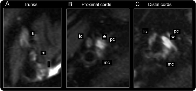
Figure 4. Lesion extension into the brachial plexus in posterior interosseous neuropathy syndrome.
Representative brachial plexus imaging findings for patient 19 are displayed. (A) At trunk level, superior (s), medial (m), and inferior (i) trunk appear normal, without increased T2 signal. (B, C) At cord levels, the posterior cord (pc; from which the radial nerve arises more distally) is severely thickened and shows high T2 signal (marked by *) as typical imaging findings of selective plexopathy. The lesion extends continuously through the posterior cord into the radial nerve. Lateral (lc) and medial cord (mc) are inconspicuous.

