

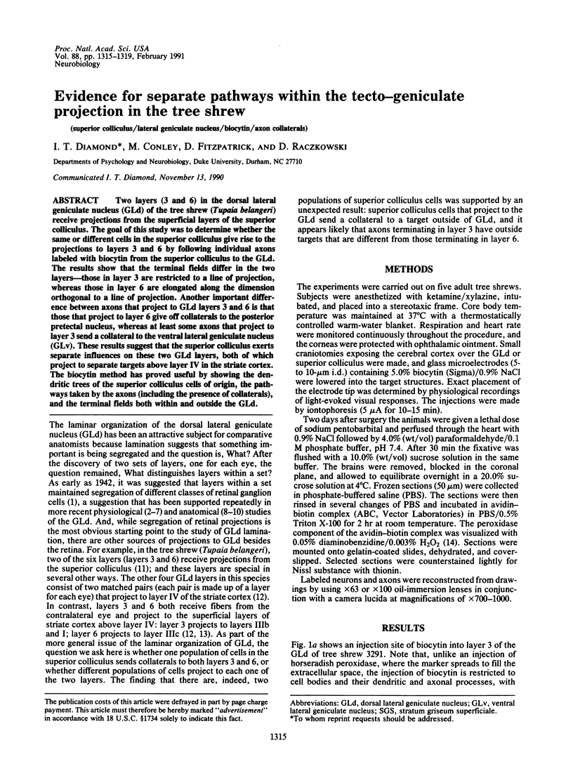
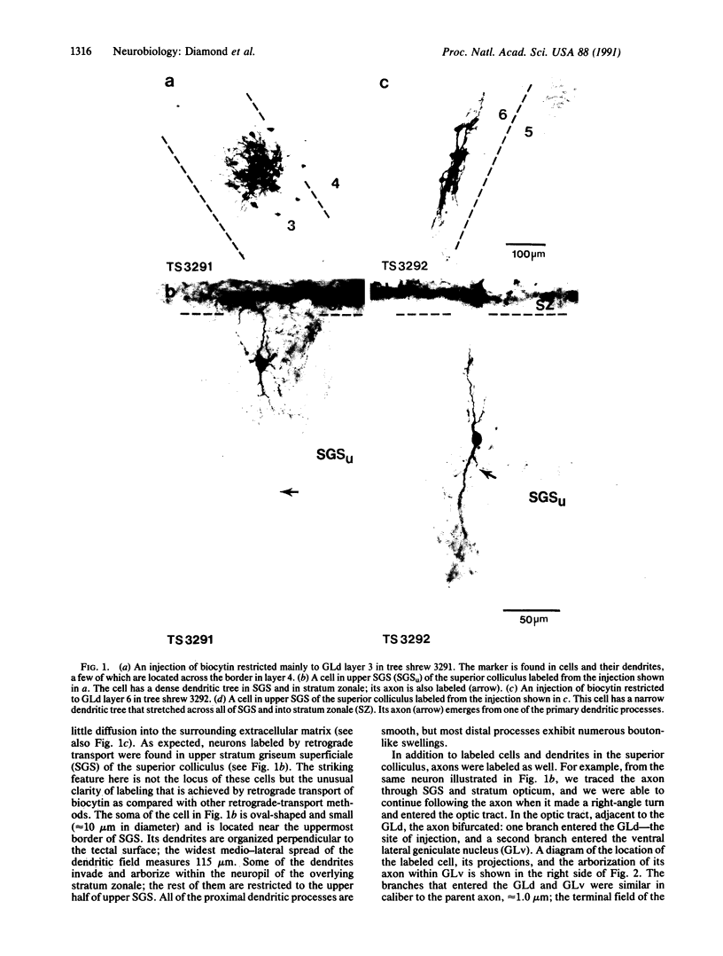
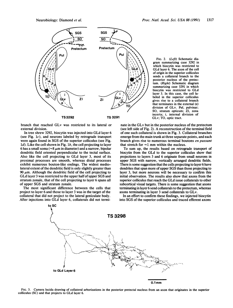
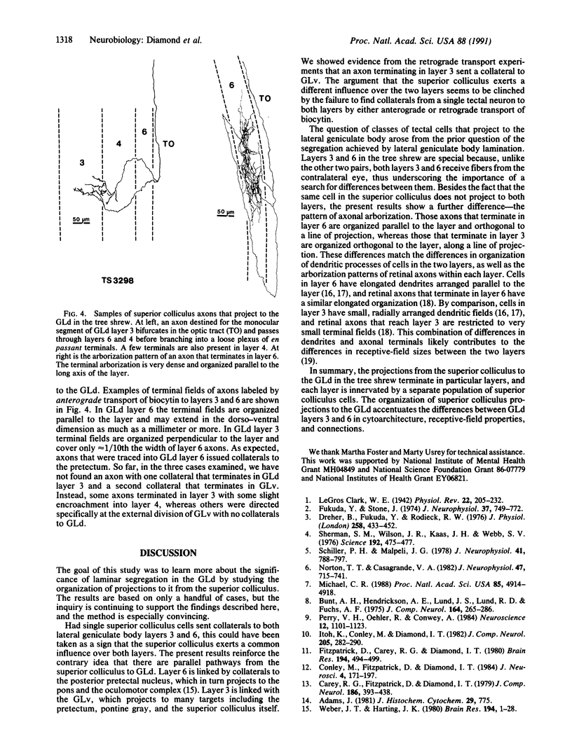
Abstract
Two layers (3 and 6) in the dorsal lateral geniculate nucleus (GLd) of the tree shrew (Tupaia belangeri) receive projections from the superficial layers of the superior colliculus. The goal of this study was to determine whether the same or different cells in the superior colliculus give rise to the projections to layers 3 and 6 by following individual axons labeled with biocytin from the superior colliculus to the GLd. The results show that the terminal fields differ in the two layers--those in layer 3 are restricted to a line of projection, whereas those in layer 6 are elongated along the dimension orthogonal to a line of projection. Another important difference between axons that project to GLd layers 3 and 6 is that those that project to layer 6 give off collaterals to the posterior pretectal nucleus, whereas at least some axons that project to layer 3 send a collateral to the ventral lateral geniculate nucleus (GLv). These results suggest that the superior colliculus exerts separate influences on these two GLd layers, both of which project to separate targets above layer IV in the striate cortex. The biocytin method has proved useful by showing the dendritic trees of the superior colliculus cells of origin, the pathways taken by the axons (including the presence of collaterals), and the terminal fields both within and outside the GLd.
Full text
PDF

Images in this article
Selected References
These references are in PubMed. This may not be the complete list of references from this article.
- Adams J. C. Heavy metal intensification of DAB-based HRP reaction product. J Histochem Cytochem. 1981 Jun;29(6):775–775. doi: 10.1177/29.6.7252134. [DOI] [PubMed] [Google Scholar]
- Brauer K., Werner L., Winkelmann E., Lüth H. J. The dorsal lateral geniculate nucleus of Tupaia glis: a Golgi, Nissl and acetylcholinesterase study. J Hirnforsch. 1981;22(1):59–74. [PubMed] [Google Scholar]
- Bunt A. H., Hendrickson A. E., Lund J. S., Lund R. D., Fuchs A. F. Monkey retinal ganglion cells: morphometric analysis and tracing of axonal projections, with a consideration of the peroxidase technique. J Comp Neurol. 1975 Dec 1;164(3):265–285. doi: 10.1002/cne.901640302. [DOI] [PubMed] [Google Scholar]
- Carey R. G., Fitzpatrick D., Diamond I. T. Layer I of striate cortex of Tupaia glis and Galago senegalensis: projections from thalamus and claustrum revealed by retrograde transport of horseradish peroxidase. J Comp Neurol. 1979 Aug 1;186(3):393–437. doi: 10.1002/cne.901860306. [DOI] [PubMed] [Google Scholar]
- Conley M., Fitzpatrick D., Diamond I. T. The laminar organization of the lateral geniculate body and the striate cortex in the tree shrew (Tupaia glis). J Neurosci. 1984 Jan;4(1):171–197. doi: 10.1523/JNEUROSCI.04-01-00171.1984. [DOI] [PMC free article] [PubMed] [Google Scholar]
- Conley M., Penny G. R., Diamond I. T. Terminations of individual optic tract fibers in the lateral geniculate nuclei of Galago crassicaudatus and Tupaia belangeri. J Comp Neurol. 1987 Feb 1;256(1):71–87. doi: 10.1002/cne.902560107. [DOI] [PubMed] [Google Scholar]
- Conway J. L., Schiller P. H. Laminar organization of tree shrew dorsal lateral geniculate nucleus. J Neurophysiol. 1983 Dec;50(6):1330–1342. doi: 10.1152/jn.1983.50.6.1330. [DOI] [PubMed] [Google Scholar]
- Dreher B., Fukada Y., Rodieck R. W. Identification, classification and anatomical segregation of cells with X-like and Y-like properties in the lateral geniculate nucleus of old-world primates. J Physiol. 1976 Jun;258(2):433–452. doi: 10.1113/jphysiol.1976.sp011429. [DOI] [PMC free article] [PubMed] [Google Scholar]
- Fitzpatrick D., Carey R. G., Diamond I. T. The projection of the superior colliculus upon the lateral geniculate body in Tupaia glis and Galago senegalensis. Brain Res. 1980 Aug 4;194(2):494–499. doi: 10.1016/0006-8993(80)91230-5. [DOI] [PubMed] [Google Scholar]
- Fukuda Y., Stone J. Retinal distribution and central projections of Y-, X-, and W-cells of the cat's retina. J Neurophysiol. 1974 Jul;37(4):749–772. doi: 10.1152/jn.1974.37.4.749. [DOI] [PubMed] [Google Scholar]
- Itoh K., Conley M., Diamond I. T. Retinal ganglion cell projections to individual layers of the lateral geniculate body in Galago crassicaudatus. J Comp Neurol. 1982 Mar 1;205(3):282–290. doi: 10.1002/cne.902050308. [DOI] [PubMed] [Google Scholar]
- Michael C. R. Retinal afferent arborization patterns, dendritic field orientations, and the segregation of function in the lateral geniculate nucleus of the monkey. Proc Natl Acad Sci U S A. 1988 Jul;85(13):4914–4918. doi: 10.1073/pnas.85.13.4914. [DOI] [PMC free article] [PubMed] [Google Scholar]
- Norton T. T., Casagrande V. A. Laminar organization of receptive-field properties in lateral geniculate nucleus of bush baby (Galago crassicaudatus). J Neurophysiol. 1982 Apr;47(4):715–741. doi: 10.1152/jn.1982.47.4.715. [DOI] [PubMed] [Google Scholar]
- Perry V. H., Oehler R., Cowey A. Retinal ganglion cells that project to the dorsal lateral geniculate nucleus in the macaque monkey. Neuroscience. 1984 Aug;12(4):1101–1123. doi: 10.1016/0306-4522(84)90006-x. [DOI] [PubMed] [Google Scholar]
- Saini K., Kretz R., Rager G. Classes of neurons in relation to the laminar organization of the lateral geniculate nucleus in the tree shrew, Tupaia belangeri. J Comp Neurol. 1987 May 1;259(1):31–49. doi: 10.1002/cne.902590104. [DOI] [PubMed] [Google Scholar]
- Schiller P. H., Malpeli J. G. Functional specificity of lateral geniculate nucleus laminae of the rhesus monkey. J Neurophysiol. 1978 May;41(3):788–797. doi: 10.1152/jn.1978.41.3.788. [DOI] [PubMed] [Google Scholar]
- Sherman S. M., Wilson J. R., Kaas J. H., Webb S. V. X- and Y-cells in the dorsal lateral geniculate nucleus of the owl monkey (Aotus trivirgatus). Science. 1976 Apr 30;192(4238):475–477. doi: 10.1126/science.816006. [DOI] [PubMed] [Google Scholar]
- Weber J. T., Harting J. K. The efferent projections of the pretectal complex: an autoradiographic and horseradish peroxidase analysis. Brain Res. 1980 Jul 21;194(1):1–28. doi: 10.1016/0006-8993(80)91315-3. [DOI] [PubMed] [Google Scholar]