Abstract
miR-146a plays important roles in cancer as it directly targets NUMB, an inhibitor of Notch signaling. miR-146a is reportedly regulated by a G>C polymorphism (SNP; rs2910164). This polymorphism affects various cancers, including colorectal cancer (CRC). However, the clinical significance of miR-146a polymorphism in CRC remains unclear. A total of 59 patients with CRC were divided into 2 groups: a CC/CG genotype (n = 32) and a GG genotype (n = 27), based on the miR-146a polymorphism. cDNA microarray analysis was performed using 59 clinical samples. Significantly enriched gene sets in each genotype were extracted using GSEA. We also investigated the association between miR-146a polymorphism and miR-146a, NUMB expression or migratory response in CRC cell lines. The CC/CG genotype was associated with significantly more synchronous liver metastasis (p = 0.007). A heat map of the two genotypes showed that the expression profiles were clearly stratified. GSEA indicated that Notch signaling and JAK/STAT3 signaling were significantly associated with the CC/CG genotype (p = 0.004 and p = 0.023, respectively). CRC cell lines with the pre-miR-146a/C revealed significantly higher miR-146a expression (p = 0.034) and higher NUMB expression and chemotactic activity. In CRC, miR-146a polymorphism is involved in liver metastasis. Identification of this polymorphism could be useful to identify patients with a high risk of liver metastasis in CRC.
Introduction
Colorectal cancer (CRC) is the third most common neoplasm worldwide and is the second leading cause of cancer-related deaths in developed countries, with the majority of deaths attributable to distant metastasis [1,2]. Despite major advancements in diagnostic and therapeutic approaches to CRC, the prognosis of patients with distant metastasis commencing with liver metastasis is still unfavorable [3,4]. Therefore, there is an urgent need to establish a novel biomarker for cancer progression and metastasis in CRC.
MicroRNAs (miRNAs) are non-coding RNAs with lengths between 21 to 25 nucleotides. They bind the 3’-untranslated region (UTR) of various target mRNAs, enhancing their degradation or their translational repression, leading to multiple biological consequences [5,6]. In several cancers, miR-146a (encoded on chromosome 5q33) is dysregulated and acts as an oncogene [7–9] or as a tumor-suppressor gene [10–14]. Meanwhile, Snail, an inducer of the epithelial to mesenchymal transition, induces miR-146a, stabilizing Wnt activity and attenuating tumorigenicity [15]. Moreover, higher levels of miR-146a expression in CRC are associated with longer survival, suggesting it acts as a tumor-suppressor gene [16].
Single nucleotide polymorphisms (SNPs) in miRNA have attracted attention because the SNPs may affect the expression and function of the miRNAs and be involved with the initiation and progression of cancer [17–19]. A common genetic variant rs2910164 located within the miR-146a precursor sequence was reported to change the stability of pri-miR by mispairing within the hairpin, followed by a reduction in the predicted ΔG from -43.1 kcal/mol to -40.3 kcal/mol [20]. The association between miR-146a polymorphism and the susceptibility to CRC has been well studied [21–24].With regard to the progression of CRC, Chae et al. demonstrated that patients with pre-miR-146a/C have a poor outcome [24], however, little is known about the clinicopathological relevance of miR-146a polymorphism in CRC. Here, we show that the association between the miR-146a polymorphism and liver metastasis in CRC occurs through Notch signaling and JAK/STAT3 signaling.
Materials and Methods
Collection of samples from CRC patients
Between December 2005 and July 2006, 59 CRC patients who underwent surgery at the National Cancer Center Hospital were enrolled in this study. This project was approved by Kyushu University Institutional Review Board for Human Genome/Gene Research and written informed consent was obtained from each patient. We collected peripheral blood mononuclear cell samples to determine the genotype of the miR-146a polymorphism. Resected tumor samples were immediately cut and stored in RNAlater (Ambion) or embedded in Tissue-Tek OCT (optimum cutting temperature) medium (Sakura, Tokyo, Japan), frozen in liquid nitrogen and kept at -80°C until RNA extraction.
CRC cell lines
Human CRC cells (RKO, HT29, WiDr, CaR1, LoVo, COLO320 and DLD1) were provided by the Japanese Cancer Research Bank (Tokyo, Japan). Cell lines were maintained in Dulbecco’s Modified Eagle’s medium supplemented with 10% fetal bovine serum and antibiotics. All cells were cultured as monolayers at 37°C in a humidified atmosphere containing 5% CO2.
Total RNA extraction
Tumor samples were used as pure cancer cells separated by laser microdissection as described in our previous report [25]. Total RNA from clinical samples and CRC cell lines were extracted using the modified acid-guanidine-phenol-chloroform method [26].
Genomic DNA extraction
Genomic DNA was extracted from peripheral blood samples from 59 cases of CRC by means of conventional methodologies, then quantified with PicoGreen (Invitrogen, Carlsbad, CA).
Quantitative reverse transcription PCR (RT-qPCR)
Quantitative analysis of miR-146a and RNU6B (internal control) was performed using specific cDNAs derived from total RNA extracted from CRC cell lines using gene-specific primers, according to the TaqMan MicroRNA Assay protocol (Assay IDs: 0000449 for has-miR-146a and 001093 for RNU6B; Applied Biosystems, Carlsbad, California, USA), as previously described [27]. The raw miR expression levels were normalized to RNU6B expression for calculation of the relative miR expression values.
RT was performed using an M-MLV reverse transcriptase kit according to the manufacturer’s protocol (Invitrogen). cDNA was generated from 8 μg total RNA in a 30 μL reaction mixture and was diluted up to 100 μL with TE. To determine the relative expression levels of NUMB, qPCR was performed with a LightCycler 480 instrument (Roche Applied Science, Basel, Switzerland) using a LightCycler 480 Probes Master kit (Roche Applied Science), according to the manufacturer’s protocols. The sequences of the primers for NUMB were as follows: sense, 5’-GTTGTCATGGGGGAGGTG-3’ and antisense, 5’-TTGCTTAAGCCTCAAATCTGC-3’. Glyceraldehyde-3-phosphate dehydrogenase (GAPDH) primers were as follows: sense, 5’-TTGGTATCGTGGAAGGACTCTCA-3’ and antisense, 5’-TGTCATATTTGGCAGGTT-3’; GAPDH served as the internal control to normalize the expression level of NUMB. The amplification conditions were as follows: 10 min at 95°C followed by 45 cycles of 10 s at 95°C and 30 s at 60°C. The expression levels were expressed as the values relative to the expression levels of Human Universal Reference Total RNA (Clontech, Palo Alto, CA, USA).
Immunoblotting analysis
Total protein was extracted from CRC cell lines with RIPA buffer. Immunoblotting was performed as described in our previous report [28]. In brief, NUMB protein was detected using rabbit polycolonal antibodies (ab14140; Abcam)?. Protein levels were normalized to the level of β-actin protein, which was detected using mouse monoclonal antibodies (Cytoskeleton, Denver, CO, USA). Blots were developed with horseradish peroxidase-linked anti-rabbit or anti-mouse immunoglobulin (Promega, Madison, WI, USA). Enhanced chemiluminescence detection reagents (Amersham Biosciences, Piscataway, NJ, USA) were used to detect antigen-antibody reactions.
Transfection of miR-146a inhibitor
Either miR-146a inhibitor or its negative control (miRVana® miRNA inhibitor, Life technologies, MH10722, hsa-miR-146a-5p) was transfected into CRC cell lines with the pre-miR-146a/C (Lovo and DLD-1) using Lipofectamine RNAiMAX Transfection Reagent (Thermo Fisher Scientific) according to manufacture’s protocols. Then, total RNA and protein were extracted for further experiments.
Migration assay
Cell migration was assessed using the BD Falcon FluoroBlok 24 Multiwell Insert System (BD Bioscience, San Jose, CA). The cells (1.0x10⁵ cells/500 μl/well) were then placed in the upper chamber of the 24-well plate with serum-free medium. The lower chamber was filled with 10% fetal bovine serum, which acts as a chemoattractant, and incubated in a humidified atmosphere (37°C and 5% CO2). After a 48 h incubation, invasive cells that migrated through the membrane were evaluated in a fluorescence plate reader at excition /emission wavelengths of 485/530 nm. Invasiveness was measured as the percentage of fluorescence of an invasive fibrosarcoma cell line (HT-1080) that served as a control. Each independent experiment was performed with at least three replicates.
PCR Amplification of Markers and DNA direct sequencing
The TaqMan probes and primers for rs2910164 were purchased from Applied Biosystems (assay ID C_15946974_10). Genotyping of clinical samples was performed with the ABI 7900HT Sequence Detection System and SDS 2.0 software (Applied Biosystems). In addition, miR-146a polymorphism of CRC cell lines was analyzed by PCR direct sequencing as described in a previous report [29,30]. The sequences of primers for a 192 bp fragment containing miR-146a polymorphism site (rs2910164) were as follows: 5’-CCGATGTGTATCCTCAGCTTTG-3’ and 5’-GCCTGAGACTCTGCCTTCTG-3’.
Expression Analysis by cDNA Microarray
We performed microarray assays on 59 CRC tissues with the Human Whole Genome Oligo DNA Microarray Kit (Agilent Technologies, Santa Clara, CA) as described in our previous report [25]. A list of expressed genes on this cDNA microarray is available online (http://www.chem.agilent.com/scripts/generic.asp?1page=5175&indcol=Y&prodcol=Y&prodcol=N&indcol=Y&prodcol=N). A hierarchical cluster analysis of all the samples was performed by Euclidean distance and Ward’s linkage algorithms.
Gene Set Enrichment Analysis (GSEA)
The association between miR-146a polymorphism and previously annotated gene expression signatures was analyzed by applying GSEA using expression profiles from cDNA microarray analysis of 59 CRC patients whose miR-146a polymorphism was provided, as previously described [28]. Gene sets extracted from the Broad Institute database and the Uniform Resource Locator of their source are as follows: KEGG_NOTCH_SIGNALING_PATHWAY (http://software.broadinstitute.org/gsea/msigdb/cards/KEGG_NOTCH_SIGNALING_PATHWAY) and V$STAT3_01 (http://software.broadinstitute.org/gsea/msigdb/cards/V$STAT3_01).
Statistical analysis
χ2 tests or Fisher’s exact test was used for comparisons between miR-146a polymorphisms and clinicopathological factors. Comparisons of miR-146a, NUMB expression or migratory response between a dominant model of miR-146a polymorphism were evaluated using Mann-Whitney’s U-test. These results were analyzed using JMP 9 software (SAS Institute, Cary, NC, USA) or R version 3.1.1 (R Core Team (2014). R: A language and environment for statistical computing. R Foundation for Statistical Computing, Vienna, Austria. URL: http://www.R-project.org/). P values less than 0.05 were considered statistically significant.
Results
Correlations between the miR-146a polymorphism and clinicopathological factors
We divided the 59 patients with CRC into 2 groups: those with the CC/CG genotype, having the pre-miR-146a/C (n = 32) and the GG genotype, not having pre-miR-146a/C, according to the dominant model of miR-146a polymorphism and compared the clinicopathological findings between the two genotypes (Table 1). Intriguingly, whereas no significant differences were noted with respect to distant metastasis, peritoneal dissemination or other factors pertaining to tumor progression, liver metastasis occurred more frequently in the CC/CG genotype than the GG genotype (p = 0.007).
Table 1. Comparative analysis of the clinicopathological findings affected by miR-146a polymorphism.
| Factor | GG genotype(n = 27) | CC/CG genotype(n = 32) | p-value |
|---|---|---|---|
| Age (years)* | 62.6 ± 9.7 | 60.3 ± 11.3 | 0.407 |
| Sex (male/female) | 13/14 | 17/15 | 0.703 |
| Location (right side/left side/rectum) | 7/9/11 | 12/8/12 | 0.606 |
| Tumor size (cm)* | 4.8 ± 1.6 | 5.6 ± 1.8 | 0.064 |
| Histological differentiation (Well/Mode) | 16/11 | 26/6 | 0.063 |
| T factor (T1, T2/T3, T4) | 5/22 | 4/28 | 0.522 |
| Lymphatic invasion (%) | 22.2 | 31.3 | 0.437 |
| Venous invasion (%) | 48.1 | 62.5 | 0.269 |
| Lymph node metastasis (%) | 59.3 | 65.6 | 0.614 |
| Distant metastasis (%) | 3.7 | 12.5 | 0.227 |
| Liver metastasis (%) | 3.7 | 31.3 | 0.007 |
| Peritoneal dissemination (%) | 0.0 | 6.3 | 0.186 |
| Stage, UICC 7th (I/II/III/IV) | 4/6/16/1 | 2/8/12/10 | 0.036 |
Abbreviations: Well, well differentiated adenocarcinoma; Mode, moderately differentiated adenocarcinoma; UICC, Union for international cancer control
*average ± standard deviation
Different cDNA expression profiles and gene set signatures found in the miR-146a polymorphism
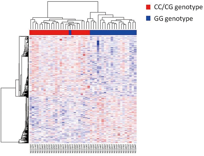
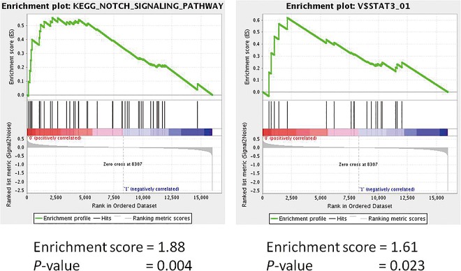
From the microarray assay, we identified 570 significantly upregulated genes and 443 significantly downregulated genes in the CC/CG genotype compared with the GG genotype. We examined a heat map of the clustering of the expression pattern of those genes in the 59 CRC cases (Fig 1). The characteristics of the expression profiles of the miR-146a polymorphism were clearly stratified. Furthermore, GSEA indicated that a number of Notch signaling pathway signatures and JAK/STAT3 signaling pathway signatures were significantly enriched in the CC/CG genotype (p = 0.004 and p = 0.023, respectively) (Fig 2).
Fig 1. Hierarchical clustering showing the expression levels of the differentially expressed genes in a comparison of CRC patients with the pre-miR-146a/C (CC/CG) genotype and without the pre-miR-146a/C (GG) genotype.
Red spots indicate upregulated and blue spots indicate downregulated probes compared with reference probes. On the top, clustering results of CRC patients are shown (dendrogram). Red bar indicates the CC/CG genotype and the blue bar indicates the GG genotype. On the left side, clustering results of the differentially expressed genes between the two genotypes are shown.
Fig 2.
Gene Set Enrichment Analysis (GSEA): Enriched gene sets for CRC patients with the C allele (CC/CG); KEGG_NOTCH_SIGNALING_PATHWAY (A) and V$STAT3_01 (B).
The influence of the miR-146a polymorphism on the expression of miR-146a and NUMB and migratory response
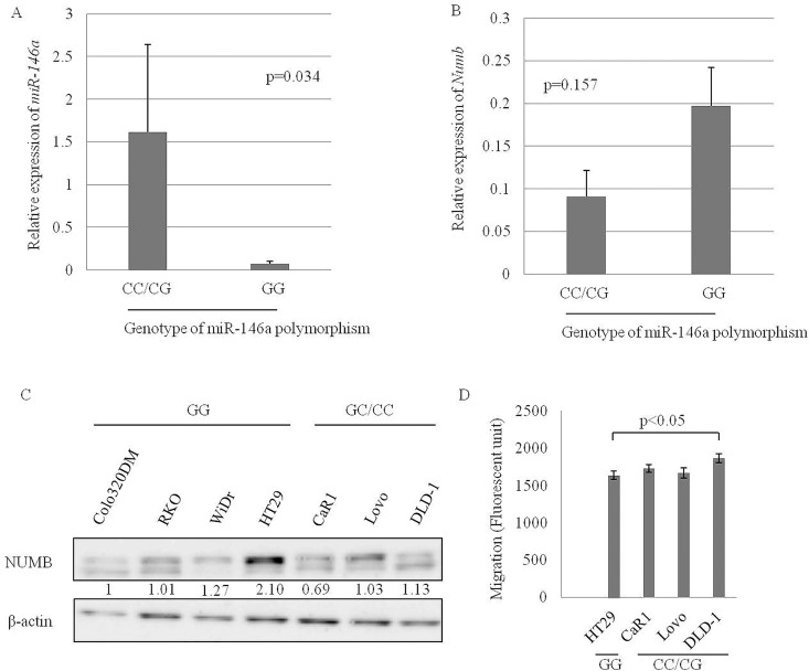
To explore the influence of miR-146a polymorphism on the expression of miR-146a and NUMB, we examined the relationship between the polymorphism and the expression of miR-146a or NUMB in 7 CRC cell lines, whose polymorphism was determined by direct sequencing (Table 2). miR-146a expression was significantly higher in 3 CRC cell lines with the pre-miR-146a/C than the 4 CRC cell lines without the pre-miR-146a/C (p = 0.034) (Fig 3A). There was no association between the miR-146a polymorphism and NUMB expression in the CRC cell lines (Fig 3B), however, immunoblotting analysis revealed that NUMB expression of CRC cell lines with pre-miR-146a/C (CaR1, Lovo and DLD-1) was lower than WiDr and HT29, CRC cell lines without pre-miR-146a/C (Fig 3C). Furthermore, DLD-1, one CRC cell line with the pre-miR-146a/C had higher chemotactic activity than the CRC cell lines without the pre-miR-146a/C (Fig 3D).
Table 2. Genotyping of miR-146a polymorphism in 7 CRC cell lines.
| CRC cell lines | Genotype of rs2910164 |
|---|---|
| RKO | GG |
| HT29 | GG |
| WiDr | GG |
| CaR1 | CC |
| LoVo | GC |
| COLO320 | GG |
| DLD1 | CC |
Abbreviations: CRC, colorectal cancer
Fig 3.
Comparison of miR-146a expression (A) and NUMB expression (B) between CRC cell lines with and without the pre-miR-146a/C. NUMB expression of CRC cell lines by immunoblotting (C). Migratory capacity of CRC cell lines with the pre-miR-146a/C compared with HT29, a representative CRC cell line without pre-miR-146a/C (D).
Inhibition of miR-146a expression increased NUMB expression in CRC
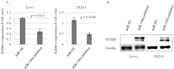
In order to confirm the influence of miR-146a expression on the NUMB expression in CRC, we investigated NUMB expression of CRC cell lines with the pre-miR-146a/C treated by miR-146a inhibitor. Knockdown of miR-146a, confirmed by RT-qPCR (Fig 4A) enhanced NUMB expression in CRC cell lines with the pre-miR-146a/C (Fig 4B).
Fig 4.
Downregulated miR-146a (A) and upregulated NUMB (B) expression of CRC cell lines with pre-miR-146a/C transfected with miR-146a inhibitor or negative control.
Discussion
Numerous previous studies have described the role of miR-146a polymorphism in patients’ susceptibility to CRC. In this study, we demonstrated that miR-146a polymorphism was also involved with liver metastasis in CRC patients. We hypothesized that the molecular mechanism of liver metastasis in CRC is caused by dramatic changes of gene signatures affected by miR-146a polymorphism. GSEA showed that Notch signaling and JAK-STAT3 signaling were significantly activated in the patients with pre-miR-146a/C. Notch signaling may be involved in cell-fate decision and promote cell survival or anti-apoptosis by mediating the expression of some anti-apoptosis or pro-survival proteins [31,32]. Notch signaling also mediates the epithelial-mesenchymal transition process, leading to metastasis [31] and has been traditionally implicated as a key mechanism for the initiation and progression in CRC [33]. Sonoshita et al. demonstrated that inhibition of Notch signaling by amino-terminal enhancer of split (Aes) suppressed the invasiveness and the intravasation of CRC cells in an orthotopic transplantation model [34]. We previously reported that Notch induced CCL2 which in turn promoted distant metastasis in vivo [35]. Another pathway HGF/c-MET/ETS-1 also plays a central role in tumor proliferation, invasion and metastasis [36,37]. However, GSEA showed no significant enrichment for the gene set of HGF/c-MET/ETS-1 pathway (data not shown). Thus, miR-146a polymorphism may promote liver metastasis in CRC via Notch signaling and JAK/STAT3 signaling.
It is well known that the common genetic variant rs2910164 is located within the miR-146a precursor sequence and that it affects the expression of mature miR-146a [29,30,38–41]. The pre-miR-146a/C genotype may reduce the stability of pri-miR by mispairing events within the hairpin [42] and it has been associated with lower expression of the miR-146a in several cancers [38–41]. We previously reported that patients with the pre-miR-146a/C genotype showed higher expression of miR-146a than those with the pre-miR-146a/G genotype in gastric cancer [30]. The effect of miR-146a polymorphism on the expression of miR-146a has been demonstrated in vitro [29]. In our study, the clinical samples whose SNP was examined were unfortunately unavailable. However, CRC cell lines with the pre-miR-146a/C genotype have significantly higher miR-146a expression than those with the pre-miR-146a/G. This discrepancy may be explained by the ethnicity, cancer type, and disease status.
NUMB protein is a key negative regulator of Notch signaling [43] and is one of the direct targets of miR-146a [15,41,44,45]. miR-146a induces an oncogenic phenotype and tumorigenesis of oral squamous cell carcinoma by directly targeting the 3’-UTR of NUMB [45]. In CRC, Snail induces miR-146a expression, which in turn targets NUMB and stabilizes β-catenin [15]. Contrary to our result with miR-146a expression, CRC cell lines with the pre-miR-146a/C genotype had the lower expression of NUMB protein and more migratory response than those with a pre-miR-146a/G genotype. NUMB suppression may be affected by alteration of miR-146a expression through this SNP, followed by increasing the migratory response via enhancing Notch signaling; however, further examination is needed to confirm the effect of miR-146a polymorphism on liver metastasis in CRC using genome editing.
In conclusion, we demonstrated that miR-146a polymorphism is associated with the tendency of CRC to metastasize to the liver. This occurs through miR-146a-mediated activation of Notch and JAK/STAT3 signaling via suppression of NUMB. Identification of miR-146a polymorphism could identify patients with high-grade CRC and be useful for follow-up with particular attention to liver metastasis.
Acknowledgments
This work was supported by the following grants and foundations: Grants-in-Aid for Scientific Research of MEXT (26461980, 15H04921, 15K10168, 15K10170). This research used computational resources of the K computer provided by the RIKEN Advanced Institute for Computational Science through the HPCI System Research project (Project ID: hp140230). Computation time was also provided by the Supercomputer System, Human Genome Center, Institute of Medical Science, University of Tokyo (http://sc.hgc.jp/shirokane.html). We appreciate the technical support of Ms. Kazumi Oda, Michiko Kasagi, Sachiko Sakuma, Noriko Mishima and Tomoko Kawano.
Data Availability
All relevant data are within the paper.
Funding Statement
This work was supported by Grants-in-Aid for Scientific Research of MEXT (26461980, 15H04921, 15K10168, 15K10170) (https://www.jsps.go.jp/english/e-grants/). The funders had no role in study design, data collection and analysis, decision to publish, or preparation of the manuscript.
References
- 1.Ferlay J, Shin HR, Bray F, Forman D, Mathers C, Parkin DM. Estimates of worldwide burden of cancer in 2008: GLOBOCAN 2008. Int J Cancer. 2010;127:2893–2917. 10.1002/ijc.25516 [DOI] [PubMed] [Google Scholar]
- 2.Washington MK. Colorectal carcinoma: selected issues in pathologic examination and staging and determination of prognostic factors. Arch Pathol Lab Med. 2008;132:1600–1607. 10.1043/1543-2165(2008)132[1600:CCSIIP]2.0.CO;2 [DOI] [PubMed] [Google Scholar]
- 3.Weitz J, Koch M, Debus J, Höhler T, Galle PR, Büchler MW. Colorectal cancer. Lancet. 2005;365:153–165. 10.1016/S0140-6736(05)17706-X [DOI] [PubMed] [Google Scholar]
- 4.O'Connell J.B., Maggard M.A., Ko C.Y.. Colon cancer survival rates with the new American Joint Committee on Cancer sixth edition staging, J Natl Cancer Inst. 2004;96:1420–1425. 10.1093/jnci/djh275 [DOI] [PubMed] [Google Scholar]
- 5.Carthew RW, Sontheimer EJ. Origins and Mechanisms of miRNAs and siRNAs. Cell. 2009;136:642–655. 10.1016/j.cell.2009.01.035 [DOI] [PMC free article] [PubMed] [Google Scholar]
- 6.Bartel DP. MicroRNAs: genomics, biogenesis, mechanism, and function. Cell. 2004;116:281–297. [DOI] [PubMed] [Google Scholar]
- 7.Wang X, Tang S, Le SY, Lu R, Rader JS, Meyers C, Zheng ZM. Aberrant expression of oncogenic and tumor-suppressive microRNAs in cervical cancer is required for cancer cell growth. PLoS One. 2008. July 2 10.1371/journal.pone.0002557 [DOI] [PMC free article] [PubMed] [Google Scholar]
- 8.Philippidou D, Schmitt M, Moser D, Margue C, Nazarov PV, Muller A, et al. Signatures of microRNAs and selected microRNA target genes in human melanoma. Cancer Res. 2010;70:4163–4173. 10.1158/0008-5472.CAN-09-4512 [DOI] [PubMed] [Google Scholar]
- 9.Sun X, Zhang J, Hou Z, Han Q, Zhang C, Tian Z. miR-146a is directly regulated by STAT3 in human hepatocellular carcinoma cells and involved in anti-tumor immune suppression. Cell Cycle. 2015;14:243–252. 10.4161/15384101.2014.977112 [DOI] [PMC free article] [PubMed] [Google Scholar]
- 10.Li Y, Vandenboom TG 2nd, Wang Z, Kong D, Ali, Philip PA, Sarkar FH. miR-146a suppresses invasion of pancreatic cancer cells. Cancer Res. 2010;70:1486–1495. 10.1158/0008-5472.CAN-09-2792 [DOI] [PMC free article] [PubMed] [Google Scholar]
- 11.Chen G, Umelo IA, Lv S, et al. miR-146a inhibits cell growth, cell migration and induces apoptosis in non-small cell lung cancer cells. PLoS One. 2013. March 26 10.1371/journal.pone.0060317 [DOI] [PMC free article] [PubMed] [Google Scholar]
- 12.Yao Q, Cao Z, Tu C, Zhao Y, Liu H, Zhang S. MicroRNA-146a acts as a metastasis suppressor in gastric cancer by targeting WASF2. Cancer Lett. 2013;335:219–224. 10.1016/j.canlet.2013.02.031 [DOI] [PubMed] [Google Scholar]
- 13.Kumaraswamy E, Wendt KL, Augustine LA, Stecklein SR, Sibala EC, Li D, et al. BRCA1 regulation of epidermal growth factor receptor (EGFR) expression in human breast cancer cells involves microRNA-146a and is critical for its tumor suppressor function. Oncogene. 2015;34:4333–4346. 10.1038/onc.2014.363 [DOI] [PMC free article] [PubMed] [Google Scholar]
- 14.Wang C, Guan S, Liu F, Chen X, Han L, Wang D, et al. Prognostic and diagnostic potential of miR-146a in oesophageal squamous cell carcinoma. Br J Cancer. 2016;114:290–297. 10.1038/bjc.2015.463 [DOI] [PMC free article] [PubMed] [Google Scholar]
- 15.Hwang WL, Jiang JK, Yang SH, Huang TS, Lan HY, Teng HW, et al. MicroRNA-146a directs the symmetric division of Snail-dominant colorectal cancer stem cells. Nat Cell Biol. 2014;16:268–280. 10.1038/ncb2910 [DOI] [PubMed] [Google Scholar]
- 16.Zeng C, Huang L, Zheng Y, Huang H, Chen L, Chi L. Expression of miR-146a in colon cancer and its significance. Nan Fang Yi Ke Da Xue Xue Bao. 2014;34:396–400. [PubMed] [Google Scholar]
- 17.Hu Z, Chen J, Tian T, Zhou X, Gu H, Xu L, et al. Genetic variants of miRNA sequences and non-small cell lung cancer survival. J Clin Invest. 2008;118:2600–2608. 10.1172/JCI34934 [DOI] [PMC free article] [PubMed] [Google Scholar]
- 18.Slaby O, Bienertova-Vasku J, Svoboda M, Vyzula R. Genetic polymorphisms and microRNAs: new direction in molecular epidemiology of solid cancer. J Cell Mol Med. 2012;16:8–21. 10.1111/j.1582-4934.2011.01359.x [DOI] [PMC free article] [PubMed] [Google Scholar]
- 19.Landau DA, Slack FJ. MicroRNAs in mutagenesis, genomic instability, and DNA repair. Semin Oncol. 2011;38:743–751. 10.1053/j.seminoncol.2011.08.003 [DOI] [PMC free article] [PubMed] [Google Scholar]
- 20.Rehmsmeier M, Steffen P, Hochsmann M, Giegerich R. Fast and effective prediction of microRNA/target duplexes. RNA. 2004;10:1507–1517. 10.1261/rna.5248604 [DOI] [PMC free article] [PubMed] [Google Scholar]
- 21.Vinci S, Gelmini S, Mancini I, Malentacchi F, Pazzagli M, Beltrami C, et al. Genetic and epigenetic factors in regulation of microRNA in colorectal cancers. Methods. 2013;59:138–146. 10.1016/j.ymeth.2012.09.002 [DOI] [PubMed] [Google Scholar]
- 22.Hezova R, Kovarikova A, Bienertova-Vasku J, Sachlova M, Redova M, Vasku A, et al. Evaluation of SNPs in miR-196-a2, miR-27a and miR-146a as risk factors of colorectal cancer. World J Gastroenterol. 2012;18:2827–2831. 10.3748/wjg.v18.i22.2827 [DOI] [PMC free article] [PubMed] [Google Scholar]
- 23.Ma L, Zhu L, Gu D, Chu H, Tong N, Chen J, et al. A genetic variant in miR-146a modifies colorectal cancer susceptibility in a Chinese population. Arch Toxicol. 2013;87:825–833. 10.1007/s00204-012-1004-2 [DOI] [PubMed] [Google Scholar]
- 24.Chae YS, Kim JG, Lee SJ, Kang BW, Lee YJ, Park JY, et al. A miR-146a polymorphism (rs2910164) predicts risk of and survival from colorectal cancer. Anticancer Res. 2013;33:3233–3239. [PubMed] [Google Scholar]
- 25.Sugimachi K, Niida A, Yamamoto K, Shimamura T, Imoto S, Iinuma H, et al. Allelic imbalance at an 8q24 oncogenic SNP is involved in activating MYC in human colorectal cancer. Ann Surg Oncol. 2014;21:515–521. [DOI] [PubMed] [Google Scholar]
- 26.Mimori K, Druck T, Inoue H, Alder H, Berk L, Mori M, et al. Cancer-specific chromosome alterations in the constitutive fragile region FRA3B. Proc Natl Acad Sci U S A. 1999;96:7456–7461. [DOI] [PMC free article] [PubMed] [Google Scholar]
- 27.Shinden Y, Iguchi T, Akiyoshi S, Ueo H, Ueda M, Hirata H, et al. miR-29b is an indicator of prognosis in breast cancer patients. Mol Clin Oncol. 2015;3:919–923. 10.3892/mco.2015.565 [DOI] [PMC free article] [PubMed] [Google Scholar]
- 28.Ueda M, Iguchi T, Nambara S, Saito T, Komatsu H, Sakimura S, et al. Overexpression of Transcription Termination Factor 1 is Associated with a Poor Prognosis in Patients with Colorectal Cancer. Ann Surg Oncol. 2015;22:1490–1498. [DOI] [PubMed] [Google Scholar]
- 29.Shen J, Ambrosone CB, DiCioccio RA, Odunsi K, Lele SB, Zhao H. A functional polymorphism in the miR-146a gene and age of familial breast/ovarian cancer diagnosis. Carcinogenesis. 2008;29:1963–1966. 10.1093/carcin/bgn172 [DOI] [PubMed] [Google Scholar]
- 30.Kogo R, Mimori K, Tanaka F, Komune S, Mori M. Clinical significance of miR-146a in gastric cancer cases. Clin Cancer Res. 2011;17:4277–4284. 10.1158/1078-0432.CCR-10-2866 [DOI] [PubMed] [Google Scholar]
- 31.Kang J, Kim E, Kim W, Seong KM, Youn H, Kim JW, et al. Rhamnetin and cirsiliol induce radiosensitization and inhibition of epithelial-mesenchymal transition (EMT) by miR-34a-mediated suppression of Notch-1 expression in non-small cell lung cancer cell lines. J Biol Chem. 2013;288:27343–27357. 10.1074/jbc.M113.490482 [DOI] [PMC free article] [PubMed] [Google Scholar]
- 32.Jia H, Yang Q, Wang T, Cao Y, Jiang QY, Ma HD, et al. Rhamnetin induces sensitization of hepatocellular carcinoma cells to a small molecular kinase inhibitor or chemotherapeutic agents. Biochim Biophys Acta. 2016;1860:1417–1430. 10.1016/j.bbagen.2016.04.007 [DOI] [PubMed] [Google Scholar]
- 33.Sancho E, Batlle E, Clevers H. Signaling pathways in intestinal development and cancer. Annu Rev Cell Dev Biol. 2004;20:695–723. 10.1146/annurev.cellbio.20.010403.092805 [DOI] [PubMed] [Google Scholar]
- 34.Sonoshita M, Aoki M, Fuwa H, Aoki K, Hosogi H, Sakai Y, et al. Suppression of colon cancer metastasis by Aes through inhibition of Notch signaling. Cancer Cell. 2011;19:125–137. 10.1016/j.ccr.2010.11.008 [DOI] [PubMed] [Google Scholar]
- 35.Yumimoto K, Akiyoshi S, Ueo H, Sagara Y, Onoyama I, Ueo H, et al. F-box protein FBXW7 inhibits cancer metastasis in a non-cell-autonomous manner. J Clin Invest. 2015;125:621–635. 10.1172/JCI78782 [DOI] [PMC free article] [PubMed] [Google Scholar]
- 36.Yu S, Yu Y, Zhao N, Cui J, Li W, Liu T. C-Met as a prognostic marker in gastric cancer: a systematic review and meta-analysis. PLoS One. 2013;8:e79137 10.1371/journal.pone.0079137 [DOI] [PMC free article] [PubMed] [Google Scholar]
- 37.Gayyed MF, Abd El-Maqsoud NM, El-Hameed El-Heeny AA, Mohammed MF. c-MET expression in colorectal adenomas and primary carcinomas with its corresponding metastases. J Gastrointest Oncol. 2015;6:618–627. 10.3978/j.issn.2078-6891.2015.072 [DOI] [PMC free article] [PubMed] [Google Scholar]
- 38.Jazdzewski K, Murray EL, Franssila K, Jarzab B, Schoenberg DR, de la Chapelle A. Common SNP in pre-miR-146a decreases mature miR expression and predisposes to papillary thyroid carcinoma. Proc Natl Acad Sci U S A. 2008;105:7269–7274. 10.1073/pnas.0802682105 [DOI] [PMC free article] [PubMed] [Google Scholar]
- 39.Xu B, Feng NH, Li PC, Tao J, Wu D, Zhang ZD, et al. A functional polymorphism in Pre-miR-146a gene is associated with prostate cancer risk and mature miR-146a expression in vivo. Prostate. 2010;70:467–472. 10.1002/pros.21080 [DOI] [PubMed] [Google Scholar]
- 40.Xu T, Zhu Y, Wei QK, Yuan Y, Zhou F, Ge YY, et al. A functional polymorphism in the miR-146a gene is associated with the risk for hepatocellular carcinoma. Carcinogenesis. 2008;29:2126–2131. 10.1093/carcin/bgn195 [DOI] [PubMed] [Google Scholar]
- 41.Forloni M, Dogra SK, Dong Y, et al. miR-146a promotes the initiation and progression of melanoma by activating Notch signaling. Elife. 2014. February 18 10.7554/eLife.01460 [DOI] [PMC free article] [PubMed] [Google Scholar]
- 42.Rehmsmeier M, Steffen P, Hochsmann M, Giegerich R. Fast and effective prediction of microRNA/target duplexes. RNA. 2004;10:1507–1517. 10.1261/rna.5248604 [DOI] [PMC free article] [PubMed] [Google Scholar]
- 43.Zhong W, Jiang MM, Weinmaster G, Jan LY, Jan YN. Differential expression of mammalian Numb, Numblike and Notch1 suggests distinct roles during mouse cortical neurogenesis. Development. 1997;124:1887–1897. [DOI] [PubMed] [Google Scholar]
- 44.Kuang W, Tan J, Duan Y, Duan J, Wang W, Jin F, et al. Cyclic stretch induced miR-146a upregulation delays C2C12 myogenic differentiation through inhibition of Numb. Biochem Biophys Res Commun. 2009;378:259–263. 10.1016/j.bbrc.2008.11.041 [DOI] [PubMed] [Google Scholar]
- 45.Hung PS, Liu CJ, Chou CS, Kao SY, Yang CC, Chang KW, et al. miR-146a enhances the oncogenicity of oral carcinoma by concomitant targeting of the IRAK1, TRAF6 and NUMB genes. PLoS One. 2013. November 26 10.1371/journal.pone.0079926 [DOI] [PMC free article] [PubMed] [Google Scholar]
Associated Data
This section collects any data citations, data availability statements, or supplementary materials included in this article.
Data Availability Statement
All relevant data are within the paper.


