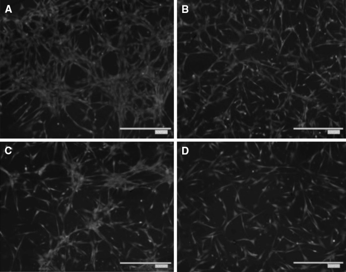
Fig. 8.
Representative images of transwell migration assay. ADMSCs were seeded on upper transwell migration chamber and the serum-free DMEM medium was added in the lower part for 24 h; (a–d control, 20, 40, 60 mg/l of IS). The cells migrated to the lower chamber and were stained with Calcein-AM as mentioned in “Materials and methods” section. The images of migrated cells were captured after stained with Calcein-AM

