Figure 4.
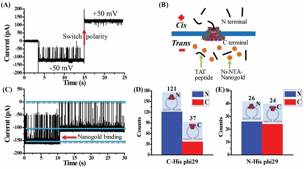
C-His phi29 connectors as control for planar lipid bilayer insertion and subsequent TAT peptide translocation. (A) While TAT peptide is premixed in both compartments (300 nM), translocation is only observed at −50 mV but not at +50 mV, as demonstrated by polarity switching. (B) Schematic of the Ni- NTA-nanogold binding assay for determining the orientation of C-His phi29 connector in the lipid membrane. (C) Representative current trace showing TAT peptide translocation through a single C-His phi29 connector and subsequent binding of Ni-NTA-nanogold (1.8 nm; 300 pM added into trans-chamber) to C-terminal His-tag of C-His phi29 connector. Applied voltage: −50 mV. Quantification of the orientation adopted by (D) C-His phi29 connector from 158 insertion events and (E) N-His phi29 connector from 50 insertion events. Cis-chamber: 1 M KCl, 5 mM HEPES, pH 7.9; trans-chamber: 0.15 M KCl, 5 mM HEPES, pH 7.9.

