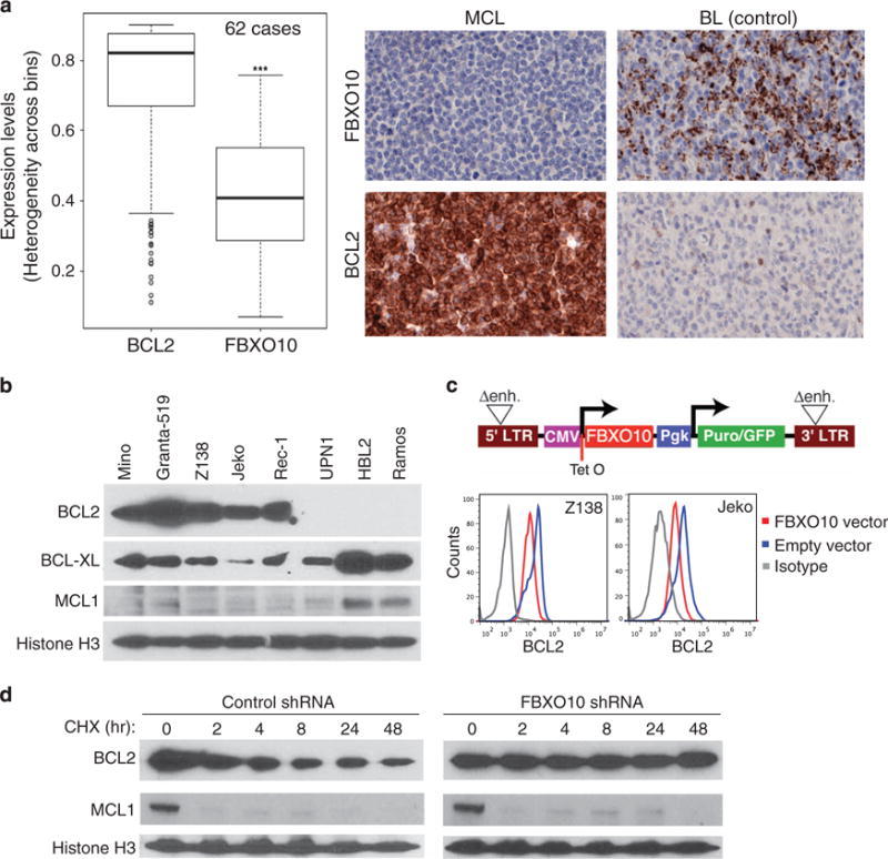
Figure 1.

High levels of BCL2 expression and defects in FBXO10-mediated proteasomal degradation in MCL cell lines. (a) A tissue microarray (TMA) of 62 cases was used for immunohistochemical staining for BCL2 and FBXO10 expression and analyzed with Inform advanced image analysis software (PerkinElmer) (***P<0.001). Representative staining images from a MCL patient are shown. Infiltrating lymphocytes of a Burkitt lymphoma (BL) served as a positive control for FBXO10 staining and malignant cells as a negative control for BCL2 staining. (b) Immunoblotting assay for expression of BCL2, BCL-XL and MCL1. Histone H3 served as a loading control. Note genomic deletion of BCL2 in UPN-1 and BCL2 amplification in Granta-519 and Z138 (ref. 34) BL cell line Ramos served as a negative control for BCL2. (c) Structure of the inducible GFP retroviral vector. Two tetracycline repressor binding sites (Tet operators) were inserted into the CMV promoter, which drives FBXO10 expression. LTR, long terminal repeat; puror/GFP, fusion of puromycin resistance and green fluorescent protein gene; Pgk, phosphoglycerate kinase promoter; Δenh., deletion of LTR promoter sequences. Intracellular BCL2 flow cytometric analysis of two MCL cell lines after 2 days of FBXO10 expression. (d) FBXO10 silencing by shRNA prolongs BCL2 half-life. Two days after shRNA induction with 20 ng/ml of doxycycline, Z138 cells were treated with 20 μg/ml of cycloheximide and analyzed by immunoblotting for indicated proteins.
