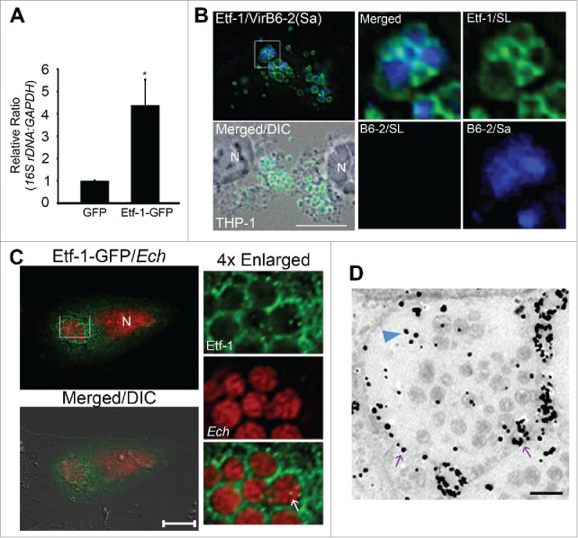
Figure 6.

Etf-1 promotes E. chaffeensis infection and traffics to E. chaffeensis inclusions. (A) HEK293 cells transfected with Etf-1-GFP or with GFP alone (control) were infected with E. chaffeensis at 1 d p.t. qPCR was performed at 2 d p.i. The 16S rDNA/GAPDH ratio for GFP-transfected HEK293 cells was set as 1. Data are presented as the mean ± standard deviation of triplicate assays. *, Significantly different (P < 0.05) by the Student t test. (B) Native Etf-1 localizes on the cytoplasmic side of E. chaffeensis inclusions. N, nucleus. The plasma membrane of infected THP-1 cells was selectively permeabilized with SLO and labeled with anti-Etf-1 (Etf-1/SL) and anti-VirB6-2 (B6-2/SL). After the first round of staining, all cell membranes were permeabilized with saponin (Sa), and the cells were stained again with anti-VirB6 (B6-2/Sa). Secondary antibodies with distinct fluorochromes were used for VirB6-2 labeling before (red) and after (blue) saponin treatment. (C) E. chaffeensis (Ech) inclusions are enveloped by Etf-1-GFP. E. chaffeensis-infected RF/6A cells were transfected with Etf-1-GFP at 1 d p.i., and treated with 0.1 μM DFP for 2 h prior to fixation at 16 h p.t. (40 p.i.). DAPI was used to stain DNA in host cell nuclei and E. chaffeensis DNA and pseudocolored in red. N, nucleus. Merged/DIC, fluorescence image merged with DIC image. Boxed area was enlarged 4-fold on the right. The white arrow indicated the presence of Etf-1-GFP inside E. chaffeensis-containing inclusions. Scale bars: 10 μm. (D) Immunogold labeling of Etf-1-GFP in E. chaffeensis-infected RF/6A cells. Silver-enhanced anti-GFP immunogold labeling of Etf-1-GFP detected on the inclusion membrane (purple arrows) or inside the inclusions (blue arrowhead). Scale bar: 2 μm.
