Abstract
Statement of the Problem:
Denture stomatitis is the common form of oral candidiasis, which is seen in the form of diffused inflammation in the areas covered by dentures. Many primary studies report the prevalence of denture stomatitis and candida albicans among patients in the Iranian population; therefore, using meta-analysis is valuable for health policy makers.
Purpose:
The purpose of the present study is to determine the prevalence of denture stomatitis and candida albicans in Iran.
Materials and Method:
Using relevant keywords, national and international databases were searched. After limiting the search strategy and deleting the duplicates, the remaining papers were screened by examining the title and abstract. In order to increase the sensitivity of search reference lists of papers were examined. Finally the index of heterogeneity between studies was defined using Cochran test (Q) and I-squared (I2). According to heterogeneity, the random effects model was used to estimate the prevalence of denture stomatitis and candida albicans in Iran.
Result:
The prevalence of denture stomatitis in 12 studies, and the prevalence of candida albicans in patients with denture stomatitis have been reported in 6 studies. The number of sample under investigated and its age range among primary studies included meta- analysis was 2271 individuals and 32.7 till 87.5 years respectively. The prevalence of denture stomatitis in preliminary studies imported to a meta-analysis varied from 1.9% to 54.6%, and its rate in Iran using the meta-analysis was estimated 28.9 % (CI 95%: 18.2-39.6). Also the overall prevalence of candida albicans in patients with denture stomatitis in Iran was estimated 60.6% (CI 95%:50.1-71.2).
Conclusion:
This study showed that the prevalence of denture stomatitis and candida albicans among patient infected denture stomatitis is relatively significant in Iran.
Keywords: Denture stomatitis , Candida albicans , Meta-analysis , Iran
Introduction
Denture stomatitis is a chronic inflammation in mucous membrane under prosthodontics which may be of local or general nature.[1] This inflammation is a common and recurrent problem in those who use denture and is accompanied by erythema, mucous membrane inflammation and sometimes pain or burning.[2]
Various studies have been reported vast spectrum of spread between 11 to 60%, in different part of the world.[3] In a study in Chile, denture stomatitis was shown as the most common oral mucosal lesions in people older than 65 years (22.3%).[4] Another study in Germany indicated a prevalence of 18.3% in 65-74 years of old population.[5] In a study made in Tehran on those elderly people over 65 who were using dentures, spread of denture stomatitis was 18.2%.[6]
However, multi-factorial causes have been reported for this long term mucosal lesion: trauma resulted from inappropriate denture, low hygiene of mouth and denture, microbes, nutritional deficiency, diabetes, immune deficiency, and some other systemic factors.[7] A review on etiology, diagnosis, and treatment of denture stomatitis indicates a combination of inflectional causes, trauma, and probable immune deficiency in host, as the etiology of the disease.[8]
Numerous studies have emphasized the main role of candida albicans as primary pathogen in creating denture stomatitis, in a way that this type of candida has initiated, stabilized and exacerbated the disease in 93% of those stricken.[9] In the study of Berdicevesky et al. the percentage of spread of candida on denture and in the mouth of those using denture was compared to a control group not using denture. The spread in the former group was 88%, while in control group, it was 52%.[10] A study on individuals using full dentures, showed 63.3% of spread of candida albicans.[11]
Oral mucosal lesions, especially denture stomatitis, have negative impacts on general health of patients. Moreover, various percentage of prevalence of this disorder as well as candida albicans has been reported in studies performed in Iran. Therefore, the present study was made to investigate the spread of denture stomatitis and frequency of candidiasis through systematic review and meta-analysis, so that, prevention and treatment planning could be done via more precise and comprehensive knowledge about this dilemma.
Materials and Method
Search Strategy
National and international journals were used to find published articles from 2000 to July, 30th 2015. National databases including Scientific Information Database (SID), Iranmedex, Magiran and Irandoc and international databases including PubMed, Google scholar, Scopus and Science direct were searched using keywords. A literature search strategy was primarily applied using the key words: Candidiasis, Candida, Denture, Colonization, Stomatitis, Candida Albicans, Prevalence, Frequency, Survey, Iran and their Persian equivalents were mainly used as search strategy. Two independent researchers carried out the search during 1st to 30th August, 2015. References made in published studies were also reviewed to increase sensitivity and to select more from studies. Search evaluation was made by one of researchers randomly, showing that no study was omitted. Sources in paper were also searched for access to those articles not published electronically, and research centers as well as experts in the field were consulted.
Selection of Studies
Full text or brief of all studies, documents, and reports resulted from advanced search were extracted. After removal of duplicates; topic, abstract, and full texts were reviewed respectively, so that, unrelated papers could be removed and the related ones could be selected. In should be noted that in order to prevent republication bias (transverse and longitudinal publication bias), examination of results for identification and removal of repetitive researches were in included in schedule.
Quality Assessment
After selection of primary studies in terms of topic and content, to assess the quality of the selected studies a previously applied checklist was used.[12] This checklist included 12 questions that were provided according to the content of STROBE checklist,[13] encompassing various aspects of methodology; determining appropriate sample size, type of study, sampling method, research population, data gathering method, variables definition and method of sample examination, means of data gathering, statistical tests, study objectives, presentation of results, in appropriate form and providing the results based on objectives. Each question was assigned one score and any study attaining at least 8 scores, has been entered into meta-analysis.[12]
Data Extraction
For each study, data were extracted based on topic, primary author, year of performance, type of study, sampling method, and sample size, language of publication, average age, denture stomatitis prevalence, and spread of candida albicans in those infected with denture stomatitis. Data were entered in excel spread sheets.
Inclusion Criteria
All Persian and English studies reporting sample size, spread of denture stomatitis and candida albicans in those suffering from denture stomatitis, were selected after gaining required score through evaluation.
Exclusion Criteria
Those studies that have not reported spread of denture stomatitis or candida albicans in people suffering from denture stomatitis, or ha not specified the sample size, and also the abstracts from conferences and seminars lacking full texts, case studies, case observing studies, clinical trials that have not provided correct estimation of prevalence, and those studies that has not gained the least quality evaluation score, were excluded.
Analysis
Stata Ver. 11 software was used to analyze data. Standard deviation of prevalence of denture stomatitis, as well as prevalence of candida albicans in those stricken by denture stomatitis in each study was calculated using binominal distribution formula. Finally, heterogeneity index[12] among studies was determined, applying Cochran’s test [Q] and I-squared. According to heterogeneity results, random or fixed model was used for estimation of denture stomatitis and candida albicans prevalence among people suffering from denture stomatitis, in Iran. Sensitivity analysis was also done to determine effective studies in terms of heterogeneity. Those factors suspicious of heterogeneity were examined through meta-regression. Estimation of initiating points of spread for denture stomatitis and candida albicans in those people suffering from denture stomatitis in Iran were calculated in forest plots with confidence interval of 95%, within which square size shows weight of each study and the lines in either side of it are indicative of confidence interval of 95%.
Results
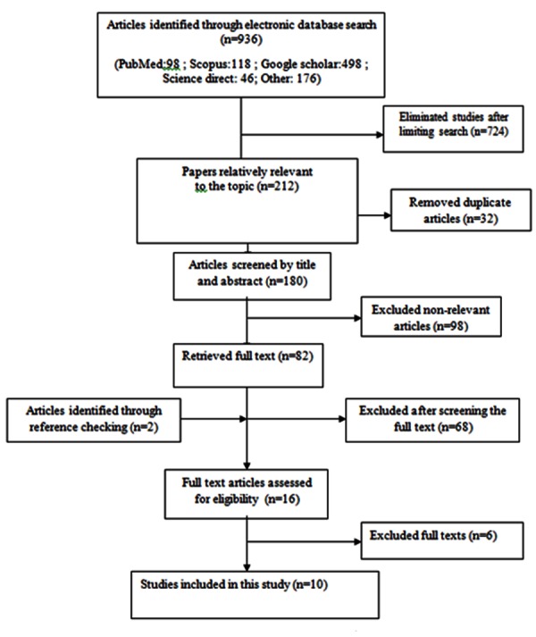
Through primary search, 1890 articles were found from national and international data bases from which 453 remained after limiting search strategy and removal of repetitive cases due to overlapping data bases. Through screening topics and abstracts, 3 cases were recognized as irrelevant. 62 remaining articles were reviewed in full text, 43 of which were irrelevant. Two articles were entered the study, after reviewing references. In continuation, evaluating quality of articles and entrance as well as exclusion criteria, 3 documents were removed and 18 articles (12 regarding prevalence of denture stomati tis and 4 concerning spread of candida albicans in patients with denture stomatitis) entered meta-analysis process (Figure 1).
Figure1.

Literature search and review flowchart for selection of primary studies
The prevalence of denture stomatitis was reported in 12 studies. Publication years were variable between 2000 and 2013. All were cross-sectional studies; respectively 6, 1, 3, and 2 cases of sampling were done in random, cluster, census, and convenience forms. Average age of sample population related to prevalence of denture varied between 32.7 and 87.5 years. Total numbers of samples under investigation in primary studies entering meta-analysis of prevalence of denture were 2271 individuals. Minimum and maximum of sample size was floating between 40 (ArbabiKalati, 2007) and 343 (Malaki, 2005).
Table 1 illustrates variables of prevalence of denture stomatitis from 1.9% (Ghasemi) with sample size of 209 individuals in Sistan and Baluchestan to 54.6% (Mozafari) with sample size of 202 persons. According to Figure 2, estimation of prevalence of denture stomatitis in Iran via random effect model was estimated 28.9% (CI 95%:18.2-39.6, I-squared=98.2%, Q=623.6, p< 0.001).
Table 1.
The characteristics of primary studies related with the prevalence of Denture Stomatitis in Iran
| Id | First author | Publication year | Age average | Sample size | Prevalence Denture |
|---|---|---|---|---|---|
| 1 | Sadeghi [14] | 2013 | 62.2 | 265 | 28.3 |
| 2 | ArbabiKalati [15] | 2007 | 32.68 | 40 | 3 |
| 3 | Matlabnejad [16] | 2002 | 68.2 | 162 | 32.9 |
| 4 | Rabiei [17] | 2009 | 87.5 | 121 | 45.6 |
| 5 | Malaki [18] | 2005 | 70.8 | 343 | 12.2 |
| 6 | Ghasemi [19] | 2004 | 80.4 | 209 | 1.91 |
| 7 | Sahebjamee [20] | 2011 | 69.7 | 141 | 38.3 |
| 8 | Atashrazm [21] | 2013 | 70 | 201 | 36 |
| 9 | Mozafari [22] | 2011 | 79.59 | 202 | 54.6 |
| 10 | Pahlavanzadeh [23] | 2012 | - | 109 | 26.6 |
| 11 | Rabiei [24] | 2013 | 72 | 203 | 52 |
| 12 | Shiravani [6] | 2002 | 65 | 275 | 18.2 |
Figure2.

Prevalence of Denture Stomatitis between the primary studies which included to Meta-analysis and total estimation of its in Iran
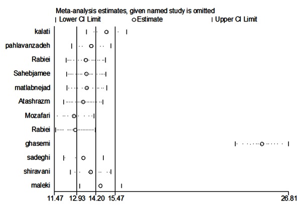
Sensitivity analysis to examine role of each primary study in heterogeneity showed that research done by Ghasemi has the most effect on heterogeneity (Figure 3). Degree of heterogeneity was decreased through removal of the mentioned study, while heterogeneity still remained significant. However, after removal of Ghasemi’s study, prevalence of denture stomatitis was estimated as 31.4% (CI 95%:21.5-41.3, I-squared= 96.7%, Q=306.2, p< 0.001). Total estimation of prevalence of denture stomatitis in Iran, in present meta-analysis is acceptable (28.9%) according to combination of all 12 articles holding entrance criteria. Publication years and average age variables, as variables suspicious of heterogeneity were examined in meta-regression model and showed no significant difference over heterogeneity (Table 2).
Figure3.

Sensitive analysis to evaluate effecting primary studies on heterogeneity
Table 2.
The characteristics of primary studies related with the prevalence of candida albicans in denture stomatitis patients in Iran
| Variables | Univariate | Multivariate | ||
|---|---|---|---|---|
| B | P | B | P | |
| Publication Year | 2.2 | 0.05 | 2.2 | 0.05 |
| Age average | 0.6 | 0.1 | 0.6 | 0.1 |
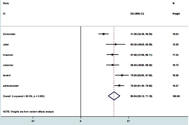
Prevalence of candida albicans in patients with denture stomatitis was reported in 6 cases. The publication year of articles entered into meta-analysis, was 2001 to 2014. All these studies were cross-sectional re searches and sampling was random in 5 cases and convenience in one. Average age of population under study in primary studies was 57.5 to 71.3. Total number of sample population was variable between 30 (Jafari) to 114 (Zomorodian) (Figure 4).
Figure4.

Prevalence of Candida Albicans in denture stomatitis patients between the primary studies which included to meta-analysis and total estimation of its in Iran
Table 3 shows variety of prevalence of candida albicans in primary studies to be 41.6% (Zomorodian) to 75% (Tavakoli). According to Figure 4, applying random effect model, prevalence of candida albicans in patients with denture stomatitis was estimated as 60.6% (CI 95%: 50.1-71.2, I-squared=82%, Q=27.7, p< 0.001).
Table 3.
Discussion
Generally, 2271 individuals with age range of 32.7 to 87.5 were considered eligible to be studied. The degree of prevalence of denture stomatitis and candida albicans was investigated among them. Data related details are given in Tables 1 and 3.
The research performed on 202 elderly people with average age of 79.59±8.88 living in retirement homes in 2011 showed maximum degree of denture prevalence in Mashhad.[22] Prevalence of denture was reported 54.6%. Minimum prevalence (1.91%) was related to a research made on 209 elderly people with average age of 80.4±4.09 referring to dental college in Zahedan, in 2004.[19]
In continuation of studies on patients with denture stomatitis, minimum prevalence (41.5%) in these people also was related to a 2011 research on 114 people with average age of 70.4±11.2, in Shiraz and maximum prevalence of this fungus was related to a 2001 research, performed on 50 people of average age 61.3±6.4 in Hamadan. The estimation of prevalence in people using denture was 75%.[25,29]
Following primary calculation, the heterogeneity index for denture prevalence (96.7%) and for candida albicans (82%) were calculated and due to high heterogeneity of results, random effect model was used in next steps.
To confirm the heterogeneity in these examinations in final and main estimation steps, the prevalence of denture stomatitis in Iranians (28.9%) with confidence interval of 18.2-39.6 and overall prevalence of candida albicans in patients with denture stomatitis (60.3%) with confidence interval of 50.1-71.2, were calculated.
Considering the heterogeneity of research findings, we had to find factor(s) creating heterogeneity through meta-regression models. Due to differences in reported prevalence across Iran, average age and publication year variables were entered into meta-regression analysis to find probable root of differences. Results showed that none of these variables had impact on reduction of heterogeneity among findings.
The estimated prevalence of denture stomatitis in the research in Greek,[30] dental college of UK31 and USA[32] were 39.6, 27, 25%, respectively. In comparison, this prevalence is more than the result of one of the study performed in Thailand (10.5%)[33] and less than the results of studies carried out in Dental College of Turkey,[34] Brazil,[35] Portugal[36] that reported the prevalence of 44, 58.2, and 45.3%, respectively. In explanation, it could be noted that prevalence and risk factors in various studies are different. Moreover, these differences might be due to various methodologies used in estimation of studies.
The methodology factors depends on age group under study, denture situation in people under study, whether samples are from dental colleges or community population, whether patients are hospitalized or not, the methods applied in evaluation of denture stomatitis, and the prevalence and statistical methods applied.[3,32,37-38]
As shown by our research, heterogeneity was not affected by age group. Moreover, it was shown that age group was not effective factor in this disease, while, some other studies have shown increase of prevalence through aging.[20]
Another effective factor in methodology is denture situation. Whether denture is fixed or removable is effective on prevalence of denture stomatitis. For example, as shown in Turkey, although just 26% of people under study used removable dentures, 18.5% of them were suffering from denture stomatitis.[39]
Another effective factor regarding differences in results of the research in comparison to other countries may be referred to the type of the sample which was taken. In justification of these differences, we may state studies made in Finland. In one of the studies, samples were gathered nationally and in another, from retirement houses respectively, reporting 48% and 35% of prevalence.[5] Even the methods applied for estimation of prevalence of denture stomatitis will cause differences, for instance, in Germany Cohort or population-based study instigated different estimations.[40]
Another factor to estimate the prevalence of denture stomatitis is application of hospitalized and non-hospitalized samples. For example, it has been shown in Finland that prevalence in hospitalized patients was different from those at home (respectively 25% against 35%).[5,41]
As mentioned before, some of these factors could be the reason for our different results from other studies. Twelve studies were similar in design, evaluation and diagnosis of lesion, but the prevalence of denture stomatitis was estimated between 17 to 77% ; in 8 studies it was reported to be higher that 45%. So, existing differences in results in comparison to other countries may be due to real differences between the current research and those performed in other countries.[42-46]
Concerning the factors creating denture stomatitis, several factors also have been mentioned, according to epidemiological studies. Demographic factors; instances of smoking, female gender and synchronic diseases which disrupt immune system action have been mentioned. Regarding using denture, unfit dentures, using denture in maxillary against mandible, compromised denture hygiene, continuous use of denture, and microbes especially candida albicans, have been cited. Of course recent studies indicate that the cause of denture stomatitis must be multi-factorial.[2]
Out study showed the prevalence of candida albicans among denture stomatitis patients was estimated to be 60.3%. It was similar to a study that performed at the Center for Oral Health Tygrbrg. In a study carried out by Budtz-Jorgensen et al. it was shown that in patients older than 70 years, c. albicans were cultivated in most denture wearers with denture stomatitis and big accumulations of hyphae existed in 77% of these patients.[9] Felix’s and King’s studies have shown that the prevalence of candida albicans among apparently healthy individuals to be 6, 2.5 % respectively.[47-48]
It should be noted that one of the causes of differences in prevalence of candidiasis in the populations under investigation is the presence of various risk factors, and variability in sensitivity as well as specificity of laboratory methods in diagnostic criterion of candidiasis. For example, some researchers just suffice to culture and others are confined just to smear preparation.[49-51]
Applying different methods for prevalence estimation leads to different results. However, as shown by researches, this fungus is capable of producing nitrosamine and carcinogens chemical substances which probably have role in mouth cancer. High spread of this fungus should be considered important. Hence, importance of periodic examination of soft tissue by oral medicine specialists or well- trained clinicians shall be emphasized by authorities and some facilities should be dedicated to these patients.[52]
The studies performed by Budts-Jorgensen and Chamani showed strong significant relationship between denture stomatitis and candida albicans, which is also proved in various studies. The factor is probably occurring along with some other factors, such as continuous use and low hygiene of denture.[9,52]
Of course, according to studies, the most important factor in the development of denture stomatitis been mentioned to be continuous use of the dentures. Age, gender, smoking, and use of denture without hygiene were factors in developing the disease that were not statistically significant. Unfortunately, in this study due to lack of information from previous studies, it was not possible to assess these factors.[30]
Finally, some limitations such as to the way different authors write their papers, and their unavailability should be noted. Though, all 12 researches applied here gained required score to enter the analysis, these papers did not refer to some major characteristics such as the degree of prevalence of sub-groups of main finding, or indirect reference made to degree of prevalence which inevitably forced the researcher to calculate it according to existing data. It seems that such shortages are caused by acceptance framework of journals, not supervising main and divergent aspects of a paper. To prevent such shortages, we have to provide appropriate infrastructures in form of reviewing acceptance frameworks of journals and paying more attention to examination of different aspects of a research subject, with the help of experienced experts.
Since there is no efficient health system especially in dental and oral settings in Iran, estimation of oral diseases prevalence just on cross sectional studies, bears some problems. Hence, one of the main benefits in this research is precise evaluation of oral lesions such as spread of denture stomatitis and candida albicans, by using systematic review and meta-analysis in the country.
Conclusion
According to above, denture stomatitis is known as a health problem, importance of which is increasing through age increase and changing life style. Although the research showed lower prevalence of denture stomatitis in comparison to other countries, the estimated value is relatively significant for Iran. Furthermore, considering the process of industrialization of Iran that will lead to increase of elderly people, it will be necessary to pay more attention to this topic. To this end, governments have to identify the most important health needs of the community through constant monitoring peoples’ dental and oral hygiene and to reduce its burden via efficient interventions.
Acknowledgement
This study was done with the approval of the Health Policy Research Center of Shiraz University of Medical Sciences, Iran for supporting this research financially.
Conflict of Interest:The authors of this manuscript certify no financial or other competing interest regarding this article.
References
- 1.Jerolimov V. Ucestalost upalnih promjena sluz-nice ispod gornje totalne proteze. Acta Stomatologica Croatica 1983; 17: 227-231. [PubMed] [Google Scholar]
- 2.Gendreau L, Loewy ZG. Epidemiology and etiology of denture stomatitis. J Prosthodont. 2011; 20: 251–260. doi: 10.1111/j.1532-849X.2011.00698.x. [DOI] [PubMed] [Google Scholar]
- 3.Arendorf TM, Walker DM. Denture stomatitis: a review. J Oral Rehabil. 1987; 14: 217–227. doi: 10.1111/j.1365-2842.1987.tb00713.x. [DOI] [PubMed] [Google Scholar]
- 4.Espinoza I, Rojas R, Aranda W, Gamonal J. Prevalence of oral mucosal lesions in elderly people in Santiago, Chile. J Oral Pathol Med. 2003; 32: 571–575. doi: 10.1034/j.1600-0714.2003.00031.x. [DOI] [PubMed] [Google Scholar]
- 5.Nevalainen MJ, Närhi TO, Ainamo A. Oral mucosal lesions and oral hygiene habits in the home-living elderly. J Oral Rehabil. 1997; 24: 332–337. doi: 10.1046/j.1365-2842.1997.d01-298.x. [DOI] [PubMed] [Google Scholar]
- 6.Motalebnezhad M, Shirvani M. Oral mucosal lesions in elderly population (Tehran Kahrizak Geriatric Institute; 2000) J Babol Univ Med Scien (JBUMS) 2002; 3: 28–33. [Google Scholar]
- 7.Scully C, Porter S. ABC of oral health. Swellings and red, white, and pigmented lesions. BMJ 2000; 321: 225–228. doi: 10.1136/bmj.321.7255.225. [DOI] [PMC free article] [PubMed] [Google Scholar]
- 8.Jeganathan S, Lin CC. Denture stomatitis--a review of the aetiology, diagnosis and management. Aust Dent J. 1992 Apr; 37: 107–114. doi: 10.1111/j.1834-7819.1992.tb03046.x. [DOI] [PubMed] [Google Scholar]
- 9.Budtz-Jörgensen E, Stenderup A, Grabowski M. An epidemiologic study of yeasts in elderly denture wearers. Community Dent Oral Epidemiol. 1975; 3: 115–119. doi: 10.1111/j.1600-0528.1975.tb00291.x. [DOI] [PubMed] [Google Scholar]
- 10.Berdicevsky I, Ben-Aryeh H, Szargel R, Gutman D. Oral candida of asymptomatic denture wearers. Int J Oral Surg. 1980; 9: 113–115. doi: 10.1016/s0300-9785(80)80047-0. [DOI] [PubMed] [Google Scholar]
- 11.Jafari A A, Fallah-Tafti A, Fattahi-bafghi A, Arzy B. Comparison the Occurrence Rate of Oral Candida Species in Edentulous Denture Wearer and Dentate Subjects. Int J Med Lab. 2014; 1: 15–21. [Google Scholar]
- 12.Moosazadeh M, Nekoei-Moghadam M, Emrani Z, Amiresmaili M. Prevalence of unwanted pregnancy in Iran: a systematic review and meta-analysis. Int J Health Plann Manage. 2014; 29: e277–e290. doi: 10.1002/hpm.2184. [DOI] [PubMed] [Google Scholar]
- 13.Vandenbroucke JP, von Elm E, Altman DG, Gøtzsche PC, Mulrow CD, Pocock SJ, Poole C, et al. Strengthening the Reporting of Observational Studies in Epidemiology (STROBE): explanation and elaboration. Int J Surg. 2014; 12: 1500–1524. doi: 10.1016/j.ijsu.2014.07.014. [DOI] [PubMed] [Google Scholar]
- 14.Sadeghi M. Prevalence and Risk Factors Associated with Denture Stomatitis in Healthy Subjects Attending a Dental School, Rafsanjan: Thesis; 2013. Avaiable from: [http://dentistry.rums.ac.ir/uploads/ 452rums.tez.pdf. ]
- 15.Arbabi Kalati F, Alavi V, Allahyari E, Maghsoodi B, Azemati S, Alipour A, Hadavi SMR. Frequency of oral mucosal disease in referral patients to dental faculty of Tabriz in 2007. Iranian Journal of Epidemiology. 2010; 6: 50–56. [Google Scholar]
- 16.Motalebnezhad M, Shirvani M. Oral mucosal lesions in elderly poulation (Tehran Kahrizak geriatric institute; 2000) J Babol Uni Med Sci. 2002;4:28–33. [Google Scholar]
- 17.Rabiei M, Kasemnezhad E, Masoudi Rad H, Shakiba M, Pourkay H. Prevalence of oral and dental disorders in institutionalised elderly people in Rasht, Iran. Gerodontology. 2010; 27: 174–177. doi: 10.1111/j.1741-2358.2009.00313.x. [DOI] [PubMed] [Google Scholar]
- 18.Malaki Z, Ghaemmaghami A, S L. Comparison of the prevalence of oral soft tissue lesions in elderly homes. J Dent Sch Shahid Beheshti Univ. 2005; 32: 663–669. [Google Scholar]
- 19.Ghasemi A. The prevalence of oral soft tissue lesions in elderly patients referred to the School of Dentistry, Zahedan. Thesis 2004. Available from: [http://www.zaums.ac.ir/index.aspx?siteid=1&pageid=7197. ]
- 20.Sahebjamee M, Basir Shabestari S, Asadi G, Neishabouri K. Predisposing Factors associated with Denture Induced Stomatitis in Complete Denture Wearers. J Dent Shiraz Univ Med Scien. 2011; 11(Supplement): 35–39. [Google Scholar]
- 21.Atashrazm P, Sadri D. Prevalence of oral mucosal lesions in a group of Iranian dependent elderly complete denture wearers. J Contemp Dent Pract. 2013; 14: 174–178. doi: 10.5005/jp-journals-10024-1295. [DOI] [PubMed] [Google Scholar]
- 22.Mozafari PM, Dalirsani Z, Delavarian Z, Amirchaghmaghi M, Shakeri MT, Esfandyari A, et al. Prevalence of oral mucosal lesions in institutionalized elderly people in Mashhad, Northeast Iran. Gerodontology. 2012; 29: e930–e934. doi: 10.1111/j.1741-2358.2011.00588.x. [DOI] [PubMed] [Google Scholar]
- 23.Pahlavanzadeh MR, Jafari AA, Ahadian H, Ghafourzadeh M, Mirzaei F. Survey the frequency of Oral Candidiasis in Denture Users Referred to Yazd School of Dentistry. J Toloo-e-behdasht. 2013; 11: 103–113. [Google Scholar]
- 24.Rabiei M, Shakiba M, Jacques V. Oral and Systemic Conditions in Elderly Population Groups in Ta-lash, North of Iran. J Dentomaxillofac Radiology, Pathology and Surgery. 2013; 2: 14–21. [Google Scholar]
- 25.Zomorodian K, Haghighi NN, Rajaee N, Pakshir K, Tarazooie B, Vojdani M, et al. Assessment of Candida species colonization and denture-related stomatitis in complete denture wearers. Med My-col. 2011; 49: 208–211. doi: 10.3109/13693786.2010.507605. [DOI] [PubMed] [Google Scholar]
- 26.Jafari AA, Fallah-Tafti A, Fattahi-Bafghi A, Arzy B. Occurrence Rate of Oral Candida Species in Edentulous Denture Wearers Dentate Subjects. Int J Med Lab. 2014; 1: 15–21. [Google Scholar]
- 27.Khozeimeh F, Bahremand T. The prevalence of chronic atrophic Candida infections in patients with denture referred to Yasouj city. Islamic Dent J. 2005; 17: 16–22. [Google Scholar]
- 28.Jafari Nodoshan A, Fallah A, Mirzaei M. The Frequency of Candida and Staphylococcus Colonization in the Oral Cavity of the Elderly. Med Lab J. 2008; 1: 27–31. [Google Scholar]
- 29.Tavakol P, Emdadi Sh. Evaluation of prevalence of oral candidiasis in patients using complete Denture wears. Dent Sch J Hamadan. 2001; 1: 87–90. [Google Scholar]
- 30.Kossioni AE. The prevalence of denture stomatitis and its predisposing conditions in an older Greek population. Gerodontology. 2011; 28: 85–90. doi: 10.1111/j.1741-2358.2009.00359.x. [DOI] [PubMed] [Google Scholar]
- 31.Zissis A, Yannikakis S, Harrison A. Comparison of denture stomatitis prevalence in 2 population groups. Int J Prosthodont. 2006; 19: 621–625. [PubMed] [Google Scholar]
- 32.Shulman JD, Rivera-Hidalgo F, Beach MM. Risk factors associated with denture stomatitis in the United States. J Oral Pathol Med. 2005; 34: 340–346. doi: 10.1111/j.1600-0714.2005.00287.x. [DOI] [PubMed] [Google Scholar]
- 33.Jeganathan S, Payne JA, Thean HP. Denture stomatitis in an elderly edentulous Asian population. J Oral Rehabil. 1997; 24: 468–472. doi: 10.1046/j.1365-2842.1997.00523.x. [DOI] [PubMed] [Google Scholar]
- 34.Kulak-Ozkan Y, Kazazoglu E, Arikan A. Oral hygiene habits, denture cleanliness, presence of yeasts andstomatitis in elderly people. J Oral Rehabil. 2002; 29: 300–304. doi: 10.1046/j.1365-2842.2002.00816.x. [DOI] [PubMed] [Google Scholar]
- 35.Freitas JB, Gomez RS, De Abreu, MH Ferreira, E Ferreira, E Relationship between the use of full dentures and mucosal alterations among elderly Brazilians. J Oral Rehabil. 2008; 35: 370–374. doi: 10.1111/j.1365-2842.2007.01782.x. [DOI] [PubMed] [Google Scholar]
- 36.Figueiral MH, Azul A, Pinto E, Fonseca PA, Branco FM, Scully C. Denture-related stomatitis: identification of aetiological and predisposing factors - a large cohort. J Oral Rehabil. 2007; 34: 448–455. doi: 10.1111/j.1365-2842.2007.01709.x. [DOI] [PubMed] [Google Scholar]
- 37.Webb BC, Thomas CJ, Willcox MD, Harty DW, Knox KW. Candida-associated denture stomatitis. Aetiology and management: a review. Part 2 Oral diseases caused by Candida species Aust Dent J 1998; 43: 160–166. doi: 10.1111/j.1834-7819.1998.tb00157.x. [DOI] [PubMed] [Google Scholar]
- 38.Wilson J. The aetiology, diagnosis and management of denture stomatitis. Br Dent J. 1998; 185: 380–384. doi: 10.1038/sj.bdj.4809821. [DOI] [PubMed] [Google Scholar]
- 39.Mumcu G, Cimilli H, Sur H, Hayran O, Atalay T. Prevalence and distribution of oral lesions: a cross-sectional study in Turkey. Oral Dis. 2005; 11: 81–87. doi: 10.1111/j.1601-0825.2004.01062.x. [DOI] [PubMed] [Google Scholar]
- 40.Reichart PA. Oral mucosal lesions in a representative cross-sectional study of aging Germans. Community Dent Oral Epidemiol. 2000; 28: 390–398. doi: 10.1034/j.1600-0528.2000.028005390.x. [DOI] [PubMed] [Google Scholar]
- 41.Peltola P, Vehkalahti MM, Wuolijoki-Saaristo K. Oral health and treatment needs of the long-term hospitalised elderly. Gerodontology. 2004; 21: 93–99. doi: 10.1111/j.1741-2358.2004.00012.x. [DOI] [PubMed] [Google Scholar]
- 42.Marchini L, Tamashiro E, Nascimento DF, Cunha VP. Self-reported denture hygiene of a sample of edentulous attendees at a University dental clinic and the relationship to the condition of the oral tissues. Gerodontology. 2004; 21: 226–228. doi: 10.1111/j.1741-2358.2004.00026.x. [DOI] [PubMed] [Google Scholar]
- 43.Baran I, Nalçaci R. Self-reported denture hygiene habits and oral tissue conditions ofcomplete denture wearers. Arch Gerontol Geriatr. 2009; 49: 237–241. doi: 10.1016/j.archger.2008.08.010. [DOI] [PubMed] [Google Scholar]
- 44.Dorey JL, Blasberg B, MacEntee MI, Conklin RJ. Oral mucosal disorders in denture wearers. J Prosthet Dent. 1985; 53: 210–213. doi: 10.1016/0022-3913(85)90111-8. [DOI] [PubMed] [Google Scholar]
- 45.Dikbas I, Koksal T, Calikkocaoglu S. Investigation of the cleanliness of dentures in a university hospital. Int J Prosthodont. 2006; 19: 294–298. [PubMed] [Google Scholar]
- 46.Khasawneh S, al-Wahadni A. Control of denture plaque and mucosal inflammation in denture wearers. J Ir Dent Assoc. 2002; 48: 132–138. [PubMed] [Google Scholar]
- 47.King GN, Healy CM, Glover MT, Kwan JT, Williams DM, Leigh IM, et al. Prevalence and risk factors associated with leukoplakia, hairyleukoplakia, erythematous candidiasis, and gingival hyperplasia in renaltransplant recipients. Oral Surg Oral Med Oral Pathol. 1994; 78: 718–726. doi: 10.1016/0030-4220(94)90086-8. [DOI] [PubMed] [Google Scholar]
- 48.Felix DH, Wray D. The prevalence of oral candidiasis in HIV-infected individuals and dental attenders in Edinburgh. J Oral Pathol Med. 1993; 22: 418–420. doi: 10.1111/j.1600-0714.1993.tb00133.x. [DOI] [PubMed] [Google Scholar]
- 49.Skoglund A, Sunzel B, Lerner UH. Comparison of three test methods used for the diagnosis of candidiasis. Scand J Dent Res. 1994; 102: 295–298. doi: 10.1111/j.1600-0722.1994.tb01472.x. [DOI] [PubMed] [Google Scholar]
- 50.Rindum JL, Stenderup A, Holmstrup P. Identification of Candida albicans types related to healthy andpathological oral mucosa. J Oral Pathol Med. 1994; 23: 406–412. doi: 10.1111/j.1600-0714.1994.tb00086.x. [DOI] [PubMed] [Google Scholar]
- 51.Holbrook WP, Rodgers GD. Candidal infections: experience in a British dental hospital. Oral Surg Oral Med Oral Pathol. 1980; 49: 122–125. doi: 10.1016/0030-4220(80)90303-5. [DOI] [PubMed] [Google Scholar]
- 52.Chamani G, Derhami A, Zarei M, Rad M. The Frequency of oral candidal infection in Kerman dental clinics. J Dent Sch. 2005; 23: 419–428. [Google Scholar]


