Abstract
The pleiotropic cytokine interleukin-6 (IL-6) has been proposed to contribute to circadian regulation of sleepiness by increasing in the blood at night. Earlier studies have reported diurnal variation of IL-6, but phase estimates are conflicting. We have therefore performed a meta-analysis on the diurnal variation of circulating IL-6. Studies were included if they reported IL-6 in plasma or serum recorded at least twice within 24 hours in the same individual. A systematic search resulted in the inclusion of 43 studies with 56 datasets, for a total of 1100 participants. Individual participant data were available from 4 datasets with a total of 56 participants. Mixed-effects meta-regression modelling confirmed that IL-6 varied across the day, the most conspicuous effect being a trough in the morning. These results stand in contrast to earlier findings of a peak in the evening or night, and suggest that diurnal variation should be taken into account in order to avoid confounding by time of day in studies of IL-6 in plasma or serum.
Introduction
Sleepiness is regulated in humans by two main processes: the circadian process, which makes us sleepier in the night, and the homeostatic process, which causes sleepiness to increase with time awake [1]. It has been proposed that interleukin-6, a pleiotropic cytokine, participates in circadian sleepiness regulation by increasing at night in the blood and inducing sleepiness through signalling in the brain [2–6]. Early studies of diurnal variation of IL-6 in humans found a peak in the night-time [7, 8], and it is this observational relationship that forms the main line of evidence for a regulatory effect of circulating IL-6 on sleepiness. However, further studies have since found peaks at different times of the day or have found no peaks at all. Fig 1 shows locations of peaks and troughs that have been estimated in the literature so far. Notably, estimates have ranged quite widely. Nonetheless, the general impression of these earlier claims is consistent with an increase of IL-6 levels in the night-time.
Fig 1. Estimates of phase reported in earlier literature.
Every count represents one claim of having located a peak (box above time-line) or a trough (box below time-line) in a dataset. Blue: Studies included in quantitative review. Green: Studies not included in quantitative review. Orange: Meta-analysis. Review papers are not included.
One previous meta-analysis of IL-6 and time of day has been reported [9] (published again in [10] and [11]). This meta-analysis primarily investigated diurnal variation of interleukin-6 in patients with rheumatoid arthritis, but also included an estimate for healthy control participants from 11 studies. Data inclusion procedures were informal; no systematic method for identifying and including data was reported. The main finding in healthy participants was an increase if IL-6 from the evening, continuing during the night, followed by a drop in the morning. The pattern in patients with rheumatoid arthritis was similar, but with a more pronounced peak in the early morning before levels started to fall.
Thus, the observational relationship between IL-6 and time of day in healthy humans has important implications for the theoretical understanding of immune-brain interactions in sleepiness regulation, but there is no consensus on estimates of phase. Therefore, we have performed a meta-analysis, aiming to investigate the diurnal variation of IL-6 in the blood.
Materials and Methods
Literature search and data acquisition
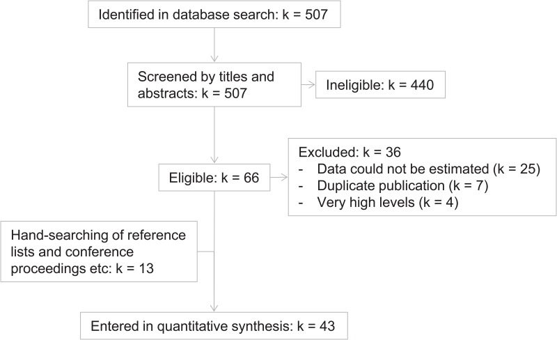
The PubMed database was searched using the terms “interleukin-6 AND (sleep OR diurnal OR circadian)”, and the limit “human”. The search was last updated on 2016-01-03. Records were reviewed by one investigator (GN). Studies were included if they reported IL-6 in plasma or serum from healthy participants with a time-course including two or more time-points within 24 hours. Fig 2 shows a flowchart of data inclusion. Table 1 shows characteristics of included studies. Table 2 lists studies that fulfilled inclusion criteria but which could nonetheless not be included. The most common reason was that data could not be estimated (k = 25). Of these 25 studies, 7 reported that data were largely or entirely under the assay detection limit. In the remaining cases, data could not be estimated because they were given as a difference score (k = 5), because they were not shown (k = 4), because time of day was not given (k = 3), or for other reasons, specified in Table 2 (k = 6). Additionally, seven studies were excluded due to duplicate publication of data, and four studies were excluded because the reported levels of IL-6 were very high and therefore judged not to represent levels consistent with physiological regulation or variation in healthy humans. Of these studies, one reported one participant, whose IL-6 levels increased ten-fold after venous catheterization [12], and we judged that this change was not representative of diurnal variation. Another study reported ten participants with mean plasma IL-6 levels of about 10-30 pg/ml over the course of two days [13]. This is about ten times higher than expected for healthy participants, raising questions about the validity of the absolute values. We judged that these measures may not accurately reflect the target outcome, and that they would unduly influence the regression model on account of the very high values. They were therefore excluded. Finally, two studies [14, 15] reported 40 and 60 participants, possibly with overlapping samples, both with mean values of about 35 and 55 pg/ml at 09:00 and 02:00, respectively. Because these levels were so much higher than expected for healthy participants, these studies were also excluded. Thirteen studies were included from sources other than the PubMed results. These were found mostly because they were cited by papers identified in the literature search, and in some cases because they were known to us on beforehand.
Fig 2. Data inclusion.
Some of the 43 included studies contained more than one dataset. The final number of datasets was 56 (see Table 1).
Table 1. Characteristics of included studies.
Where studies reported several datasets, these are specified separately.
| 1st Author | Year | Ref. | nsubjects | ntimepoints | Regression weight, % | Notes |
|---|---|---|---|---|---|---|
| Sothern | 1995 | [17] | 11 | 8 | 0.99 | a |
| Späth-Schwalbe | 1998 | [41] | 16 | 4 | 1.01 | |
| Dugué | 1998 | [42] | 22 | 3 | 1.21 | |
| Vgontzas | 1999 | [24] | 8 | 24 | 1.24 | |
| Ündar | 1999 | [37] | 10 | 7 | 0.84 | |
| Späth-Schwalbe | 2000 | [43] | 18 | 3 | 0.99 | |
| Redwine | 2000 | [44] | 31 | 15 | 3.80 | b |
| Haack | 2000 | [45] | 20 | 9 | 1.90 | |
| Vgontzas | 2000 | [46] | 12; 11 | 2; 2 | 0.54; 0.49 | c |
| Baker | 2001 | [47] | 8 | 6 | 0.62 | |
| Haack | 2001 | [48] | 10 | 10 | 1.00 | |
| Haack | 2002 | [21] | 12 | 25 | 1.99 | |
| Vgontzas | 2002 | [35] | 11 | 24 | 1.71 | |
| Vgontzas | 2003 | [34] | 15; 13 | 48; 48 | 3.29; 2.85 | |
| Alberti | 2003 | [49] | 20 | 2 | 0.49 | |
| Irwin | 2004 | [50] | 15 | 4 | 0.95 | |
| Vgontzas | 2004 | [51] | 25 | 48 | 5.49 | |
| Szczudlik | 2004 | [52] | 17 | 4 | 1.08 | |
| Alesci | 2005 | [33] | 9 | 22 | 1.34 | |
| Mehra | 2006 | [26] | 150; 23; 12; 35; 30 | 2; 2; 2; 2; 2 | 6.72; 1.03; 0.54; 1.56; 1.34 | d |
| Burgos | 2006 | [38] | 11 | 8 | 0.99 | |
| Vgontzas | 2007 | [53] | 20; 20 | 48; 48 | 4.39; 4.39 | e |
| Frey | 2007 | [54] | 19 | 15 | 2.33 | b |
| Lindahl | 2007 | [55] | 14 | 4 | 0.89 | |
| Miles | 2008 | [40] | 51 | 5 | 3.61 | |
| Vgontzas | 2008 | [56] | 15; 13 | 48; 48 | 3.29; 2.85 | |
| Knudsen | 2008 | [39] | 15 | 8 | 1.14 | a, f |
| Benedict | 2009 | [57] | 17 | 15 | 2.09 | |
| Peeling | 2009 | [58] | 8 | 5 | 0.57 | |
| Phillips | 2010 | [59] | 7 | 3; 3 | 0.38; 0.38 | g |
| Rief | 2010 | [60] | 60; 52 | 2; 2 | 2.69; 2.33 | |
| Gill | 2010 | [61] | 14 | 13 | 1.60 | |
| Chennaoui | 2011 | [62] | 12 | 6 | 0.93 | |
| Pledge | 2011 | [63] | 6 | 4 | 0.76 | |
| Crispim | 2012 | [64] | 6; 7; 9 | 7; 7; 7 | 0.50; 0.59; 0.75 | |
| Voderholzer | 2012 | [65] | 16 | 6 | 1.24 | |
| Grigoleit | 2012 | [66] | 10 | 7 | 0.84 | |
| Abedelmalek | 2013 | [67] | 12 | 2 | 0.54 | |
| Pejovic | 2013 | [68] | 30 | 24 | 4.65 | |
| Lekander | 2013 | [69] | 9 | 40 | 1.30 | a |
| Agorastos | 2014 | [36] | 11 | 24 | 1.71 | |
| Kritikou | 2014 | [70] | 18; 21 | 24; 24 | 2.79, 3.26 | |
| Karshikoff | 2015 | [18] | 21 | 4 | 1.31 | a, h |
| Total | 1100 | 789 | 100 |
a: Individual participant data were available. b: Some data were given in time relative to sleep onset or wake-up, and were re-coded using mean chronological time as a best approximation. c: Averaged over 3 consecutive days. d: 358 of 385 participants were included in analyses of IL-6. Final n for each sub-group was not given, and was therefore conservatively coded as the lowest possible n in each sub-group. Error bars were denoted as standard deviation, but were coded as standard errors because they were incredibly small for standard deviations. e: Each dataset was said to have 50% of the total participants (n = 41), and both were conservatively coded as n = 20. f: 15 of 16 participants could be identified in the graph. g: The same 7 participants were included twice with a 10-week interval, yielding two different data sets. h: We have previously published data from this study [71].
Table 2. Characteristics of excluded studies.
| 1st author | Year | Ref. | nsubjects | Reason for exclusion |
|---|---|---|---|---|
| Lemmer | 1992 | [72] | 12 | Data could not be estimated (below detection limit) |
| Gudewill | 1992 | [7] | 12 | Data could not be estimated (given as counts) |
| Pollmächer | 1993 | [73] | 15 | Data could not be estimated (not shown; were “close to assay detection limit”) |
| Bauer | 1994 | [8] | 5 | Data could not be estimated (given as arbitrary units) |
| Dinges | 1994 | [25] | 20 | Data could not be estimated (not shown) |
| Arvidson | 1994 | [74] | 10 | Data could not be estimated (below detection limit) |
| Seiler | 1994 | [20] | 6 | Data could not be estimated (clock time not given, and too low resolution) |
| Seiler | 1995 | [75] | 6 | Data same as in [20] |
| Sothern | 1995 | [32] | 10 | Data same as in [17] |
| Pollmächer | 1996 | [76] | 20 | Data could not be estimated (given as difference between treatments) |
| Korth | 1996 | [77] | 20 | Data could not be estimated (too low resolution) |
| Crofford | 1997 | [78] | 5 | Data could not be estimated (largely below detection limit) |
| Gudmundsson | 1997 | [12] | 1 | Very high levels |
| Born | 1997 | [13] | 10 | Very high levels |
| Lissoni | 1998 | [79] | 10 | Data could not be estimated (largely below detection limit) |
| Bornstein | 1998 | [80] | 9 | Data could not be estimated (not shown) |
| Hermann | 1998 | [81] | 10 | Data could not be estimated (too low resolution) |
| Kanabrocki | 1999 | [31] | 11 | Data same as in [17] |
| Genesca | 2000 | [82] | 8 | Data could not be estimated (largely below detection limit) |
| Mastorakos | 2000 | [83] | 5 | Data same as in [78] |
| Mullington | 2000 | [84] | 19 | Data could not be estimated (given as change between conditions), also possibly same as in [77] |
| Johansson | 2000 | [85] | 18 | Data could not be estimated (largely below detection limit) |
| Shaw | 2001 | [86] | 10 | Data could not be estimated (largely below detection limit) |
| Lange | 2002 | [87] | 18 | Data could not be estimated (given as z-transformed change between time points) |
| Domínguez-Rodríguez | 2003 | [14] | 40 | Very high levels |
| Domínguez-Rodríguez | 2004 | [15] | 60 | Very high levels and possibly overlapping with [14] |
| Haack | 2007 | [88] | 18 | Data could not be estimated (given as change between conditions) |
| Eisenberger | 2009 | [89] | 16 | Data could not be estimated (too low resolution) |
| Eisenberger | 2010 | [90] | 16 | Data same as in [89] |
| Haimovich | 2010 | [91] | 2 | Data could not be estimated (given as change between conditions) |
| Sauvet | 2010 | [92] | 12 | Data same as in [62] |
| Grigoleit | 2011 | [93] | 34 | Data could not be estimated (control condition not shown) |
| Miles | 2012 | [94] | 30 | Data same as in [40] |
| Schrepf | 2014 | [95] | 28 | Data could not be estimated (time of sampling not shown) |
| Wegner | 2014 | [96] | 18 | Data could not be estimated (too low resolution) |
| Scott | 2015 | [97] | 14 | Data could not be estimated (clock time not given) |
| Total | 468 |
The total number of participants does not count twice participants included in duplicate reports.
Data were estimated from published tables or from graphs using GetData Graph Digitizer, version 2.25.0.25 (getdata-graph-digitizer.com). Error bars were assumed to represent standard errors unless otherwise indicated. Time of day was coded, as well as sleep or wake, time asleep, and time awake. For studies reporting participants kept in the lab overnight, data obtained at the time-point when the lights-out period began were coded as awake and data from the time-point when the lights-out period ended were coded as asleep. When times for falling asleep and waking up were not recorded or not reported, we assumed that they were 23:00 and 07:00. When applicable, time from catheter insertion was also coded. Unless otherwise specified, serial sampling with more than two samples within the same 24-hour period was assumed to have been performed using an indwelling catheter inserted at the first sampling time point. Data recorded during sleep deprivation were not included. IL-6 data were ln-transformed to better approximate a normal distribution. For datasets where individual participant data were not available, transformation was performed as described in [16].
Individual participant data were available from 4 datasets, which were coded separately (see Table 1). In these datasets, data points below assay detection limits (meaning lowest known point of assay linear range) were conservatively re-coded to the value of the detection limit. In Sothern 1995 [17], 7 values out of 88 (8%), ranging from 0.5 to 0.96 pg/ml, were re-coded to 1 pg/ml. In Karshikoff 2015 [18], 23 values out of 83 (28%), ranging from 0.01 to 0.88 pg/ml, were re-coded to 0.9 pg/ml.
Ethical approval was not required. The study protocol was not registered. All data and the full analysis code are freely available at [19].
Meta-analysis
To investigate the diurnal time course of IL-6 in plasma and possible moderator variables, we used hierarchical mixed-effects models. This approach allows for more complex model fitting and is expected to have higher statistical power compared to fitting models separately in each dataset and then analysing summary measures such as acrophase and amplitude. Diurnal variation was investigated by fitting cosinor functions with periods of 24, 12, and 6 hours. Time from catheterisation was included with a random slope for each data set in order to account for the proposed effect that catheterization induces higher values in blood drawn from the catheter [20, 21]. Effects of sleep were investigated exploratively with a binary factor for sleep/wake, as well as time asleep and time awake. Datasets were weighted by the number of participants multiplied by the square root of the number of time-points in each study. Models were compared using likelihood ratio tests. Analyses were performed using R version 3.2.0 [22] with the nlme package [23].
Results
Diurnal variation of IL-6
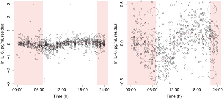
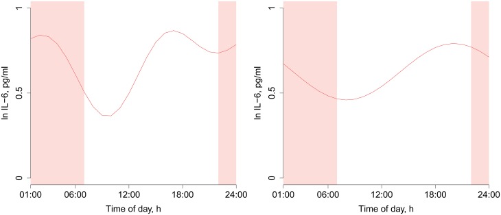
First, we fitted a null model including only time from catheterization and a random intercept for each dataset. Fig 3 shows residuals after these effects have been accounted for, suggesting that there remains variation to explain. The distribution of these residuals suggests a morning trough in IL-6 levels (Fig 3). Next, we compared a model with a 24 h cosinor function to the null model. The 24 h cosinor model fit better (log likelihood -483.5 vs -528.8, p < 0.0001, Fig 4). We then added another cosinor function with 12 h period. We did this for two reasons. The first reason was that addition of shorter periods allows a better estimation of non-sinusoidal effects, albeit at a cost of higher risk of overfitting. The second reason was that 12 h periods have been proposed by earlier investigators [24], and we considered that these claims should be tested. The model with both 24 h and 12 h cosinor functions fit better than the model with only the 24 h period (log likelihood -460.1 vs -483.5, p < 0.0001, Fig 4). Next, we exploratively investigated the addition of yet another cosinor with a 6 h period, but that did not improve model fit (log likelihood -457.7 vs -460.1, p = 0.09, prediction not shown).
Fig 3. Residuals from null model.
Data points sized by regression weight. In the null model, a random intercept for each study and a linear effect of time from catheterization have been included. Therefore, these residuals show the putative diurnal variation to be modeled. To explore this variation, we fitted a weighted LOESS curve (red line). This curve shows a trough in the morning. Note that the shape of the LOESS curve depends on the smoothing parameter. It is therefore possible to generate different LOESS curves from the same data, and not all of them show a peak in the early afternoon. The LOESS curve was fitted on three repeated days of the same data, and the curve for the second day shown, to ensure that the estimates would meet at 00:00 and 24:00. Time from 22:00 to 07:00 is shaded to indicate the night. Left: All data points shown. Right: Y axis range restricted to increase resolution.
Fig 4. Predicted diurnal time courses from meta-regression models.
Left: Best-fitting model including cosinor functions with 24 and 12 h periods. Right: Model including only 24 h period. Time from 22:00 to 07:00 is shaded to indicate the night.
Attempting to disentangle diurnal variation from effects of sleep, we investigated the addition of model effects for sleep (asleep/awake), time asleep (hours since sleep onset), and time awake (hours since wake onset). Starting with the best-fitting model including 24 and 12 h periods, we found that the addition of sleep, time asleep and time awake, or all three variables, did not improve model fit (log likelihoods -459.9, -459.2, and -459.0, respectively, vs -460.1, with p values 0.80, 0.36, and 0.36). Finally, we investigated the addition of sleep, time asleep and time awake, and all three variables, to the model with 24 h period. When comparing these models to the best-fitting model with 24 and 12 h periods, we saw worse fit (log likelihoods -475.5, -471.5, and -471.5, vs -460.1, with p values ≤ 0.0001). When comparing to the model with 24 h period only, we found that the addition of sleep, time asleep and time awake, or all three variables, yielded better-fitting models (log likelihoods -475.5, -471.5, and -471.5, vs -483.5, p values 0.0003, < 0.0001, and < 0.0001).
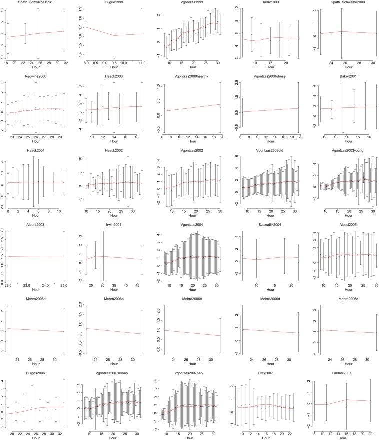
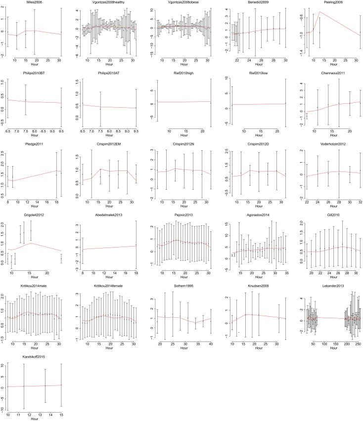
The best-fitting model with 24 and 12 h periods had a conspicuous trough between 09:00 and 10:00 in the morning and a second less pronounced trough close to 22:00, and two peaks located close to 17:00 and 02:00 (Fig 4). Since diurnal rhythms are commonly investigated using cosinor functions with 24 h periods, we show predictions from this simpler model too (Fig 4). This model estimated bathyphase (lowest point) at 08:05 and acrophase (highest point) at 20:05, with an amplitude of 0.166. All the included datasets, with predictions from the best-fitting model, are shown in Figs 5 and 6. Individual participant data are shown in Fig 7, for those four studies from which individual participant data were available.
Fig 5. Data and fitted time courses, showing the first 30 out of 56 datasets.
Data are shown as estimated from original publications, with error bars showing standard deviations. Y axes show ln IL-6 (pg/ml) throughout. Hours are in chronological time where 1 is 01:00 on the first day. Red lines show predictions from the best-fitting model.
Fig 6. Data and fitted time courses, showing the last 26 out of 56 datasets.
Data are shown as estimated from original publications, with error bars showing standard deviations. Y axes show ln IL-6 (pg/ml) throughout. Hours are in chronological time where 1 is 01:00 on the first day. Red lines show predictions from the best-fitting model.
Fig 7. Individual participant data.
Individual participant data were available from four datasets and are shown here mainly for the purpose of illustrating the high degree of variability within individuals. To illustrate summary effects within each dataset, thick lines show loess functions fitted to each dataset. Time from 22:00 to 07:00 is shaded to indicate the night. For Lekander 2013, two sets of measurements, made with a few days’ interval, have been plotted over the same time course, in red and blue respectively.
Assessment of risk of bias
Our literature review identified 36 eligible studies which could not be included. Not counting participants with duplicate data, these studies reported 468 participants, compared to the 1100 included in our meta-analysis. Of the ineligible studies, seven, most of which were published in the 1990:s, were unable to find reliable data because most values were close to or below assay detections limits (Table 2). From the point of view of bias, this is not a major problem since assay detection limits were unrelated to time of day for sampling. More concerning are data that were not reported because no effect was found. Only one paper explicitly stated that data were not shown for this reason [25]. Additionally, an unknown number of studies with eligible data have never been published. It is likely that unreported studies were more likely not to have found significant effects. However, with regard to estimating diurnal phase, we suspect that earlier investigators have been happy to report effects regardless of the location of peaks and troughs, reflected in the wide variety of published estimates (Fig 1). Therefore, even though the first reports of diurnal effects reported peaks in the night, we suspect that the studies included here were not strongly biased towards reporting effects at any particular time of day. Furthermore, to the extent that earlier reports may have been influenced by a prevailing theory, the effect most often referred to is a night-time peak. Since our meta-analysis did not find a night-time peak, we think it is unlikely that a bias in favor of this particular effect will have had a major influence on our model estimates.
Included datasets comprise both data from studies that were designed to measure diurnal or circadian variation, and data from studies that incidentally happened to fulfil our inclusion criteria. The latter group generally had fewer time points for measurements. Since this meta-analysis uses mixed-effects meta-regression by time of day, and is hence not based on single summary measures of included studies, it is not possible to investigate heterogeneity and bias by usual means such as a funnel plot. The results did not strongly depend on any single study. The most influential study was Mehra et al. [26], with a total regression weight of 11.19% over five different datasets.
Since this analysis concerns an observational relationship, confounding from a variety of sources cannot be ruled out. Light exposure and photoperiod were not controlled nor recorded except in laboratory studies, and season or time of the year were not reported frequently enough to justify inclusion in coding. Similarly, physical activity can affect levels of IL-6, but any instructions to participants about physical activity, and measures of their behavior, were for the most part not reported in included studies. Our assumption that participants in datasets not specifying sleep times on average slept 23:00-07:00 would limit the ability to estimate effects of sleep, but that is beyond the scope of this paper, and as the sleep variable was not included in the best-fitting model, the assumption only indirectly affects the risk of bias in diurnal variation. Since the sleep variable made very little difference when added to the best-fitting model, it is unlikely that a different assumption would lead to a different result.
Based on the above considerations, we judge that the risk of bias due to selective publishing and data inclusion is probably moderate to low.
Discussion
Our meta-analysis confirmed that circulating IL-6 shows diurnal variation. The most marked effect was a morning trough. The best-fitting model included a 24 h and a 12 h cosinor component. The LOESS curve shown in Fig 3 suggests a relatively flat curve from the afternoon to the late night, and the better fit obtained by adding the 12 h component may reflect the rather steep change occurring between the morning and the afternoon. For this reason, we are reluctant to consider the best-fitting model as proof that there are two distinct peaks and/or troughs during the 24 h day.
Our analytical approach treats sleep as a confounder to be eliminated, in order to best estimate diurnal variation. The effect of sleep on circulating IL-6 is an interesting question in its own right, but is better addressed by experimental sleep deprivation studies, which were not investigated here. A recent meta-analysis of the effect of sleep deprivation on IL-6 included 12 studies and found no significant effect [27].
One previous meta-analysis [9] (reported again in [10] and [11]) has investigated diurnal variation of circulating IL-6. As discussed in the introduction, this earlier meta-analysis aimed primarily to describe diurnal variation in patients with rheumatoid arthritis, and no systematic method to find data from healthy participants was described. Compared to this earlier meta-analysis, we have used a more systematic approach and we include more data (k = 56 datasets compared to k = 11). The present findings contrast markedly with those of the earlier meta-analysis, as that study located a morning peak at approx. 06:00 in healthy controls (Fig 1) as well as in patients with rheumatoid arthritis, while we report a morning trough at about 08:00-09:00 in healthy humans. This raises the hypothesis that diurnal variation is different between healthy humans and patients with rheumatoid arthritis, possibly correlating in patients to the diurnal time course of symptoms such as joint stiffness and pain, which tend to be worse in the morning.
The shape of the diurnal curve estimated in this meta-analysis is not suggestive of a mechanism where the immune system secretes more IL-6 into the blood at night in order to promote sleepiness. While the present results do not disprove this putative mechanism, the lack of a night-time peak, compared to the afternoon, suggests that other regulatory mechanisms are dominant.
The estimated morning trough is rather close after the time of day when cortisol levels peak, and also close to the diurnal trough of monocyte and lymphocyte concentrations in peripheral blood [28–30]. The data investigated here cannot speak directly to the relationship between diurnal variation of IL-6 to cortisol and white blood cell concentrations, and further studies will be required to elucidate whether any direct links exist.
The diurnal variation estimated here is large enough to pose a risk of confounding if sampling is performed without regard to time of day, and we therefore recommend that time of day should be taken in to consideration in studies recording IL-6 in plasma or serum from healthy humans. As far as we are aware, IL-6 is the the only cytokine to date to be subject to a meta-analysis of diurnal variation. Further research is required to determine conclusively whether other cytokines also show diurnal variation.
Supporting Information
(DOC)
Data Availability
All data and the full analysis code are freely available at: https://github.com/GNilsonne/IL6_diurnal.
Funding Statement
The authors received no specific funding for this work.
References
- 1. Borbély AA. Processes underlying sleep regulation. Hormone Research. 1998;49(3-4):114–117. 10.1159/000023156 [DOI] [PubMed] [Google Scholar]
- 2. Irwin M. Effects of sleep and sleep loss on immunity and cytokines. Brain, behavior, and immunity. 2002;16(5):503–512. 10.1016/S0889-1591(02)00003-X [DOI] [PubMed] [Google Scholar]
- 3. Bryant PA, Trinder J, Curtis N. Sick and tired: Does sleep have a vital role in the immune system? Nature reviews Immunology. 2004;4(6):457–467. 10.1038/nri1369 [DOI] [PubMed] [Google Scholar]
- 4. Vgontzas AN, Bixler EO, Lin HM, Prolo P, Trakada G, Chrousos GP. IL-6 and its circadian secretion in humans. Neuroimmunomodulation. 2005;12(3):131–140. 10.1159/000084844 [DOI] [PubMed] [Google Scholar]
- 5. Rohleder N, Aringer M, Boentert M. Role of interleukin-6 in stress, sleep, and fatigue. Annals of the New York Academy of Sciences. 2012;1261:88–96. 10.1111/j.1749-6632.2012.06634.x [DOI] [PubMed] [Google Scholar]
- 6. Gamaldo CE, Shaikh AK, McArthur JC. The sleep-immunity relationship. Neurologic clinics. 2012;30(4):1313–1343. 10.1016/j.ncl.2012.08.007 [DOI] [PubMed] [Google Scholar]
- 7. Gudewill S, Pollmächer T, Vedder H, Schreiber W, Fassbender K, Holsboer F. Nocturnal plasma levels of cytokines in healthy men. European Archives of Psychiatry and Clinical Neuroscience. 1992;242(1):53–56. 10.1007/BF02190343 [DOI] [PubMed] [Google Scholar]
- 8. Bauer J, Hohagen F, Ebert T, Timmer J, Ganter U, Krieger S, et al. Interleukin-6 serum levels in healthy persons correspond to the sleep-wake cycle. The clinical investigator. 1994;72(4):315–315. 10.1007/BF00180048 [DOI] [PubMed] [Google Scholar]
- 9. Straub RH, Cutolo M. Circadian rhythms in rheumatoid arthritis: Implications for pathophysiology and therapeutic management. Arthritis & Rheumatism. 2007;56(2):399–408. 10.1002/art.22368 [DOI] [PubMed] [Google Scholar]
- 10. Cutolo M, Straub RH, Buttgereit F. Circadian rhythms of nocturnal hormones in rheumatoid arthritis: translation from bench to bedside. Annals of the Rheumatic Diseases. 2008;67(7):905–908. 10.1136/ard.2008.088955 [DOI] [PubMed] [Google Scholar]
- 11. Cutolo M, Straub RH. Circadian rhythms in arthritis: hormonal effects on the immune/inflammatory reaction. Autoimmunity Reviews. 2008;7(3):223–228. 10.1016/j.autrev.2007.11.019 [DOI] [PubMed] [Google Scholar]
- 12. Gudmundsson A, Ershler WB, Goodman B, Lent SJ, Barczi S, Carnes M. Serum Concentrations of Interleukin-6 Are Increased When Sampled Through an Indwelling Venous Catheter. Clinical Chemistry. 1997;43(11):2199–2201. [PubMed] [Google Scholar]
- 13. Born J, Lange T, Hansen K, Mölle M, Fehm HL. Effects of sleep and circadian rhythm on human circulating immune cells. The Journal of Immunology. 1997;158(9):4454–4464. [PubMed] [Google Scholar]
- 14. Domínguez Rodríguez A, Abreu González P, García MJ, de la Rosa A, Vargas M, Marrero F. [Circadian variations in proinflammatory cytokine concentrations in acute myocardial infarction]. Revista española de cardiología. 2003;56(6):555–560. [DOI] [PubMed] [Google Scholar]
- 15. Dominguez-Rodriguez A, Abreu-Gonzalez P, Garcia M, Ferrer J, de la Rosa A, Vargas M, et al. Light/dark patterns of interleukin-6 in relation to the pineal hormone melatonin in patients with acute myocardial infarction. Cytokine. 2004;26(2):89–93. 10.1016/j.cyto.2004.01.003 [DOI] [PubMed] [Google Scholar]
- 16. Higgins JPT, White IR, Anzures-Cabrera J. Meta-analysis of skewed data: Combining results reported on log-transformed or raw scales. Statistics in Medicine. 2008;27(29):6072–6092. 10.1002/sim.3427 [DOI] [PMC free article] [PubMed] [Google Scholar]
- 17. Sothern RB, Roitman-Johnson B, Kanabrocki EL, Yager JG, Fuerstenberg RK, Weatherbee JA, et al. Circadian characteristics of interleukin-6 in blood and urine of clinically healthy men. In vivo (Athens, Greece). 1995;9(4):331–339. [PubMed] [Google Scholar]
- 18. Karshikoff B, Lekander M, Soop A, Lindstedt F, Ingvar M, Kosek E, et al. Modality and sex differences in pain sensitivity during human endotoxemia. Brain, Behavior, and Immunity. 2015;46:35–43. 10.1016/j.bbi.2014.11.014 [DOI] [PubMed] [Google Scholar]
- 19. Nilsonne G, Ingre M. IL6_diurnal: Relese for publication. Zenodo. [Google Scholar]
- 20. Seiler W, Müller H, Hiemke C. Interleukin-6 in plasma collected with an indwelling cannula reflects local, not systemic, concentrations. Clinical Chemistry. 1994;40(9):1778–1779. [PubMed] [Google Scholar]
- 21. Haack M, Kraus T, Schuld A, Dalal M, Koethe D, Pollmächer T. Diurnal variations of interleukin-6 plasma levels are confounded by blood drawing procedures. Psychoneuroendocrinology. 2002;27(8):921–931. 10.1016/S0306-4530(02)00006-9 [DOI] [PubMed] [Google Scholar]
- 22. R Core Team. R: A Language and Environment for Statistical Computing. Vienna, Austria: R Foundation for Statistical Computing; 2015. Available from: http://www.R-project.org/. [Google Scholar]
- 23.Pinheiro J, Bates D, DebRoy S, Sarkar D, R Core Team. nlme: Linear and Nonlinear Mixed Effects Models; 2015. Available from: http://CRAN.R-project.org/package=nlme.
- 24. Vgontzas AN, Papanicolaou DA, Bixler EO, Lotsikas A, Zachman K, Kales A, et al. Circadian interleukin-6 secretion and quantity and depth of sleep. The Journal of clinical endocrinology and metabolism. 1999;84(8):2603–2607. 10.1210/jcem.84.8.5894 [DOI] [PubMed] [Google Scholar]
- 25. Dinges DF, Douglas SD, Zaugg L, Campbell DE, McMann JM, Whitehouse WG, et al. Leukocytosis and natural killer cell function parallel neurobehavioral fatigue induced by 64 hours of sleep deprivation. Journal of Clinical Investigation. 1994;93(5):1930–1939. 10.1172/JCI117184 [DOI] [PMC free article] [PubMed] [Google Scholar]
- 26. Mehra R, Storfer-Isser A, Kirchner H, et al. Soluble interleukin 6 receptor: A novel marker of moderate to severe sleep-related breathing disorder. Archives of Internal Medicine. 2006;166(16):1725–1731. 10.1001/archinte.166.16.1725 [DOI] [PubMed] [Google Scholar]
- 27. Irwin MR, Olmstead R, Carroll JE. Sleep Disturbance, Sleep Duration, and Inflammation: A Systematic Review and Meta-Analysis of Cohort Studies and Experimental Sleep Deprivation. Biological Psychiatry. 10.1016/j.biopsych.2015.05.014 [DOI] [PMC free article] [PubMed] [Google Scholar]
- 28. Lasselin J, Rehman J, Åkerstedt T, Lekander A, Axelsson J. Effect of long-term sleep restriction and subsequent recovery sleep on the diurnal rhythms of white blood cell subpopulations. Brain, Behavior, and Immunity. 10.1016/j.bbi.2014.10.004 [DOI] [PubMed] [Google Scholar]
- 29. Sennels HP, Jørgensen HL, Hansen AS, Goetze JP, Fahrenkrug A. Diurnal variation of hematology parameters in healthy young males: The Bispebjerg study of diurnal variations Scandinavian Journal of Clinical and Laboratory Investigation. 10.3109/00365513.2011.602422 [DOI] [PubMed] [Google Scholar]
- 30. Ackermann K, Revell VL, Lao O, Rombouts EJ, Skene DJ, Kayser M. Diurnal Rhythms in Blood Cell Populations and the Effect of Acute Sleep Deprivation in Healthy Young Men Sleep. 10.5665/sleep.1954 [DOI] [PMC free article] [PubMed] [Google Scholar]
- 31. Kanabrocki EL, Sothern RB, Messmore HL, Roitman-Johnson B, McCormick JB, Dawson S, et al. Circadian interrelationships among levels of plasma fibrinogen, blood platelets, and serum interleukin-6. Clinical and applied thrombosis/hemostasis: official journal of the International Academy of Clinical and Applied Thrombosis/Hemostasis. 1999;5(1):37–42. [DOI] [PubMed] [Google Scholar]
- 32. Sothern RB, Roitman-Johnson B, Kanabrocki EL, Yager JG, Roodell MM, Weatherbee JA, et al. Circadian characteristics of circulating interleukin-6 in men. Journal of Allergy and Clinical Immunology. 1995;95(5):1029–1035. 10.1016/S0091-6749(95)70104-4 [DOI] [PubMed] [Google Scholar]
- 33. Alesci S, Martinez PE, Kelkar S, Ilias I, Ronsaville DS, Listwak SJ, et al. Major depression is associated with significant diurnal elevations in plasma interleukin-6 levels, a shift of its circadian rhythm, and loss of physiological complexity in its secretion: clinical implications. The Journal of clinical endocrinology and metabolism. 2005;90(5):2522–2530. 10.1210/jc.2004-1667 [DOI] [PubMed] [Google Scholar]
- 34. Vgontzas AN, Zoumakis M, Bixler EO, Lin HM, Prolo P, Vela-Bueno A, et al. Impaired nighttime sleep in healthy old versus young adults is associated with elevated plasma interleukin-6 and cortisol levels: physiologic and therapeutic implications. The Journal of clinical endocrinology and metabolism. 2003;88(5):2087–2095. 10.1210/jc.2002-021176 [DOI] [PubMed] [Google Scholar]
- 35. Vgontzas AN, Zoumakis M, Papanicolaou DA, Bixler EO, Prolo P, Lin HM, et al. Chronic insomnia is associated with a shift of interleukin-6 and tumor necrosis factor secretion from nighttime to daytime. Metabolism. 2002;51(7):887–892. 10.1053/meta.2002.33357 [DOI] [PubMed] [Google Scholar]
- 36. Agorastos A, Hauger RL, Barkauskas DA, Moeller-Bertram T, Clopton PL, Haji U, et al. Circadian rhythmicity, variability and correlation of interleukin-6 levels in plasma and cerebrospinal fluid of healthy men. Psychoneuroendocrinology. 2014;44:71–82. 10.1016/j.psyneuen.2014.02.020 [DOI] [PubMed] [Google Scholar]
- 37. Undar L, Ertuğrul C, Altunbaş H, Akça S. Circadian variations in natural coagulation inhibitors protein C, protein S and antithrombin in healthy men: a possible association with interleukin-6. Thrombosis and haemostasis. 1999;81(4):571–575. [PubMed] [Google Scholar]
- 38. Burgos I, Richter L, Klein T, Fiebich B, Feige B, Lieb K, et al. Increased nocturnal interleukin-6 excretion in patients with primary insomnia: A pilot study. Brain, Behavior, and Immunity. 2006;20(3):246–253. 10.1016/j.bbi.2005.06.007 [DOI] [PubMed] [Google Scholar]
- 39.Knudsen LS, Christensen IJ, Lottenburger T, Svendsen MN, Nielsen HJ, Nielsen L, et al. type [; 2008]Available from: http://informahealthcare.com/doi/abs/10.1080/13547500701615017%20.
- 40. Miles MP, Andring JM, Pearson SD, Gordon LK, Kasper C, Depner CM, et al. Diurnal variation, response to eccentric exercise, and association of inflammatory mediators with muscle damage variables. Journal of Applied Physiology. 2008;104(2):451–458. 10.1152/japplphysiol.00572.2007 [DOI] [PubMed] [Google Scholar]
- 41. Späth-Schwalbe E, Hansen K, Schmidt F, Schrezenmeier H, Marshall L, Burger K, et al. Acute effects of recombinant human interleukin-6 on endocrine and central nervous sleep functions in healthy men. The Journal of clinical endocrinology and metabolism. 1998;83(5):1573–1579. 10.1210/jcem.83.5.4795 [DOI] [PubMed] [Google Scholar]
- 42. Dugué B, Leppänen E. Short-term variability in the concentration of serum interleukin-6 and its soluble receptor in subjectively healthy persons. Clinical chemistry and laboratory medicine: CCLM / FESCC. 1998;36(5):323–325. 10.1515/CCLM.1998.054 [DOI] [PubMed] [Google Scholar]
- 43. Späth-Schwalbe E, Lange T, Perras B, Lorenz Fehm H, Born J. Interferon-alpha acutely impairs sleep in healthy humans. Cytokine. 2000;12(5):518–521. 10.1006/cyto.1999.0587 [DOI] [PubMed] [Google Scholar]
- 44. Redwine L, Hauger RL, Gillin JC, Irwin M. Effects of sleep and sleep deprivation on interleukin-6, growth hormone, cortisol, and melatonin levels in humans. The Journal of clinical endocrinology and metabolism. 2000;85(10):3597–3603. [DOI] [PubMed] [Google Scholar]
- 45. Haack M, Reichenberg A, Kraus T, Schuld A, Yirmiya R, Pollmächer T. EFFECTS OF AN INTRAVENOUS CATHETER ON THE LOCAL PRODUCTION OF CYTOKINES AND SOLUBLE CYTOKINE RECEPTORS IN HEALTHY MEN. Cytokine. 2000;12(6):694–698. 10.1006/cyto.1999.0665 [DOI] [PubMed] [Google Scholar]
- 46. Vgontzas AN, Papanicolaou DA, Bixler EO, Hopper K, Lotsikas A, Lin HM, et al. Sleep apnea and daytime sleepiness and fatigue: relation to visceral obesity, insulin resistance, and hypercytokinemia. The Journal of clinical endocrinology and metabolism. 2000;85(3):1151–1158. 10.1210/jcem.85.3.6484 [DOI] [PubMed] [Google Scholar]
- 47. Baker DG, Ekhator NN, Kasckow JW, Hill KK, Zoumakis E, Dashevsky BA, et al. Plasma and cerebrospinal fluid interleukin-6 concentrations in posttraumatic stress disorder. Neuroimmunomodulation. 2001;9(4):209–217. 10.1159/000049028 [DOI] [PubMed] [Google Scholar]
- 48. Haack M, Schuld A, Kraus T, Pollmächer T. Effects of Sleep on Endotoxin-Induced Host Responses in Healthy Men. Psychosomatic Medicine. 2001;63(4):568–578. 10.1097/00006842-200107000-00008 [DOI] [PubMed] [Google Scholar]
- 49. Alberti A, Sarchielli P, Gallinella E, Floridi A, Floridi A, Mazzotta G, et al. Plasma cytokine levels in patients with obstructive sleep apnea syndrome: a preliminary study. Journal of Sleep Research. 2003;12(4):305–311. 10.1111/j.1365-2869.2003.00361.x [DOI] [PubMed] [Google Scholar]
- 50. Irwin M, Rinetti G, Redwine L, Motivala S, Dang J, Ehlers C. Nocturnal proinflammatory cytokine-associated sleep disturbances in abstinent African American alcoholics. Brain, Behavior, and Immunity. 2004;18(4):349–360. 10.1016/j.bbi.2004.02.001 [DOI] [PubMed] [Google Scholar]
- 51. Vgontzas AN, Zoumakis E, Bixler EO, Lin HM, Follett H, Kales A, et al. Adverse effects of modest sleep restriction on sleepiness, performance, and inflammatory cytokines. The Journal of clinical endocrinology and metabolism. 2004;89(5):2119–2126. 10.1210/jc.2003-031562 [DOI] [PubMed] [Google Scholar]
- 52. Szczudlik A, Dziedzic T, Bartus S, Slowik A, Kieltyka A. Serum interleukin-6 predicts cortisol release in acute stroke patients. Journal of endocrinological investigation. 2004;27(1):37–41. 10.1007/BF03350908 [DOI] [PubMed] [Google Scholar]
- 53. Vgontzas AN, Pejovic S, Zoumakis E, Lin HM, Bixler EO, Basta M, et al. Daytime napping after a night of sleep loss decreases sleepiness, improves performance, and causes beneficial changes in cortisol and interleukin-6 secretion. American Journal of Physiology—Endocrinology and Metabolism. 2007;292(1):E253–E261. 10.1152/ajpendo.00651.2005 [DOI] [PubMed] [Google Scholar]
- 54. Frey DJ, Fleshner M, Wright KP Jr. The effects of 40 hours of total sleep deprivation on inflammatory markers in healthy young adults. Brain, Behavior, and Immunity. 2007;21(8):1050–1057. 10.1016/j.bbi.2007.04.003 [DOI] [PubMed] [Google Scholar]
- 55. Lindahl MS, Olovsson M, Nyberg S, Thorsen K, Olsson T, Sundström Poromaa I. Increased cortisol responsivity to adrenocorticotropic hormone and low plasma levels of interleukin-1 receptor antagonist in women with functional hypothalamic amenorrhea. Fertility and Sterility. 2007;87(1):136–142. 10.1016/j.fertnstert.2006.06.029 [DOI] [PubMed] [Google Scholar]
- 56. Vgontzas AN, Zoumakis E, Bixler EO, Lin HM, Collins B, Basta M, et al. Selective effects of CPAP on sleep apnoea-associated manifestations. European Journal of Clinical Investigation. 2008;38(8):585–595. 10.1111/j.1365-2362.2008.01984.x [DOI] [PMC free article] [PubMed] [Google Scholar]
- 57. Benedict C, Scheller J, Rose-John S, Born J, Marshall L. Enhancing influence of intranasal interleukin-6 on slow-wave activity and memory consolidation during sleep. The FASEB Journal. 2009;23(10):3629–3636. 10.1096/fj.08-122853 [DOI] [PubMed] [Google Scholar]
- 58. Peeling P, Dawson B, Goodman C, Landers G, Wiegerinck ET, Swinkels DW, et al. Effects of exercise on hepcidin response and iron metabolism during recovery. International journal of sport nutrition and exercise metabolism. 2009;19(6):583–597. 10.1123/ijsnem.19.6.583 [DOI] [PubMed] [Google Scholar]
- 59. PHILLIPS MD, FLYNN MG, MCFARLIN BK, STEWART LK, TIMMERMAN KL. Resistance Training at Eight-Repetition Maximum Reduces the Inflammatory Milieu in Elderly Women. [Miscellaneous Article]. Medicine & Science in Sports & Exercise February 2010. 2010;42(2):314–325. 10.1249/MSS.0b013e3181b11ab7 [DOI] [PubMed] [Google Scholar]
- 60. Rief W, Mills PJ, Ancoli-Israel S, Ziegler MG, Pung MA, Dimsdale JE. Overnight changes of immune parameters and catecholamines are associated with mood and stress. Psychosomatic medicine. 2010;72(8):755–762. 10.1097/PSY.0b013e3181f367e2 [DOI] [PMC free article] [PubMed] [Google Scholar]
- 61. Gill J, Luckenbaugh D, Charney D, Vythilingam M. Sustained Elevation of Serum Interleukin-6 and Relative Insensitivity to Hydrocortisone Differentiates Posttraumatic Stress Disorder with and Without Depression. Biological Psychiatry. 2010;68(11):999–1006. 10.1016/j.biopsych.2010.07.033 [DOI] [PubMed] [Google Scholar]
- 62. Chennaoui M, Sauvet F, Drogou C, Van Beers P, Langrume C, Guillard M, et al. Effect of one night of sleep loss on changes in tumor necrosis factor alpha (TNF-alpha) levels in healthy men. Cytokine. 2011;56(2):318–324. 10.1016/j.cyto.2011.06.002 [DOI] [PubMed] [Google Scholar]
- 63. Pledge D, Grosset JF, Onambélé-Pearson GL. Is there a morning-to-evening difference in the acute IL-6 and cortisol responses to resistance exercise? Cytokine. 2011;55(2):318–323. 10.1016/j.cyto.2011.05.005 [DOI] [PubMed] [Google Scholar]
- 64. Crispim CA, Padilha HG, Zimberg IZ, Waterhouse J, Dattilo M, Tufik S, et al. Adipokine Levels Are Altered by Shiftwork: A Preliminary Study. Chronobiology International. 2012;29(5):587–594. 10.3109/07420528.2012.675847 [DOI] [PubMed] [Google Scholar]
- 65. Voderholzer U, Fiebich BL, Dersch R, Feige B, Piosczyk H, Kopasz M, et al. Effects of Sleep Deprivation on Nocturnal Cytokine Concentrations in Depressed Patients and Healthy Control Subjects. The Journal of Neuropsychiatry and Clinical Neurosciences. 2012;24(3):354–366. 10.1176/appi.neuropsych.11060142 [DOI] [PubMed] [Google Scholar]
- 66. Grigoleit JS, Kullmann JS, Winkelhaus A, Engler H, Wegner A, Hammes F, et al. Single-trial conditioning in a human taste-endotoxin paradigm induces conditioned odor aversion but not cytokine responses. Brain, Behavior, and Immunity. 2012;26(2):234–238. 10.1016/j.bbi.2011.09.001 [DOI] [PubMed] [Google Scholar]
- 67. Abedelmalek S, Chtourou H, Aloui A, Aouichaoui C, Souissi N, Tabka Z. Effect of time of day and partial sleep deprivation on plasma concentrations of IL-6 during a short-term maximal performance. European Journal of Applied Physiology. 2013;113(1):241–248. 10.1007/s00421-012-2432-7 [DOI] [PubMed] [Google Scholar]
- 68. Pejovic S, Basta M, Vgontzas AN, Kritikou I, Shaffer ML, Tsaoussoglou M, et al. Effects of recovery sleep after one work week of mild sleep restriction on interleukin-6 and cortisol secretion and daytime sleepiness and performance. American journal of physiology Endocrinology and metabolism. 2013;305(7):E890–896. 10.1152/ajpendo.00301.2013 [DOI] [PMC free article] [PubMed] [Google Scholar]
- 69. Lekander M, Andreasson AN, Kecklund G, Ekman R, Ingre M, Akerstedt T, et al. Subjective health perception in healthy young men changes in response to experimentally restricted sleep and subsequent recovery sleep. Brain, Behavior, and Immunity. 2013;34:43–46. 10.1016/j.bbi.2013.06.005 [DOI] [PubMed] [Google Scholar]
- 70. Kritikou I, Basta M, Vgontzas AN, Pejovic S, Liao D, Tsaoussoglou M, et al. Sleep apnoea, sleepiness, inflammation and insulin resistance in middle-aged males and females. European Respiratory Journal. 2014;43(1):145–155. 10.1183/09031936.00126712 [DOI] [PubMed] [Google Scholar]
- 71.Nilsonne G. Endotoxin_RestingState_2015: Release for publication; 2015. Available from: 10.5281/zenodo.35770. [DOI]
- 72. Lemmer B, Schwuléra U, Thrun A, Lissner R. Circadian rhythm of soluble interleukin-2 receptor in healthy individuals. European cytokine network. 1992;3(3):335–336. [PubMed] [Google Scholar]
- 73. Pollmächer T, Schreiber W, Gudewill S, Vedder H, Fassbender K, Wiedemann K, et al. Influence of endotoxin on nocturnal sleep in humans. The American journal of physiology. 1993;264(6 Pt 2):R1077–1083. [DOI] [PubMed] [Google Scholar]
- 74. Arvidson NG, Gudbjörnsson B, Elfman L, Rydén AC, Tötterman TH, Hällgren R. Circadian rhythm of serum interleukin-6 in rheumatoid arthritis. Annals of the Rheumatic Diseases. 1994;53(8):521–524. 10.1136/ard.53.8.521 [DOI] [PMC free article] [PubMed] [Google Scholar]
- 75. Seiler W, Müller H, Hiemke C. Diurnal variations of plasma interleukin-6 in man: methodological implications of continuous use of indwelling cannulae. Annals of the New York Academy of Sciences. 1995;762:468–470. 10.1111/j.1749-6632.1995.tb32370.x [DOI] [PubMed] [Google Scholar]
- 76. Pollmächer T, Mullington J, Korth C, Schreiber W, Hermann D, Orth A, et al. Diurnal Variations in the Human Host Response to Endotoxin. Journal of Infectious Diseases. 1996;174(5):1040–1045. 10.1093/infdis/174.5.1040 [DOI] [PubMed] [Google Scholar]
- 77. Korth C, Mullington J, Schreiber W, Pollmächer T. Influence of endotoxin on daytime sleep in humans. Infection and Immunity. 1996;64(4):1110 [DOI] [PMC free article] [PubMed] [Google Scholar]
- 78. Crofford LJ, Kalogeras KT, Mastorakos G, Magiakou MA, Wells J, Kanik KS, et al. Circadian relationships between interleukin (IL)-6 and hypothalamic-pituitary-adrenal axis hormones: failure of IL-6 to cause sustained hypercortisolism in patients with early untreated rheumatoid arthritis. The Journal of clinical endocrinology and metabolism. 1997;82(4):1279–1283. 10.1210/jcem.82.4.3852 [DOI] [PubMed] [Google Scholar]
- 79. Lissoni P, Rovelli F, Brivio F, Brivio O, Fumagalli L. Circadian secretions of IL-2, IL-12, IL-6 and IL-10 in relation to the light/dark rhythm of the pineal hormone melatonin in healthy humans. Natural immunity. 1998;16(1):1–5. 10.1159/000069464 [DOI] [PubMed] [Google Scholar]
- 80. Bornstein SR, Licinio J, Tauchnitz R, Engelmann L, Negrão AB, Gold P, et al. Plasma leptin levels are increased in survivors of acute sepsis: associated loss of diurnal rhythm, in cortisol and leptin secretion. The Journal of clinical endocrinology and metabolism. 1998;83(1):280–283. 10.1210/jcem.83.1.4610 [DOI] [PubMed] [Google Scholar]
- 81. Hermann DM, Mullington J, Hinze-Selch D, Schreiber W, Galanos C, Pollmächer T. Endotoxin-induced changes in sleep and sleepiness during the day. Psychoneuroendocrinology. 1998;23(5):427–437. 10.1016/S0306-4530(98)00030-4 [DOI] [PubMed] [Google Scholar]
- 82. Genesca J, Segura R, Gonzalez A, Catalan R, Marti R, Torregrosa M, et al. Nitric oxide may contribute to nocturnal hemodynamic changes in cirrhotic patients. The American Journal of Gastroenterology. 2000;95(6):1539–1544. 10.1111/j.1572-0241.2000.02092.x [DOI] [PubMed] [Google Scholar]
- 83. Mastorakos G, Ilias I. Relationship between interleukin-6 (IL-6) and hypothalamic-pituitary-adrenal axis hormones in rheumatoid arthritis. Zeitschrift für Rheumatologie. 2000;59 Suppl 2:II/75–79. 10.1007/s003930070023 [DOI] [PubMed] [Google Scholar]
- 84. Mullington J, Korth C, Hermann DM, Orth A, Galanos C, Holsboer F, et al. Dose-dependent effects of endotoxin on human sleep. American Journal of Physiology—Regulatory, Integrative and Comparative Physiology. 2000;278(4):R947–R955. [DOI] [PubMed] [Google Scholar]
- 85. Johansson A, Carlström K, Ahrén B, Cederquist K, Krylborg E, Forsberg H, et al. Abnormal cytokine and adrenocortical hormone regulation in myotonic dystrophy. The Journal of clinical endocrinology and metabolism. 2000;85(9):3169–3176. 10.1210/jcem.85.9.6794 [DOI] [PubMed] [Google Scholar]
- 86. Shaw JA, Chin-Dusting JPF, Kingwell BA, Dart AM. Diurnal Variation in Endothelium-Dependent Vasodilatation Is Not Apparent in Coronary Artery Disease. Circulation. 2001;103(6):806–812. 10.1161/01.CIR.103.6.806 [DOI] [PubMed] [Google Scholar]
- 87. Lange T, Marshall L, Späth-Schwalbe E, Fehm HL, Born J. Systemic immune parameters and sleep after ultra-low dose administration of IL-2 in healthy men. Brain, Behavior, and Immunity. 2002;16(6):663–674. 10.1016/S0889-1591(02)00018-1 [DOI] [PubMed] [Google Scholar]
- 88. Haack M, Sanchez E, Mullington JM. Elevated inflammatory markers in response to prolonged sleep restriction are associated with increased pain experience in healthy volunteers. Sleep. 2007;30(9):1145–1152. [DOI] [PMC free article] [PubMed] [Google Scholar]
- 89. Eisenberger NI, Inagaki TK, Rameson LT, Mashal NM, Irwin MR. An fMRI Study of Cytokine-Induced Depressed Mood and Social Pain: The Role of Sex Differences. NeuroImage. 2009;47(3):881–890. 10.1016/j.neuroimage.2009.04.040 [DOI] [PMC free article] [PubMed] [Google Scholar]
- 90. Eisenberger NI, Inagaki TK, Mashal NM, Irwin MR. Inflammation and social experience: an inflammatory challenge induces feelings of social disconnection in addition to depressed mood. Brain, Behavior, and Immunity. 2010;24(4):558–563. 10.1016/j.bbi.2009.12.009 [DOI] [PMC free article] [PubMed] [Google Scholar]
- 91. Haimovich B, Calvano J, Haimovich A, Calvano SE, Coyle SMM, Lowry SF. In vivo endotoxin synchronizes and suppresses clock gene expression in human peripheral blood leukocytes *. Critical Care Medicine March 2010. 2010;38(3):751–758. 10.1097/CCM.0b013e3181cd131c [DOI] [PMC free article] [PubMed] [Google Scholar]
- 92. Sauvet F, Leftheriotis G, Gomez-Merino D, Langrume C, Drogou C, Van Beers P, et al. Effect of acute sleep deprivation on vascular function in healthy subjects. Journal of applied physiology (Bethesda, Md: 1985). 2010;108(1):68–75. 10.1152/japplphysiol.00851.2009 [DOI] [PubMed] [Google Scholar]
- 93. Grigoleit JS, Kullmann JS, Wolf OT, Hammes F, Wegner A, Jablonowski S, et al. Dose-Dependent Effects of Endotoxin on Neurobehavioral Functions in Humans. PLoS ONE. 2011;6(12):e28330 10.1371/journal.pone.0028330 [DOI] [PMC free article] [PubMed] [Google Scholar]
- 94. MILES M, KELLER J, KORDICK L, KIDD J. Basal, Circadian, and Acute Inflammation in Normal versus Overweight Men. [Miscellaneous Article]. Medicine & Science in Sports & Exercise December 2012. 2012;44(12):2290–2298. 10.1249/MSS.0b013e318267b209 [DOI] [PubMed] [Google Scholar]
- 95. Schrepf A, O’Donnell M, Luo Y, Bradley CS, Kreder K, Lutgendorf S. Inflammation and inflammatory control in interstitial cystitis/bladder pain syndrome: Associations with painful symptoms. PAIN. [DOI] [PMC free article] [PubMed] [Google Scholar]
- 96. Wegner A, Elsenbruch S, Maluck J, Grigoleit JS, Engler H, Jäger M, et al. Inflammation-induced hyperalgesia: Effects of timing, dosage, and negative affect on somatic pain sensitivity in human experimental endotoxemia. Brain, Behavior, and Immunity. [DOI] [PubMed] [Google Scholar]
- 97. Scott HA, Latham JR, Callister R, Pretto JJ, Baines K, Saltos N, et al. Acute exercise is associated with reduced exhaled nitric oxide in physically inactive adults with asthma. Annals of Allergy, Asthma & Immunology. 2015;114(6):470–479. 10.1016/j.anai.2015.04.002 [DOI] [PubMed] [Google Scholar]
Associated Data
This section collects any data citations, data availability statements, or supplementary materials included in this article.
Supplementary Materials
(DOC)
Data Availability Statement
All data and the full analysis code are freely available at: https://github.com/GNilsonne/IL6_diurnal.


