Abstract
Left–right (L-R) asymmetry of the internal organs of vertebrates is presaged by domains of asymmetric gene expression in the lateral plate mesoderm (LPM) during somitogenesis. Ciliated L-R coordinators (LRCs) are critical for biasing the initiation of asymmetrically expressed genes, such as nodal and pitx2, to the left LPM. Other midline structures, including the notochord and floorplate, are then required to maintain these asymmetries. Here we report an unexpected role for the zebrafish EGF-CFC gene one-eyed pinhead (oep) in the midline to promote pitx2 expression in the LPM. Late zygotic oep (LZoep) mutants have strongly reduced or absent pitx2 expression in the LPM, but this expression can be rescued to strong levels by restoring oep in midline structures only. Furthermore, removing midline structures from LZoep embryos can rescue pitx2 expression in the LPM, suggesting the midline is a source of an LPM pitx2 repressor that is itself inhibited by oep. Reducing lefty1 activity in LZoep embryos mimics removal of the midline, implicating lefty1 in the midline-derived repression. Together, this suggests a model where Oep in the midline functions to overcome a midline-derived repressor, involving lefty1, to allow for the expression of left side-specific genes in the LPM.
This article is part of the themed issue ‘Provocative questions in left–right asymmetry’.
Keywords: Nodal, Lefty, one-eyed pinhead, EGF-CFC, asymmetry, zebrafish
1. Introduction
(a). Nodal signalling in left–right patterning
Left–right (L–R) patterning in the vertebrate embryo emerges as genes become asymmetrically expressed during somitogenesis. Genes encoding components of the Nodal signalling pathway, including the nodal ligand and the downstream target pitx2, are expressed in the left lateral plate mesoderm (LPM) but remain absent from the right side (reviewed in [1–5]). Nodal is a transforming growth factor-beta (TGF-β) superfamily member that signals through serine/threonine kinase receptors to phosphorylate Smad2 and Smad3 (reviewed in [6,7]). This requires EGF-CFC proteins, such as that encoded by the zebrafish gene one-eyed pinhead (oep), which act as co-receptors for Nodal ligands [8–11]. Smad2 and Smad3 then interact with the co-Smad, Smad4, as well as transcription factors, such as FoxHI, to induce transcription of downstream targets [12–18]. These targets include the asymmetrically expressed genes pitx2, nodal itself and the lefty repressors that restrict the extent of Nodal signalling. Owing to the feedback nature of the pathway, lefty expression denotes both areas where Nodal signalling has occurred and areas where Nodal signalling is now being repressed (reviewed in [6,7]). Lefty is thought to repress Nodal signals by physically associating with both Nodal itself as well as with EGF-CFC co-receptors including Oep [8,10,11].
Prior to the emergence of nodal expression in the left LPM, nodal is expressed at the midline around so-called left–right coordinators (LRCs). These are the ventral node in mouse embryos [19], the gastroceol roof plate in Xenopus laevis [20], and Kupffer's vesicle (KV) in zebrafish and Medaka fish [21–23]. Some evidence, including phenotypes of hypomorphic- or tissue-specific knock-out alleles of Nodal in the mouse, suggests that Nodal ligand emanating from this LRC domain is required for initiation of Nodal expression in the LPM [24–28]. In zebrafish, knockdown of the Nodal orthologue southpaw (spaw) results in a loss of spaw expression in the LPM, although transcription of spaw around the zebrafish LRC is unaffected [29]. Together, this suggests that LRC–Nodal activity induces expression of LPM–nodal. Subsequently, Nodal spreads throughout the left LPM and induces other asymmetrically expressed genes.
Further support for an essential role of Nodal signalling in inducing asymmetric expression in the LPM comes from the analysis of mutations in the EGF-CFC genes oep in zebrafish and Cryptic/Cfc1 in mouse. These genes are expressed bilaterally in the LPM, and in the embryonic midline and its precursors [30,31]. Cryptic mutants lack expression of Nodal, Lefty2, and Pitx2 in the LPM, and have organ laterality defects including right pulmonary isomerism and randomization of heart looping [32,33]. Similarly, zebrafish oep is required for L–R patterning. Maternal-zygotic oep (MZoep) mutants lack most mesendoderm [9]. This early requirement for oep in Nodal signalling is rescued by oep mRNA injection into 1-cell stage embryos, generating ‘late zygotic’ oep (LZoep) embryos [9,33]. These embryos are fully viable and develop to adulthood. All major structures and organs, ranging from midline and central nervous system to heart and pancreas, appear morphologically normal. However, as oep activity in later development is not provided by injected mRNA, LZoep mutants have L–R patterning defects [33]; they do not express nodal/cyclops or pitx2 in the LPM nor in an additional asymmetric expression domain in the dorsal diencephalon [33–35]. LZoep mutants also display a randomization of heart looping and pancreas positioning in the viscera, and of parapineal positioning in the brain [33,34]. The importance of EGF-CFC genes in L–R development also extends to humans, as CFC1 mutations are associated with human L–R patterning defects [36].
How Nodal signals are transferred from LRC to LPM is not fully understood. One possibility is that Nodal ligand directly travels from the lateral edges of the LRC to the LPM, a process that requires sulfated glycosaminoglycans (sGAGs; [27,37]). Moreover, Ca2+ signals are also observed around LRCs [38–40], and these signals can spread laterally towards the LPM via gap junctions [41,42]. Indeed, it has been hypothesized that Ca2+ signals aid in the transduction of Nodal ligand from LRC to LPM by inducing the secretion of sGAGs [41,43,44].
(b). Roles for midline structures in initiating and maintaining L–R asymmetries
Although the striking domain of asymmetric gene expression occurs in the LPM, various midline structures are critical for both initiating and maintaining these asymmetries. In vertebrates, a major model for how L–R asymmetries are initiated involves an asymmetric fluid flow within LRCs, driven by the rotation of polarized motile cilia [19,20,23,45–53]. In support of this model, a plethora of mutants that exhibit cilia motility abnormalities also display defects of L–R patterning (for just a few examples see [54–58]). How asymmetric fluid flow is sensed by the embryo to drive downstream asymmetries in gene expression around LRCs is not fully understood, but the process likely involves both sensory cilia and the polycystin transmembrane proteins Pkd1l1 and Pkd2 [39,59–67]. Downstream of fluid flow and Polycystin function, R > L asymmetries in Dand5 (also called charon in zebrafish and Medaka, Cerl2 in mouse and Coco in Xenopus), a member of the DAN family of Nodal inhibitors, emerge at the lateral edges of LRCs [28,68–75]. This results in more active Nodal emanating from the left side of LRCs and, presumably as a result, the activation of Nodal preferentially in the left LPM. As such, asymmetries in Dand5 are critical for establishing unilateral left-sided Nodal expression in various vertebrate embryos.
Once asymmetric gene expression is established in the LPM, midline structures are also critical for maintaining asymmetries. Initial analysis of zebrafish mutants, in which an intact notochord fails to form, revealed defects in asymmetric gene expression in the LPM with subsequent abnormalities in cardiac asymmetry [76–78]. Similar results were obtained upon extirpation of midline structures in Xenopus [76,79]. In embryos without intact midline structures, gene expression normally restricted to the left side becomes bilaterally expressed in the left and right LPM. Based on these results, the midline (predominantly notochord and floorplate) was proposed to act as a barrier in L–R patterning. In the barrier model, the midline would act to prevent Nodal activity within the left LPM from accessing the right LPM, which remains capable of expressing nodal. The barrier activities of the midline would be perturbed upon loss of midline structures, allowing Nodal signals from the left access to the right LPM, resulting in bilateral expression of left side-specific genes.
Bilateral expression of pitx2 and other genes in the LPM is also seen upon loss of lefty1 function in mouse and zebrafish [80,81]. lefty1 is expressed in the floorplate of mouse embryos and in the notochord in zebrafish [82–84] and has been proposed to act as the molecular component of the midline that restricts Nodal signalling to the left side. In mouse, Lefty1 is induced in the midline by Nodal signals from the left LPM, and may thus serve as a barrier to Nodal signals preventing their access to the right LPM [85]. Many results pertaining to the barrier/repressor function of the midline were synthesized into a self-enhancement and lateral-inhibition (SELI) model in the mouse embryo [86]. In this model, Nodal is induced in both the left and right LPM, though at a much lower level in the latter. Thus, the critical threshold for robust activation and spreading (the self-enhancement) is not reached in the right LPM as it is in the left. Left LPM Nodal induces Lefty in both the left LPM and midline and these repressive signals raise the threshold for Nodal enhancement in the right LPM (the lateral inhibition), thereby maintaining unilateral expression. In the absence of Lefty repressors, Nodal is more strongly induced in the right LPM and, moreover, Nodal signals might also spread from the left LPM to the right LPM, ultimately resulting in bilateral expression of Nodal signals even in the presence of correct initial symmetry breaking at the LRC.
In addition to the floorplate/notochord lefty barrier, two additional ‘midline barriers’ that also involve repression of Nodal signals have been described in zebrafish [80]. Zebrafish expression of Nodal/Spaw in the LPM induces its own expression, but in contrast with mouse, does not induce the expression of lefty in the LPM. Instead, lefty1 expression in the notochord is driven by Spaw as it propagates anteriorly within the LPM at these stages [87,88]). lefty2 is induced within the left side of the heart field once Spaw reaches the anterior left LPM [80]. Removal of lefty2 from embryos allows spaw expression from the left anterior LPM to proceed across the region anterior to the notochord, into the right LPM, where it proceeds towards the posterior. This demonstrates that lefty2 expression in the heart prevents spaw expression from spreading from the left to the right LPM. Similarly, bone morphogenetic protein (BMP) signalling is required to prevent Spaw in the left LPM from inducing spaw expression in a domain posterior to KV. In the absence of BMP signalling, spaw is ectopically expressed throughout the posterior LPM around the tailbud, and from there spreads to the right LPM. Thus, along with lefty1 expression in the notochord, these two ‘barriers’ confine nodal/spaw expression to the left LPM from an area lateral to KV to an area lateral to the anterior notochord in zebrafish [80].
Current models suggest that the midline is essentially passive, responding to Nodal signals from the LPM by expressing lefty1 and then preventing Nodal signals from activating in the right LPM. However, lefty1 expression in the posterior notochord in zebrafish begins prior to the expression of nodal in the LPM, demonstrating that this domain of lefty1, whose function is currently unknown, cannot be initiated by Nodal signals from the LPM. Based on the results reported here, we propose that this early domain of lefty1 contributes to establishing the threshold required for Nodal signals to initiate and propagate in the LPM. We demonstrate an active role for the Nodal co-receptor Oep in repressing early Lefty activity in the midline, prior to the emergence of asymmetric gene expression, to allow for expression of the Nodal target pitx2 in the LPM.
2. Material and methods
(a). Zebrafish
Danio rerio strains were raised under standard conditions at 28°C. We used the PWT strain for experiments as this strain was found to have a very low level of abnormal asymmetric gene expression [60]. Transplanted and injected embryos were raised at 28°C until shield stage and then placed at 25°C overnight. The next morning, embryos were returned to 28°C until they reached 18–20 somites, when they were fixed for in situ analysis. All embryos were staged according to [89]. The oeptz257 mutant was used in this study [90–93].
(b). RNA and morpholino injections
Capped mRNA transcripts were synthesized using Ambion mMessage mMachine™ kits and quantitated by UV spectrophotometry. oep mRNA was transcribed from plasmid pJZoepFlag1–2 linearized with SmaI [31]. Morpholino antisense oligos were obtained from GeneTools, LLC and resuspended to a stock concentration of 50 µg µl−1 in dH2O. The ntla morpholino sequence we used in these studies is described in [94]. The lefty1 morpholino used is described in [95]. RNA and morpholinos were diluted in 5 mg ml−1 phenol red dissolved in 0.2 M KCl to concentrations that would deliver the desired amount in 100–500 pl injection volumes. Injection volumes were calibrated by micrometer as described [9]. RNA and morpholinos were microinjected into dechorionated embryos at the 1–2 cell stage. Concentrations injected per embryo were as follows: oep RNA 50 pg, ntla morpholino 200 or 400 pg, lefty1 morpholino 250 pg and control morpholino 250 pg. Note, these experiments were conducted in early 2000s and the preparation of morpholinos at that time allowed for clean phenocopies at very low concentrations. Today, we typically use 2 ng or more of these same morpholinos to produce the same effect. To generate LZoep embryos, oep RNA was injected into 1–2 cell stage embryos derived from crossing adults homozygous for the oeptz257 mutation.
(c). Shield transplantations and ablations
To label donor cells, donor embryos were injected with lysine fixable fluorescein dextran 10 000 MW dye (Molecular Probes) resuspended at 5% w/v in 5 mg ml−1 phenol red dissolved in 0.2 M KCl. For transplantation, donor embryos were injected with 500 pl fluorescein dextran alone or containing the appropriate amount of RNA or morpholino as described above. Shield transplantations and ablations were done as described [96]. Transplantations and ablations were performed in either 1× Danieau as described [96], or in Hank's balanced salt solution. Any transplanted embryos that did not develop normally were omitted from analysis. For example, embryos with abnormal bends or kinks in the axis, abnormal extension of the axis, or embryos with any degree of cyclopia were omitted. In embryos containing shield transplants, labelling of some dorsal spinal cord neurons was occasionally observed [97], but any embryos with substantial labelling outside of axial mesoderm derivatives were omitted from the analysis. Shield-ablated embryos were analysed by in situ hybridization for both pitx2 [33] and for sonic hedgehog (shh) [98] transcripts. Only shield-ablated embryos that seemed to develop normally (see above), but yet had gaps of shh expression in the notochord and floorplate, were included in the analysis.
(d). In situ hybridization and microscopy
In situ hybridization was performed using standard techniques. Transplanted cells were detected with anti-fluorescein antibodies conjugated to HRP (Roche) and developed using DAB substrate and hydrogen peroxide. Plasmids used for in situ: pitx2 [33] and sonic hedgehog (shh) [98]. Embryos were mounted in Permount® (Fisher) and photographed on a Zeiss Axioplan microscope equipped with a Zeiss AxioCam digital camera. Images were adjusted for brightness and contrast with Adobe Photoshop 5.5.
3. Results
(a). Abnormal expression of pitx2 in LZoep embryos
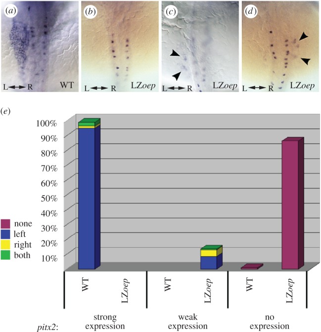
oep mutants in which the early requirement for oep is rescued by oep mRNA injection at the 1-cell stage—generating late-zygotic oep (LZoep) mutants—develop L–R asymmetry defects. In these embryos, the asymmetric expression of pitx2, spaw, cyclops and lefty2 is predominantly absent in the LPM and dorsal diencephalon of 18–22 somite LZoep embryos ([33–35] and R.D.B., data not shown; figure 1). While wild-type levels of pitx2 are never observed, further characterization reveals that a small percentage of LZoep embryos express weak levels of pitx2 in the LPM (n = 33/246; 13.5%; figure 1c,d,e). In these embryos, pitx2 was found in small patches of cells within areas of the LPM where pitx2 would normally be expressed. This expression was not restricted to a particular area along the anterior–posterior axis. Weak expression of pitx2 was never observed in the dorsal diencephalon of LZoep embryos (data not shown). The weak expression observed in LZoep embryos was not restricted to the left LPM, but was also observed on the right (n = 11/33) or bilaterally in the left and right LPM (n = 1/33; figure 1e). These observations suggest that there are two defects in asymmetric gene expression in LZoep embryos. First, pitx2 expression is not induced to wild-type levels in the LPM. Second, in those embryos that do exhibit LPM pitx2 expression, proper establishment of the asymmetric L–R pattern of pitx2 does not occur.
Figure 1.
pitx2 expression is abnormal in LZoep embryos. Dorsal views of pitx2 expression in wild-type (a) and LZoep (b–d) embryos at the 18 somite stage. pitx2 is expressed in presumed Rohon-Beard neurons in the spinal cord that form columns on either side of the midline. The LPM lies on both sides of the midline and normally expresses pitx2 robustly on the left (a). In LZoep, pitx2 expression is either absent from the LPM (b) or is expressed in a few clumps of cells (arrowheads in c,d). Anterior is up; left (L) and right (R) are indicated. (e) Bar graph representing pitx2 expression in wild-type (n = 535) and LZoep (n = 246) embryos. In this and the following graphs, the percentage of embryos expressing pitx2 is indicated by bars. Embryos were placed into one of three categories: strong expression (as in a), weak expression (as in c,d) or no expression (as in b). Each bar is colour coded to illustrate the sidedness of expression. Left LPM expression alone is marked in dark blue. Yellow denotes the population where expression was found only in the right LPM. Green denotes embryos where expression was found in both the left and right LPM. Non-expressing embryos are denoted by the maroon bar in the ‘no expression’ category. Both the strength of pitx2 expression and the side of pitx2 expression were significantly different between wild-type and LZoep embryos (p < 0.00001 in both cases; χ2-test applied).
(b). oep in the midline of LZoep embryos can restore pitx2 expression
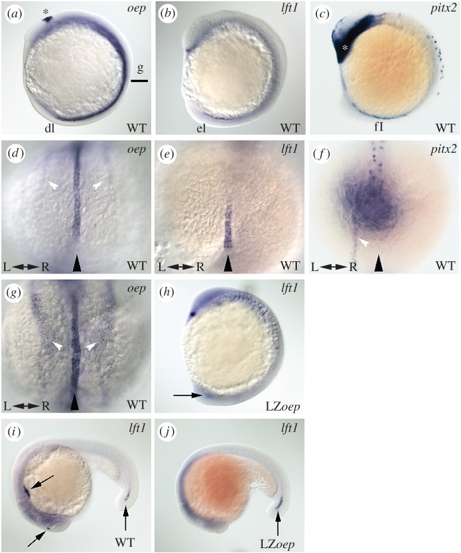
The loss of pitx2 LPM expression in LZoep embryos is owing to insufficient oep activity. To determine the spatial requirement for oep in pitx2 expression in the LPM, we generated genetic chimeras by transplantation. Asymmetric pitx2 expression in the LPM is first observed at 10 somites in wild-type embryos (figure 2f) and is present through 26 h post fertilization (hpf). During somitogenesis, oep is expressed in the left and right LPM, in the developing dorsal diencephalon and in derivatives of axial mesoderm including the midline notochord, overlapping with lefty1 expression ([31]; figure 2d,g). As Oep is required for Nodal signalling, and pitx2 is a known target of Nodal signalling, it is conceivable that oep is predominantly required in the LPM to allow Nodal ligand to induce pitx2 to be expressed at wild-type levels. To test this possibility, we first transplanted wild-type cells into 1000-cell stage LZoep embryos. Transplantation at this early stage places wild-type cells in a scattered fashion throughout many tissues in the embryo. LZoep embryos with wild-type cells in the LPM were obtained, but rescue of pitx2 levels was not observed (n = 10). Large numbers of cells can be placed more accurately into the LPM by performing margin transplants at the 50% epiboly stage. Despite having large numbers of wild-type cells in the LPM, but not in other tissues, 10/14 showed no pitx2 expression in the LPM and 4/14 showed weak expression of pitx2 similar to LZoep controls. This suggests that providing Oep in the LPM does not allow for Nodal signalling to occur in an embryo otherwise lacking Oep.
Figure 2.
Expression of one-eyed pinhead, lefty1 and pitx2. (a–c) Lateral views of wild-type embryos at the 10 somite stage. Anterior is up; dorsal to the right. Lines and lower case letters mark the level of the embryo shown in (d–g). (d–f) posterior views and (g) dorsal view of the embryos in (a–c). Anterior is up; left (L) and right (R) are indicated. oep is expressed in the dorsal diencephalon (asterisk in a), in the midline (black arrowhead in d,g) and in the LPM (white arrowheads d,g). At this stage, oep is expressed in thin stripes in the posterior LPM (d) and more broadly in more anterior parts of the LPM (g). lft1 is expressed in a small portion of the anterior prechordal plate (dot of expression in the head region of b) and in the posterior midline (black arrowhead in e). The posterior notochord expression of lft1 is observed even earlier at the 3 somite stage, prior to when the first asymmetric expression of pitx2 is observed. pitx2 is expressed in head mesoderm (asterisk in c) and in presumptive Rohon-Beard neurons in the spinal cord. The first asymmetric expression of pitx2 is evident at this stage (white arrowhead in f) to the left of the midline (black arrowhead). Note the view in (f) is slightly displaced to those in (d,e) to better view the asymmetric expression of pitx2. The dark shading in (f) is from the expression in head mesoderm (asterisk in a) directly opposite the plane of view. (h) Expression of lft1 in an LZoep embryo at the 10 somite stage. lft1 expression can vary from weak to undetectable (10/13; as shown in (h), indicated by black arrow), to almost wild-type in intensity (3/13). As lft is induced by Nodal, the loss of late Nodal signalling in LZoep embryos may prevent the full induction of lft1 observed in wild-type embryos. Accordingly, more embryos express lft1 at the 3 somite stage (6/12 wild-type levels; 6/12 weaker levels). (i) Lateral view of lft1 expression in an 18–20 somite stage wild-type embryo. Arrows mark expression domains in the diencephalon, the heart field and the posterior notochord [82,84]. (j) Lateral view of lft1 expression in an LZoep embryo at the 18–20 somite stage. Note only the expression domain in the posterior notochord (black arrow) is observed in LZoep embryos (5/8 wild-type to reduced levels; 3/8 no obvious expression).
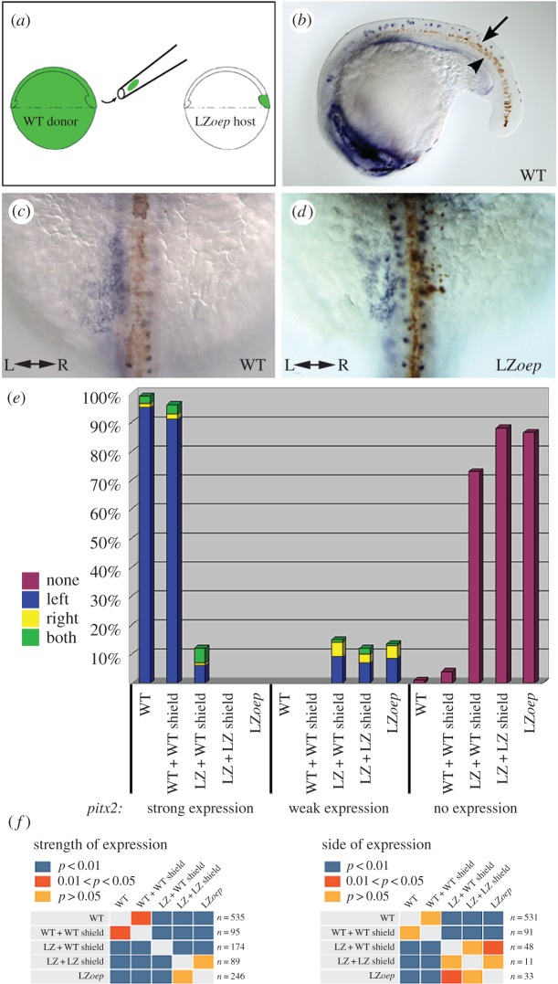
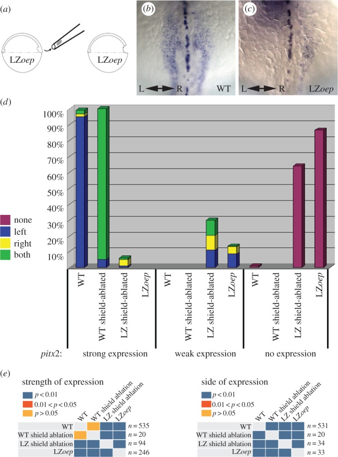
As oep is also expressed in the midline during the stages when pitx2 is first expressed in the LPM (figure 2d,g), oep may be required in the midline to affect pitx2 expression. We tested this hypothesis by placing wild-type cells into the midline of LZoep embryos using shield transplantation (figure 3a) [96,97]. The zebrafish shield is equivalent to the organizer in Xenopus and gives rise to axial mesoderm and floor plate. In these experiments, the shield of the host embryos was removed and replaced by the shield from the donor. This type of transplantation places donor cells throughout the floor plate and axial mesoderm derivatives, including the hatching gland, head mesoderm and notochord (figure 3b). We did not observe transplanted cells in the dorsal forerunner cells or in KV with this procedure (data not shown). Transplantation of a wild-type shield into a wild-type host does not strongly affect the levels or the asymmetric expression of pitx2 in the LPM (figure 3c,e,f), indicating that this form of transplantation does not robustly impact normal L–R patterning per se. While strong levels of pitx2 were never observed in LZoep controls, wild-type shields transplanted into LZoep embryos were capable of restoring pitx2 expression to strong levels, similar to wild-type, in the LPM in a proportion of embryos (figure 3d,e,f). To further ensure that this result is not simply a consequence of the transplantation technique, shields from LZoep embryos were transplanted into LZoep hosts. These transplanted embryos expressed weak levels of pitx2 similar to LZoep controls, but strong levels of expression were not observed (figure 3e). These results suggest that restoring oep expression in the midline can promote pitx2 expression in the LPM in LZoep embryos. Given that pitx2 is a bona fide Nodal transcriptional target, and Nodal requires EGF-CFC proteins to function, we assume any pitx2 expression in the LPM requires that residual Oep protein supplied via injection is still present.
Figure 3.
Shield transplantation experiments in wild-type and LZoep embryos. (a) Illustration of the experiment. A needle is used to remove the shield from a donor embryo labelled with fluorescein dextran tracer dye. The shield from the host embryo is removed and replaced with the shield from the donor. (b) Lateral view of a wild-type embryo after a wild-type shield transplant. pitx2 expression is in blue, labelled cells from the donor shield are in brown. Note that the floorplate (black arrow) and the notochord (black arrowhead) can be labelled with this technique. Labelled cells were not observed in the dorsal forerunner cells or in KV (data not shown). Dorsal views of pitx2 expression in a wild-type embryo with a wild-type shield transplant (c) and an LZoep embryo with a wild-type shield transplant (d) at the 18 somite stage. Anterior is up; left (L) and right (R) are indicated. (e) Bar graph representing pitx2 expression in shield transplanted embryos. The genotype of the host is listed first, followed by the genotype of the donor. Expression categories and colour codes are described in figure 1. (f) Statistical significance of the experiments was determined using χ2 and Fisher's exact tests. Comparisons of strength of pitx2 expression and the sidedness of pitx2 expression were conducted. For sidedness, only embryos that displayed expression were included in the analysis, reflected by the change in n values. Blue denotes p-values of less than 0.01, orange denotes p-values between 0.01 and 0.05, and light orange denotes p-values greater than 0.05.
To determine if a particular region of the midline must express oep to activate pitx2 in the LPM, we monitored where transplanted cells were located. We did not find any correlation between the position of wild-type cells in the midline at the 18 somite stage and rescue of pitx2 expression in the LPM of LZoep embryos. For example, some LZoep embryos with strong expression of pitx2 had wild-type cells only in the most anterior floorplate cells, while others had wild-type cells throughout all axial mesoderm derivatives (data not shown). Intriguingly, while placing wild-type cells into the midline of LZoep hosts could restore pitx2 expression to strong levels in some cases, it did not correct the randomization of pitx2 expression seen in LZoep embryos (see §4).
(c). Blocking ntla activity in LZoep embryos can restore pitx2 expression
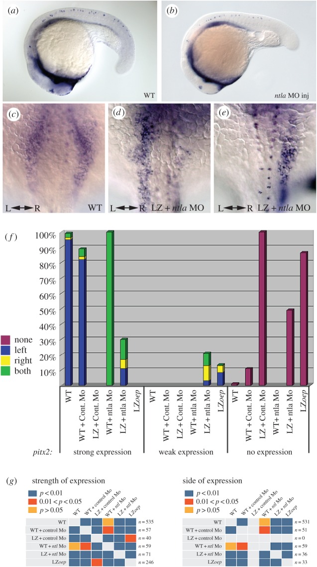
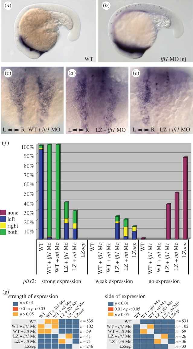
Our transplantation experiments suggest that the midline-localized oep has an active role in promoting pitx2 expression in the LPM. This is in apparent contrast with models that support a more passive role for the midline (see §1). To further test the function of the midline in L–R patterning, we removed midline structures from LZoep embryos using morpholino antisense oligonucleotides (MO) against no tail (ntla; the zebrafish Brachyury/T orthologue) [99]. ntla mutants and morphants lack the notochord, have expanded floorplate, have a substantially reduced KV and display bilateral expression of pitx2 and other left side-specific genes [29,34,35,77,78,100–103]. Injection of the ntla MO in wild-type or LZoep embryos results in 100% of the injected embryos displaying the morphological ntla phenotype. In wild-type embryos injected with ntla MO, pitx2 is expressed bilaterally as is seen in ntla mutants (figure 4c). Surprisingly, injection of ntla MO into LZoep embryos can restore expression of pitx2 in the LPM to strong levels in many cases; while LZoep mutants never exhibited strong LPM pitx2, 30% of LZoep mutants injected with ntla MO showed strong levels (figure 4d–g). Unlike injection of ntla MO, injection of a control MO does not dramatically alter pitx2 gene expression levels in wild-type or LZoep embryos (figure 4f,g). The most parsimonious explanation for these results is that the notochord and/or KV in LZoep embryos represses pitx2 transcription in the LPM, and that this repression can be released by blocking ntla function and removing midline structures.
Figure 4.
no tail (ntla) morpholino (MO) injections in wild-type and LZoep embryos. Lateral views of 20 somite stage wild-type embryos injected with a control MO (a) or the ntla MO (b). Note the shortening of posterior structures in the ntla MO-injected embryo. Dorsal views of pitx2 expression in ntla MO-injected wild-type (c) and LZoep (d,e) embryos at the 18 somite stage. Anterior is up; left (L) and right (R) are indicated. (f) Bar graph representing pitx2 expression in ntla MO-injected embryos. Genotype of injected embryos is listed first, followed by the injected MO. Expression categories and colour codes are described in figure 1. (g) Statistical significance of experiments conducted as described in figure 3.
(d). Midline ablations in LZoep embryos can restore pitx2 expression
Loss of ntla affects the notochord and the structure of KV [100,103]. To test if the phenotype of ntla MO-injected LZoep embryos is owing to loss of the notochord specifically, we removed portions of the midline by shield ablation. Portions of the shield, and cells on either side of the shield, were removed by suction with a needle (figure 5a; [96]). The damaged shield region only partially regenerates, resulting in embryos that often have gaps in the notochord and floorplate as can be detected by in situ hybridization of the midline marker sonic hedgehog (shh). To select against embryos with non-axial mesoderm tissue losses, only embryos that appeared to develop normally, but had gaps in midline shh expression, were included in the analysis (see §2).
Figure 5.
Shield ablations in wild-type and LZoep embryos. (a) Illustration of shield ablation experiment. A needle is used to remove the shield, and cells on either side of the shield. Dorsal views of pitx2 and shh expression in shield-ablated wild-type (b) and LZoep (c) embryos at the 18 somite stage. Anterior is up; left (L) and right (R) are indicated. Damage to the midline in (b,c) is observed as gaps in the normally continuous shh expression in the midline. Note that these images are focused on the shh expression in the embryos, thus the pitx2 expression in the Rohan-Beard neurons is not as visible. (d) Bar graph representing pitx2 expression in shield-ablated embryos. Expression categories and colour codes are described in figure 1. (e) Statistical significance of experiments conducted as described in figure 3.
When shields were ablated in wild-type embryos, the majority of embryos expressed pitx2 bilaterally in the LPM, indicating that damage to the midline affects pitx2 expression as expected (figure 5b,d). Shield ablation in LZoep embryos increased the number of embryos that weakly expressed pitx2 and restored pitx2 expression in the LPM to strong levels in some embryos, results that were statistically significantly different from non-ablated LZoep embryos (figure 5c–e). Compared with ntla MO injection, the extent of midline removal is considerably less in shield-ablated LZoep embryos and, accordingly, the proportion of LZoep embryos expressing strong levels of pitx2 was lower. Taken together, the effects of ntla MO and shield ablation in LZoep embryos demonstrate that disruption of the notochord can restore LPM pitx2 expression to strong levels in LZoep embryos.
(e). Blocking lefty1 activity in LZoep embryos can restore pitx2 expression
lefty1, the feedback inhibitor of Nodal, is expressed in the notochord of zebrafish embryos [82,84], and knockdown of lefty1 results in bilateral expression of Nodal targets in the LPM [80,104]. We note that zebrafish lefty1 (lft1) is expressed during early somitogenesis in the posterior midline, just anterior to KV, prior to the initiation of asymmetric gene expression in the LPM (this study and see [84]). At the 3 somite stage, 11/11 wild-type embryos expressed lft1 in the posterior notochord region. Asymmetric pitx2 expression begins at the 10 somite stage in the posterior LPM, just lateral to the lft1 expression domain in the midline (figure 2e,f). We also detect lft1 expression in the midline of LZoep embryos at early and late somitogenesis stages, but sometimes at lower levels (figure 2h,j).
To test if lft1 plays a role in the midline-dependent repression we observe, we injected MO against lft1 into wild-type and LZoep embryos. Injection of low levels of the lft1 MO into wild-type embryos does not affect morphogenesis through somitogenesis (figure 6a,b; also see [80,104]), but results in predominantly bilateral expression of pitx2 (figure 6c,f). This pattern of expression is statistically no different from that observed upon midline removal using the ntla MO (figure 6f,g). Indeed, injection of lft1 MO into LZoep embryos can restore pitx2 expression to strong levels in the LPM, strikingly similar to what is observed when the midline is ablated in LZoep embryos (figure 6d–g). These results indicate that lft1 is required for the repression of LPM pitx2 expression in LZoep mutants.
Figure 6.
lft1 MO injections in wild-type and LZoep embryos. Lateral views of 18–20 somite stage LZoep embryos injected with a control MO (a) or the lft1 MO (b). lft1-injected embryos developed without apparent defects, but were often slightly delayed compared with uninjected controls. Dorsal views of pitx2 expression in lft1 MO-injected wild-type (c) or LZoep (d,e) embryos at the 18 somite stage. Anterior is up; left (L) and right (R) are indicated. (f) Bar graph representing pitx2 expression in MO-injected embryos. Genotype of injected embryos is listed first, followed by the injected MO. The ntla MO results from figure 4 are shown here to aid in comparison with the lft1 MO results. Expression categories and colour codes are described in figure 1. (g) Statistical significance of experiments conducted as described in figure 3.
(f). oep in the midline of LZoep embryos does not rescue randomization of pitx2 expression
The results above show that the major phenotype of LZoep embryos, the strong reduction of pitx2 expression in the LPM, can be rescued by providing oep in the midline, ablating the midline or by blocking lft1 function. To determine if the second phenotype in LZoep mutants, the randomized induction of pitx2, is also rescued in these treatments, we monitored the L–R distribution of pitx2 expression in all experiments. We find that manipulations that rescued pitx2 expression to strong levels did not restore expression solely in the left LPM but also resulted in right LPM expression (figures 3e, 4f and 5d). Additionally, manipulations that resulted in bilateral pitx2 expression in wild-type embryos did not consistently result in bilateral expression in LZoep. For instance, knockdown of ntla results in bilateral pitx2 expression in WT embryos but randomized expression in those LZoep embryos that express any LPM pitx2 (figure 4f). This suggests that there is an additional role for Oep in establishing or promoting asymmetric gene expression that is not rescued in our experiments.
4. Discussion
Since the first experiments that implicated the notochord in L–R patterning, the importance of an intact midline in this process has emerged from mutant analysis, gene knockdown experiments and midline extirpations. The results presented here suggest both an activating role and a repressive role for the midline in promoting transcription of pitx2 in the LPM in zebrafish. In LZoep embryos, the loss of strong pitx2 expression levels in the LPM can be rescued by introducing wild-type cells into the midline. This suggests that oep in the midline can actively promote pitx2 expression in the LPM. Furthermore, we show that removal of midline structures from LZoep embryos, either by shield ablations or injection of ntla MO, can also restore pitx2 expression to strong levels in the LPM. This suggests that the midline represses pitx2 in the LPM. These results can be reconciled by a model in which oep in the midline functions to overcome a midline-derived repressor of LPM Nodal activity.
(a). A model for Lefty and Oep function prior to asymmetric gene expression during the establishment of L–R asymmetry: the midline as an ‘activator’ and ‘repressor’
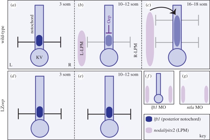
Based on our results, and previously proposed models for the establishment of L–R asymmetry, we propose the following model for L–R patterning in zebrafish (figure 7a–c). Both the left and right LPM have the potential to express left side-specific genes such as pitx2, which typically occurs at the 10 somite stage. lft1 expression (dark blue; figure 7a) is induced in the posterior notochord prior to 10 somites, most likely by an earlier Nodal signalling event. Indeed, at the 3 somite stage, robust lft1 can be observed in this domain. Lefty1 from this midline domain increases the amount of Nodal ligand required to induce LPM-nodal (i.e. raises the threshold required for robust Nodal activation) and thus represses pitx2 transcription (black inhibition arrows; figure 7a). This occurs symmetrically and prevents LPM Nodal activation prematurely, before the symmetry-breaking event at KV has occurred. An oep-dependent process in the midline weakens repression by Lefty1 (grey inhibition arrows; figure 7b). As Lefty can bind to Oep, and overexpression of Oep can suppress the effects of overexpressing Lefty [8], we suggest that physical interaction between Lefty1 and Oep at the midline sequesters some Lefty1, thereby reducing the effective amount of Lefty1 able to repress LPM Nodal signals. This antagonism of Lefty1 by Oep would thus allow Nodal signals to activate transcription in the LPM (light purple; figure 7b).
Figure 7.
Model depicting antagonism of Lefty by Oep prior to the emergence of asymmetric gene expression in the zebrafish LPM. All renderings are of the notochord and surrounding LPM. The notochord terminates into KV (oval). (a) lft1 is expressed in the posterior notochord before asymmetric gene expression is detected (dark blue). We postulate that Lefty acts to repress Nodal signalling to the LPM (black repression arrows) and thus prevents activation of Nodal target genes in the LPM prior to the L–R biasing event that occurs at KV. (b) An oep-dependent signalling event overcomes/weakens the midline repressor, Lefty (weaker repression depicted by grey repression arrows). This weakened repressor can still prevent Nodal activity from occurring in the LPM initially. A biasing mechanism, flow generated by monocilia in KV, leads to downregulation of the Nodal inhibitor charon around KV (not depicted) and allows Nodal signalling to initiate in the left LPM at around the 10 somite stage (light purple). (c) Nodal autoregulation then leads to the robust expression of left side-specific genes including pitx2. Nodal signalling to the right LPM continues to be repressed by the higher level of Charon on the right of KV in cooperation with Lefty from the midline. (d) In LZoep embryos, the early posterior notochord lft1 repressor is active and not weakened owing to the later absence of Oep. (e) Nodal signalling does not reach the threshold to overcome the repressors and robust transcription in the LPM does not take place. (f) In lft1 MO-injected LZoep embryos, the early posterior notochord repressor is removed, thus the threshold for Nodal to signal to the left and the right LPM is lower, and both sides can initiate transcription of left side-specific genes. (g) In ntla MO-injected LZoep embryos, midline structures are absent and thus, there is no midline repressor. Nodal signalling can thus initiate transcription of left side-specific genes bilaterally even in LZoep embryos as Oep is not required to overcome the repressor.
While the early interaction between oep and lft1 that our experiments have uncovered is important for allowing Nodal signals to initiate in the LPM, it may be less critical for setting up the asymmetry of these signals. After all, the early domain of lft1 is L–R symmetrical and we would expect this to dampen Nodal signalling in both the left and right LPM equally. Instead, asymmetries in LPM Nodal activity are generated downstream of asymmetric flow in KV and the resulting left-sided downregulation of the Nodal repressor charon. Flow-dependent downregulation of charon and oep-dependent dampening of Lefty in the midline would thus cooperate to generate a signalling threshold in which Nodal signals are able to robustly activate in the left LPM around the 10 somite stage but not in the right LPM. Subsequently, Nodal signals spread anteriorly in the left LPM and induce further lft1 expression in the notochord (figure 7c), which then acts to maintain asymmetry by preventing Nodal signals from activating and spreading in the right LPM.
This model can explain the phenotypes we observe in LZoep embryos as well as in the various treatments (ablations, transplants and MO injections) performed in this study. In LZoep embryos (figure 7d–g), lft1 is still present in the posterior notochord (figure 7d). This expression of lft1 is likely owing to residual Oep protein from the 1-cell oep mRNA injection at the time when lft1 is induced, several hours before pitx2 is initiated in the LPM. However, in these same embryos, later oep function is greatly reduced. This late reduction in Oep in LZoep mutants, therefore, results in more active Lefty repressor in the posterior notochord; thus, Nodal signals are repressed at a level that prevents them from initiating target gene expression in the LPM (figure 7e). Removing the repressor by loss of the midline (ntla MO (figure 7g) or shield ablation) or by reducing lft1 itself (figure 7f) is sufficient to allow for signalling to occur in the LPM of LZoep mutants because these treatments replace the requirement of Oep to repress Lefty. Importantly, these treatments also suggest that there is sufficient functioning Oep in the LPM of LZoep embryos to allow for robust Nodal activity; thus, the lost or weakened pitx2 expression in LZoep is not exclusively a consequence of loss of Oep in the LPM. This agrees with our finding that adding Oep-positive cells to the LPM of LZoep mutants cannot rescue high levels of LPM pitx2 expression. Furthermore, although the potential for rescue is clear, we suggest that the rescue of LPM pitx2 in LZoep mutants, either after wild-type shield transplants or injection of ntla MO, is only partial because the LPM still has limited functional Oep remaining from the early mRNA oep injection.
This model is consistent with the timing and location of lft1, pitx2 and oep expression. Zebrafish lft1 is expressed in the posterior notochord preceding pitx2 and spaw expression in the LPM. pitx2 expression is observed later (beginning around the 10 somite stage) in the posterior-most LPM, just lateral to the posterior notochord and KV (figure 2). lft1 expression in the midline later extends from the posterior notochord to the anterior, induced by Nodal from the LPM [87,88]. Our findings provide a previously unappreciated role for early lft1 in establishing LPM Nodal signals.
Moreover, the model explains the results of our midline ablation experiments in wild-type and LZoep embryos. Midline ablations result in bilateral expression of pitx2 in wild-type embryos and can restore pitx2 to strong levels in LZoep mutants. In our model, the source of the repressor (the early domain of lft1), the notochord, is removed in these experiments, allowing Nodal signals to activate pitx2 in the LPM strongly and bilaterally. It is worth noting that the biasing mechanism occurring at KV should be unaffected by these treatments as our transplants did not contribute to the dorsal forerunner or KV populations. Nevertheless, we likely see bilateral pitx2 expression because loss of the early lft1 repressor allows Nodal activity at both the left and right LPM to induce Nodal targets, regardless of subtle differences in Nodal levels caused by the biasing mechanism at KV. However, it is worth noting that KV function may not strictly be required for Nodal signalling in the LPM. In ntla mutants or embryos injected with ntla MO, ciliated cells are present, but KV does not form. Moreover, spaw expression around KV is lost in ntla mutants, suggesting that the activating signal for Nodal in the LPM in this mutant must come from another source that remains elusive at this point. It is possible that this LPM-spaw activation in the absence of KV-spaw occurs because lft1 and charon, which both encode Spaw repressors, are also not expressed in ntla mutants, and therefore, the threshold for LPM-spaw activation is likely markedly reduced. Nevertheless, as KV-spaw is absent in ntla mutants, this observation calls into question the current major model that LRC Nodal signals induce the LPM Nodal cascade. In the mouse, node-derived Nodal is thought travel to the LPM and initiate Nodal expression there; indeed, Nodal mutants fail to activate LPM Nodal expression [24]. However, it is yet to be determined whether node-derived Nodal protein alone is sufficient to induce LPM Nodal in the endogenous context or whether node-Nodal is able to active the LPM Nodal cascade in the absence of inhibitors like Cerl-2. It is worth noting that, unlike what we report in zebrafish, it is thought that LPM Nodal signals are responsible for inducing floor-plate expression of Lefty1 [85], and therefore, removing Lefty1 from Nodal mutants would likely have no impact on whether the Nodal cascade is activated in the LPM.
(b). The role of midline Oep in generating the asymmetry of LPM Nodal signals
In addition to the role oep plays in promoting gene transcription in the LPM, our results also suggest another role for oep in establishing proper L–R pattern. The weak expression seen in some LZoep embryos is not restricted to the left side, suggesting that the loss of oep leads to a randomization of the L–R pattern. We do not know where oep is required in this process, as oep in the midline did not rescue randomization in LZoep mutants. As a result, this is more difficult to reconcile, but may be a consequence of the unique background that LZoep embryos provide. The ability for Nodal to signal is likely waning during somitogenesis as Oep provided by RNA injection diminishes. LZoep embryos with weak pitx2 expression, and the fraction of embryos that respond with strong pitx2 expression in our experiments, may encompass those with more residual Oep. Thus, it is possible that one-sided expression, versus bilateral, occurs in our experiments where the midline is removed in LZoep embryos, owing to the amount of Oep available in the LPM for the response. Furthermore, L–R patterning can be easily disrupted by situations that alter the timing and expression of repressors and activators. In LZoep embryos without additional treatments, the randomization of pitx2 expression could likewise be owing to the indeterminate levels of Oep present, which may alter the timing of response to signals from the LRC.
As a final note, in addition to the midline, oep is also expressed in the LPM and diencephalon. The shield transplantations and midline ablations reported here did not restore pitx2 expression in the diencephalon of LZoep mutants (data not shown), suggesting that oep might be required in the diencephalon for expression to occur. We have previously shown that repression of left side-specific genes also occurs in the diencephalon [34], thus it is intriguing to speculate that oep also plays a role in overcoming this repression.
In summary, the results in this paper support a model wherein midline Oep functions to repress lft1, expressed in the notochord, thereby lowering the threshold and thus promoting activation of the Nodal cascade in the LPM.
Supplementary Material
Acknowledgements
This work was conducted when R.D.B. was a postdoctoral fellow in the laboratory of Alexander F. Schier, and I thank Alex for his mentorship and support. I thank members of the Schier and Yelon laboratories for discussions; Derek Stemple for teaching me the shield transplantation procedure; and S. Zimmerman, T. Bruno and N. Dillon for fish care.
Ethics
This work was conducted at the Skirball Institute of Biomolecular Medicine at New York University Langone Medical Center. Established zebrafish protocols were approved and adhered to in accordance with the Institutional Animal Care and Use Committee of New York University Langone Medical Center.
Data accessibility
The datasets supporting this article have been uploaded as part of the electronic supplementary material.
Authors' contributions
R.D.B. performed the experiments. R.D.B. and D.T.G interpreted the data, wrote and revised the manuscript, and approved of the final submission.
Competing interests
We have no competing interests
Funding
R.D.B. was supported in her postdoctoral position by a Damon Runyon Fellowship DRG-1491 from the Damon Runyon Cancer Research Foundation and by a postdoctoral fellowship from the American Heart Association Heritage Affiliate. Left–right patterning work in her laboratory is currently supported by a National Institute of Child Health and Development grant 2R01HD048584. D.T.G. is supported by a postdoctoral fellowship from the American Heart Association Founders Affiliate.
References
- 1.Burdine RD, Schier AF. 2000. Conserved and divergent mechanisms in left–right axis formation. Genes Dev. 14, 763–776. [PubMed] [Google Scholar]
- 2.Levin M. 2005. Left–right asymmetry in embryonic development: a comprehensive review. Mech. Dev. 122, 3–25. ( 10.1016/j.mod.2004.08.006) [DOI] [PubMed] [Google Scholar]
- 3.Nakamura T, Hamada H. 2012. Left–right patterning: conserved and divergent mechanisms. Development 139, 3257–3262. ( 10.1242/dev.061606) [DOI] [PubMed] [Google Scholar]
- 4.Ramsdell AF. 2005. Left–right asymmetry and congenital cardiac defects: getting to the heart of the matter in vertebrate left–right axis determination. Dev. Biol. 288, 1–20. ( 10.1016/j.ydbio.2005.07.038) [DOI] [PubMed] [Google Scholar]
- 5.Shiratori H, Hamada H. 2014. TGFβ signaling in establishing left–right asymmetry. Semin. Cell Dev. Biol. 32, 80–84. ( 10.1016/j.semcdb.2014.03.029) [DOI] [PubMed] [Google Scholar]
- 6.Sampath K, Robertson EJ. 2016. Keeping a lid on nodal: transcriptional and translational repression of nodal signalling. Open Biol. 6, 150200 ( 10.1098/rsob.150200) [DOI] [PMC free article] [PubMed] [Google Scholar]
- 7.Schier AF. 2009. Nodal morphogens. Cold Spring Harb. Perspect. Biol. 1, a003459 ( 10.1101/cshperspect.a003459) [DOI] [PMC free article] [PubMed] [Google Scholar]
- 8.Cheng SK, Olale F, Brivanlou AH, Schier AF. 2004. Lefty blocks a subset of TGFβ signals by antagonizing EGF-CFC coreceptors. PLoS Biol. 2, E30 ( 10.1371/journal.pbio.0020030) [DOI] [PMC free article] [PubMed] [Google Scholar]
- 9.Gritsman K, Zhang J, Cheng S, Heckscher E, Talbot WS, Schier AF. 1999. The EGF-CFC protein one-eyed pinhead is essential for nodal signaling. Cell. 97, 121–132. ( 10.1016/S0092-8674(00)80720-5) [DOI] [PubMed] [Google Scholar]
- 10.Reissmann E, et al. 2001. The orphan receptor ALK7 and the Activin receptor ALK4 mediate signaling by Nodal proteins during vertebrate development. Genes Dev. 15, 2010–2022. ( 10.1101/gad.201801) [DOI] [PMC free article] [PubMed] [Google Scholar]
- 11.Yeo C, Whitman M. 2001. Nodal signals to Smads through Cripto-dependent and Cripto-independent mechanisms. Mol. Cell. 7, 949–957. ( 10.1016/S1097-2765(01)00249-0) [DOI] [PubMed] [Google Scholar]
- 12.Hoodless PA, et al. 2001. FoxH1 (Fast) functions to specify the anterior primitive streak in the mouse. Genes Dev. 15, 1257–1271. ( 10.1101/gad.881501) [DOI] [PMC free article] [PubMed] [Google Scholar]
- 13.Slagle CE, Aoki T, Burdine RD. 2011. Nodal-dependent mesendoderm specification requires the combinatorial activities of FoxH1 and Eomesodermin. PLoS Genet. 7, e1002072 ( 10.1371/journal.pgen.1002072) [DOI] [PMC free article] [PubMed] [Google Scholar]
- 14.Yeo CY, Chen X, Whitman M. 1999. The role of FAST-1 and Smads in transcriptional regulation by activin during early Xenopus embryogenesis. J. Biol. Chem. 274, 26 584–26 590. ( 10.1074/jbc.274.37.26584) [DOI] [PubMed] [Google Scholar]
- 15.Yoon SJ, Wills AE, Chuong E, Gupta R, Baker JC. 2011. HEB and E2A function as SMAD/FOXH1 cofactors. Genes Dev. 25, 1654–1661. ( 10.1101/gad.16800511) [DOI] [PMC free article] [PubMed] [Google Scholar]
- 16.Zhou S, Zawel L, Lengauer C, Kinzler KW, Vogelstein B. 1998. Characterization of human FAST-1, a TGFβ and activin signal transducer. Mol. Cell. 2, 121–127. ( 10.1016/S1097-2765(00)80120-3) [DOI] [PubMed] [Google Scholar]
- 17.Pogoda HM, Solnica-Krezel L, Driever W, Meyer D. 2000. The zebrafish forkhead transcription factor FoxH1/Fast1 is a modulator of nodal signaling required for organizer formation. Curr. Biol. 10, 1041–1049. ( 10.1016/S0960-9822(00)00669-2) [DOI] [PubMed] [Google Scholar]
- 18.Sirotkin HI, Gates MA, Kelly PD, Schier AF, Talbot WS. 2000. fast1 is required for the development of dorsal axial structures in zebrafish. Curr. Biol. 10, 1051–1054. ( 10.1016/S0960-9822(00)00679-5) [DOI] [PubMed] [Google Scholar]
- 19.Nonaka S, Tanaka Y, Okada Y, Takeda S, Harada A, Kanai Y, Kido M, Hirokawa N. 1998. Randomization of left–right asymmetry due to loss of nodal cilia generating leftward flow of extraembryonic fluid in mice lacking KIF3B motor protein. Cell 95, 829–837. ( 10.1016/S0092-8674(00)81705-5) [DOI] [PubMed] [Google Scholar]
- 20.Schweickert A, Weber T, Beyer T, Vick P, Bogusch S, Feistel K, Blum M. 2007. Cilia-driven leftward flow determines laterality in Xenopus. Curr. Biol. 17, 60–66. ( 10.1016/j.cub.2006.10.067) [DOI] [PubMed] [Google Scholar]
- 21.Essner JJ, Amack JD, Nyholm MK, Harris EB, Yost HJ. 2005. Kupffer's vesicle is a ciliated organ of asymmetry in the zebrafish embryo that initiates left–right development of the brain, heart and gut. Development 132, 1247–1260. ( 10.1242/dev.01663) [DOI] [PubMed] [Google Scholar]
- 22.Essner JJ, Vogan KJ, Wagner MK, Tabin CJ, Yost HJ, Brueckner M. 2002. Conserved function for embryonic nodal cilia. Nature 418, 37–38. ( 10.1038/418037a) [DOI] [PubMed] [Google Scholar]
- 23.Okada Y, Takeda S, Tanaka Y, Belmonte JC, Hirokawa N. 2005. Mechanism of nodal flow: a conserved symmetry breaking event in left–right axis determination. Cell 121, 633–644. ( 10.1016/j.cell.2005.04.008) [DOI] [PubMed] [Google Scholar]
- 24.Brennan J, Norris DP, Robertson EJ. 2002. Nodal activity in the node governs left–right asymmetry. Genes Dev. 16, 2339–2344. ( 10.1101/gad.1016202) [DOI] [PMC free article] [PubMed] [Google Scholar]
- 25.Norris DP, Brennan J, Bikoff EK, Robertson EJ. 2002. The Foxh1-dependent autoregulatory enhancer controls the level of Nodal signals in the mouse embryo. Development 129, 3455–3468. [DOI] [PubMed] [Google Scholar]
- 26.Lowe LA, Yamada S, Kuehn MR. 2001. Genetic dissection of nodal function in patterning the mouse embryo. Development 128, 1831–1843. [DOI] [PubMed] [Google Scholar]
- 27.Oki S, Hashimoto R, Okui Y, Shen MM, Mekada E, Otani H, Saijoh Y, Hamada H. 2007. Sulfated glycosaminoglycans are necessary for Nodal signal transmission from the node to the left lateral plate in the mouse embryo. Development 134, 3893–3904. ( 10.1242/dev.009464) [DOI] [PubMed] [Google Scholar]
- 28.Kawasumi A, Nakamura T, Iwai N, Yashiro K, Saijoh Y, Belo JA, Shiratori H, Hamada H. 2011. Left–right asymmetry in the level of active Nodal protein produced in the node is translated into left–right asymmetry in the lateral plate of mouse embryos. Dev. Biol. 353, 321–330. ( 10.1016/j.ydbio.2011.03.009) [DOI] [PMC free article] [PubMed] [Google Scholar]
- 29.Long S, Ahmad N, Rebagliati M. 2003. The zebrafish nodal-related gene southpaw is required for visceral and diencephalic left–right asymmetry. Development 130, 2303–2316. ( 10.1242/dev.00436) [DOI] [PubMed] [Google Scholar]
- 30.Shen MM, Wang H, Leder P. 1997. A differential display strategy identifies Cryptic, a novel EGF-related gene expressed in the axial and lateral mesoderm during mouse gastrulation. Development 124, 429–442. [DOI] [PubMed] [Google Scholar]
- 31.Zhang J, Talbot WS, Schier AF. 1998. Positional cloning identifies zebrafish one-eyed pinhead as a permissive EGF-related ligand required during gastrulation. Cell 92, 241–251. ( 10.1016/S0092-8674(00)80918-6) [DOI] [PubMed] [Google Scholar]
- 32.Gaio U, et al. 1999. A role of the cryptic gene in the correct establishment of the left–right axis. Curr. Biol. 9, 1339–1342. ( 10.1016/S0960-9822(00)80059-7) [DOI] [PubMed] [Google Scholar]
- 33.Yan YT, Gritsman K, Ding J, Burdine RD, Corrales JD, Price SM, Talbot WS, Schier AF, Shen MM. 1999. Conserved requirement for EGF-CFC genes in vertebrate left–right axis formation. Genes Dev. 13, 2527–2537. ( 10.1101/gad.13.19.2527) [DOI] [PMC free article] [PubMed] [Google Scholar]
- 34.Concha ML, Burdine RD, Russell C, Schier AF, Wilson SW. 2000. A Nodal signaling pathway regulates the laterality of neuroanatomical asymmetries in the zebrafish forebrain. Neuron 28, 399–409. ( 10.1016/S0896-6273(00)00120-3) [DOI] [PubMed] [Google Scholar]
- 35.Liang JO, et al. 2000. Asymmetric nodal signaling in the zebrafish diencephalon positions the pineal organ. Development 127, 5101–5112. [DOI] [PubMed] [Google Scholar]
- 36.Bamford RN, et al. 2000. Loss-of-function mutations in the EGF-CFC gene CFC1 are associated with human left–right laterality defects. Nat. Genet. 26, 365–369. ( 10.1038/81695) [DOI] [PubMed] [Google Scholar]
- 37.Marjoram L, Wright C. 2011. Rapid differential transport of Nodal and Lefty on sulfated proteoglycan-rich extracellular matrix regulates left–right asymmetry in Xenopus. Development 138, 475–485. ( 10.1242/dev.056010) [DOI] [PMC free article] [PubMed] [Google Scholar]
- 38.Francescatto L, Rothschild SC, Myers AL, Tombes RM. 2010. The activation of membrane targeted CaMK-II in the zebrafish Kupffer's vesicle is required for left–right asymmetry. Development 137, 2753–2762. ( 10.1242/dev.049627) [DOI] [PubMed] [Google Scholar]
- 39.McGrath J, Somlo S, Makova S, Tian X, Brueckner M. 2003. Two populations of node monocilia initiate left–right asymmetry in the mouse. Cell 114, 61–73. ( 10.1016/S0092-8674(03)00511-7) [DOI] [PubMed] [Google Scholar]
- 40.Yuan S, Zhao L, Brueckner M, Sun Z. 2015. Intraciliary calcium oscillations initiate vertebrate left–right asymmetry. Curr. Biol. 25, 556–567. ( 10.1016/j.cub.2014.12.051) [DOI] [PMC free article] [PubMed] [Google Scholar]
- 41.Viotti M, Niu L, Shi SH, Hadjantonakis AK. 2012. Role of the gut endoderm in relaying left–right patterning in mice. PLoS Biol. 10, e1001276 ( 10.1371/journal.pbio.1001276) [DOI] [PMC free article] [PubMed] [Google Scholar]
- 42.Saund RS, Kanai-Azuma M, Kanai Y, Kim I, Lucero MT, Saijoh Y. 2012. Gut endoderm is involved in the transfer of left–right asymmetry from the node to the lateral plate mesoderm in the mouse embryo. Development 139, 2426–2435. ( 10.1242/dev.079921) [DOI] [PMC free article] [PubMed] [Google Scholar]
- 43.Beyer T, Thumberger T, Schweickert A, Blum M. 2012. Connexin26-mediated transfer of laterality cues in Xenopus. Biol. Open. 1, 473–481. ( 10.1242/bio.2012760) [DOI] [PMC free article] [PubMed] [Google Scholar]
- 44.Norris DP. 2012. Cilia, calcium and the basis of left–right asymmetry. BMC Biol. 10, 102 ( 10.1186/1741-7007-10-102) [DOI] [PMC free article] [PubMed] [Google Scholar]
- 45.Supatto W, Fraser SE, Vermot J. 2008. An all-optical approach for probing microscopic flows in living embryos. Biophys. J. 95, L29–L31. ( 10.1529/biophysj.108.137786) [DOI] [PMC free article] [PubMed] [Google Scholar]
- 46.Amack JD, Wang X, Yost HJ. 2007. Two T-box genes play independent and cooperative roles to regulate morphogenesis of ciliated Kupffer's vesicle in zebrafish. Dev. Biol. 310, 196–210. ( 10.1016/j.ydbio.2007.05.039) [DOI] [PubMed] [Google Scholar]
- 47.Kreiling JA, Williams G, Creton R. 2007. Analysis of Kupffer's vesicle in zebrafish embryos using a cave automated virtual environment. Dev. Dyn. 236, 1963–1969. ( 10.1002/dvdy.21191) [DOI] [PubMed] [Google Scholar]
- 48.Nonaka S, Shiratori H, Saijoh Y, Hamada H. 2002. Determination of left–right patterning of the mouse embryo by artificial nodal flow. Nature 418, 96–99. ( 10.1038/nature00849) [DOI] [PubMed] [Google Scholar]
- 49.Nonaka S, Yoshiba S, Watanabe D, Ikeuchi S, Goto T, Marshall WF, Hamada H, Stemple D. 2005. De novo formation of left–right asymmetry by posterior tilt of nodal cilia. PLoS Biol. 3, e268 ( 10.1371/journal.pbio.0030268) [DOI] [PMC free article] [PubMed] [Google Scholar]
- 50.Kramer-Zucker AG, Olale F, Haycraft CJ, Yoder BK, Schier AF, Drummond IA. 2005. Cilia-driven fluid flow in the zebrafish pronephros, brain and Kupffer's vesicle is required for normal organogenesis. Development 132, 1907–1921. ( 10.1242/dev.01772) [DOI] [PubMed] [Google Scholar]
- 51.Okabe N, Xu B, Burdine RD. 2008. Fluid dynamics in zebrafish Kupffer's vesicle. Dev. Dyn. 237, 3602–3612. ( 10.1002/dvdy.21730) [DOI] [PMC free article] [PubMed] [Google Scholar]
- 52.Borovina A, Superina S, Voskas D, Ciruna B. 2010. Vangl2 directs the posterior tilting and asymmetric localization of motile primary cilia. Nat. Cell Biol. 12, 407–412. ( 10.1038/ncb2042) [DOI] [PubMed] [Google Scholar]
- 53.Hashimoto M, et al. 2010. Planar polarization of node cells determines the rotational axis of node cilia. Nat. Cell Biol. 12, 170–176. ( 10.1038/ncb2020) [DOI] [PubMed] [Google Scholar]
- 54.Supp DM, Witte DP, Potter SS, Brueckner M. 1997. Mutation of an axonemal dynein affects left–right asymmetry in inversus viscerum mice. Nature 389, 963–966. ( 10.1038/40140) [DOI] [PMC free article] [PubMed] [Google Scholar]
- 55.Jaffe KM, et al. 2016. c21orf59/kurly controls both cilia motility and polarization. Cell Rep. 14, 1841–1849. ( 10.1016/j.celrep.2016.01.069) [DOI] [PMC free article] [PubMed] [Google Scholar]
- 56.Hjeij R, et al. 2014. CCDC151 mutations cause primary ciliary dyskinesia by disruption of the outer dynein arm docking complex formation. Am. J. Hum. Genet. 95, 257–274. ( 10.1016/j.ajhg.2014.08.005) [DOI] [PMC free article] [PubMed] [Google Scholar]
- 57.Tarkar A, et al. 2013. DYX1C1 is required for axonemal dynein assembly and ciliary motility. Nat. Genet. 45, 995–1003. ( 10.1038/ng.2707) [DOI] [PMC free article] [PubMed] [Google Scholar]
- 58.Narasimhan V, et al. 2015. Mutations in CCDC11, which encodes a coiled-coil containing ciliary protein, causes situs inversus due to dysmotility of monocilia in the left–right organizer. Hum. Mutat. 36, 307–318. ( 10.1002/humu.22738) [DOI] [PubMed] [Google Scholar]
- 59.Pennekamp P, Karcher C, Fischer A, Schweickert A, Skryabin B, Horst J, Blum M, Dworniczak B. 2002. The ion channel polycystin-2 is required for left–right axis determination in mice. Curr. Biol. 12, 938–943. ( 10.1016/S0960-9822(02)00869-2) [DOI] [PubMed] [Google Scholar]
- 60.Schottenfeld J, Sullivan-Brown J, Burdine RD. 2007. Zebrafish curly up encodes a Pkd2 ortholog that restricts left-side-specific expression of southpaw. Development 134, 1605–1615. ( 10.1242/dev.02827) [DOI] [PubMed] [Google Scholar]
- 61.Bisgrove BW, Snarr BS, Emrazian A, Yost HJ. 2005. Polaris and Polycystin-2 in dorsal forerunner cells and Kupffer's vesicle are required for specification of the zebrafish left–right axis. Dev. Biol. 287, 274–288. ( 10.1016/j.ydbio.2005.08.047) [DOI] [PubMed] [Google Scholar]
- 62.Obara T, et al. 2006. Polycystin-2 immunolocalization and function in zebrafish. J. Am. Soc. Nephrol. 17, 2706–2718. ( 10.1681/ASN.2006040412) [DOI] [PMC free article] [PubMed] [Google Scholar]
- 63.Field S, et al. 2011. Pkd1l1 establishes left–right asymmetry and physically interacts with Pkd2. Development 138, 1131–1142. ( 10.1242/dev.058149) [DOI] [PMC free article] [PubMed] [Google Scholar]
- 64.Kamura K, Kobayashi D, Uehara Y, Koshida S, Iijima N, Kudo A, Yokoyama T, Takeda H. 2011. Pkd1l1 complexes with Pkd2 on motile cilia and functions to establish the left–right axis. Development 138, 1121–1129. ( 10.1242/dev.058271) [DOI] [PubMed] [Google Scholar]
- 65.Grimes DT, et al. 2016. Genetic analysis reveals a hierarchy of interactions between polycystin-encoding genes and genes controlling cilia function during left–right determination. PLoS Genet. 12, e1006070 ( 10.1371/journal.pgen.1006070) [DOI] [PMC free article] [PubMed] [Google Scholar]
- 66.Yoshiba S, et al. 2012. Cilia at the node of mouse embryos sense fluid flow for left–right determination via Pkd2. Science 338, 226–231. ( 10.1126/science.1222538) [DOI] [PMC free article] [PubMed] [Google Scholar]
- 67.Boskovski MT, Yuan S, Pedersen NB, Goth CK, Makova S, Clausen H, Brueckner M, Khokha MK. 2013. The heterotaxy gene GALNT11 glycosylates Notch to orchestrate cilia type and laterality. Nature 504, 456–459. ( 10.1038/nature12723) [DOI] [PMC free article] [PubMed] [Google Scholar]
- 68.Hashimoto H, et al. 2004. The Cerberus/Dan-family protein Charon is a negative regulator of Nodal signaling during left–right patterning in zebrafish. Development 131, 1741–1753. ( 10.1242/dev.01070) [DOI] [PubMed] [Google Scholar]
- 69.Hojo M, et al. 2007. Right-elevated expression of charon is regulated by fluid flow in medaka Kupffer's vesicle. Dev. Growth Differ. 49, 395–405. ( 10.1111/j.1440-169X.2007.00937.x) [DOI] [PubMed] [Google Scholar]
- 70.Vonica A, Brivanlou AH. 2007. The left–right axis is regulated by the interplay of Coco, Xnr1 and derriere in Xenopus embryos. Dev. Biol. 303, 281–294. ( 10.1016/j.ydbio.2006.09.039) [DOI] [PubMed] [Google Scholar]
- 71.Oki S, Kitajima K, Marques S, Belo JA, Yokoyama T, Hamada H, Meno C. 2009. Reversal of left–right asymmetry induced by aberrant Nodal signaling in the node of mouse embryos. Development 136, 3917–3925. ( 10.1242/dev.039305) [DOI] [PubMed] [Google Scholar]
- 72.Schweickert A, Vick P, Getwan M, Weber T, Schneider I, Eberhardt M, Beyer T, Pachur A, Blum M. 2010. The nodal inhibitor Coco is a critical target of leftward flow in Xenopus. Curr. Biol. 20, 738–743. ( 10.1016/j.cub.2010.02.061) [DOI] [PubMed] [Google Scholar]
- 73.Lopes SS, Lourenco R, Pacheco L, Moreno N, Kreiling J, Saude L. 2010. Notch signalling regulates left–right asymmetry through ciliary length control. Development 137, 3625–3632. ( 10.1242/dev.054452) [DOI] [PubMed] [Google Scholar]
- 74.Marques S, Borges AC, Silva AC, Freitas S, Cordenonsi M, Belo JA. 2004. The activity of the Nodal antagonist Cerl-2 in the mouse node is required for correct L/R body axis. Genes Dev. 18, 2342–2347. ( 10.1101/gad.306504) [DOI] [PMC free article] [PubMed] [Google Scholar]
- 75.Inacio JM, Marques S, Nakamura T, Shinohara K, Meno C, Hamada H, Belo JA. 2013. The dynamic right-to-left translocation of Cerl2 is involved in the regulation and termination of Nodal activity in the mouse node. PLoS ONE 8, e60406 ( 10.1371/journal.pone.0060406) [DOI] [PMC free article] [PubMed] [Google Scholar]
- 76.Danos MC, Yost HJ. 1996. Role of notochord in specification of cardiac left–right orientation in zebrafish and Xenopus. Dev. Biol. 177, 96–103. ( 10.1006/dbio.1996.0148) [DOI] [PubMed] [Google Scholar]
- 77.Rebagliati MR, Toyama R, Fricke C, Haffter P, Dawid IB. 1998. Zebrafish nodal-related genes are implicated in axial patterning and establishing left–right asymmetry. Dev. Biol. 199, 261–272. ( 10.1006/dbio.1998.8935) [DOI] [PubMed] [Google Scholar]
- 78.Sampath K, Rubinstein AL, Cheng AM, Liang JO, Fekany K, Solnica-Krezel L, Korzh V, Halpern ME. 1998. Induction of the zebrafish ventral brain and floorplate requires cyclops/nodal signalling. Nature 395, 185–189. ( 10.1038/26020) [DOI] [PubMed] [Google Scholar]
- 79.Lohr JL, Danos MC, Yost HJ. 1997. Left–right asymmetry of a nodal-related gene is regulated by dorsoanterior midline structures during Xenopus development. Development 124, 1465–1472. [DOI] [PubMed] [Google Scholar]
- 80.Lenhart KF, Lin SY, Titus TA, Postlethwait JH, Burdine RD. 2011. Two additional midline barriers function with midline lefty1 expression to maintain asymmetric Nodal signaling during left–right axis specification in zebrafish. Development 138, 4405–4410. ( 10.1242/dev.071092) [DOI] [PMC free article] [PubMed] [Google Scholar]
- 81.Meno C, et al. 1998. Lefty-1 is required for left–right determination as a regulator of lefty-2 and nodal. Cell 94, 287–297. ( 10.1016/S0092-8674(00)81472-5) [DOI] [PubMed] [Google Scholar]
- 82.Bisgrove BW, Essner JJ, Yost HJ. 1999. Regulation of midline development by antagonism of lefty and nodal signaling. Development 126, 3253–3262. [DOI] [PubMed] [Google Scholar]
- 83.Meno C, Saijoh Y, Fujii H, Ikeda M, Yokoyama T, Yokoyama M, Toyoda Y, Hamada H. 1996. Left–right asymmetric expression of the TGFβ-family member lefty in mouse embryos. Nature 381, 151–155. ( 10.1038/381151a0) [DOI] [PubMed] [Google Scholar]
- 84.Thisse C, Thisse B. 1999. Antivin, a novel and divergent member of the TGFβ superfamily, negatively regulates mesoderm induction. Development 126, 229–240. [DOI] [PubMed] [Google Scholar]
- 85.Yamamoto M, et al. 2003. Nodal signaling induces the midline barrier by activating Nodal expression in the lateral plate. Development 130, 1795–1804. ( 10.1242/dev.00408) [DOI] [PubMed] [Google Scholar]
- 86.Nakamura T, Mine N, Nakaguchi E, Mochizuki A, Yamamoto M, Yashiro K, Meno C, Hamada H. 2006. Generation of robust left–right asymmetry in the mouse embryo requires a self-enhancement and lateral-inhibition system. Dev. Cell 11, 495–504. ( 10.1016/j.devcel.2006.08.002) [DOI] [PubMed] [Google Scholar]
- 87.Wang X, Yost HJ. 2008. Initiation and propagation of posterior to anterior (PA) waves in zebrafish left–right development. Dev. Dyn. 237, 3640–3647. ( 10.1002/dvdy.21771) [DOI] [PMC free article] [PubMed] [Google Scholar]
- 88.Xu B. 2010. Systematic analysis of asymmetric Nodal signaling in the development of zebrafish Left–Right Patterning. Thesis Department of Molecular Biology, Princeton University, Princeton, NJ 08540.
- 89.Kimmel CB, Ballard WW, Kimmel SR, Ullmann B, Schilling TF. 1995. Stages of embryonic development of the zebrafish. Dev. Dyn. 203, 253–310. ( 10.1002/aja.1002030302) [DOI] [PubMed] [Google Scholar]
- 90.Brand M, et al. 1996. Mutations affecting development of the midline and general body shape during zebrafish embryogenesis. Development 123, 129–142. [DOI] [PubMed] [Google Scholar]
- 91.Hammerschmidt M, et al. 1996. Mutations affecting morphogenesis during gastrulation and tail formation in the zebrafish, Danio rerio. Development 123, 143–151. [DOI] [PubMed] [Google Scholar]
- 92.Schier AF, Neuhauss SC, Helde KA, Talbot WS, Driever W. 1997. The one-eyed pinhead gene functions in mesoderm and endoderm formation in zebrafish and interacts with no tail. Development 124, 327–342. [DOI] [PubMed] [Google Scholar]
- 93.Strahle U, Jesuthasan S, Blader P, Garcia-Villalba P, Hatta K, Ingham PW. 1997. one-eyed pinhead is required for development of the ventral midline of the zebrafish (Danio rerio) neural tube. Genes Funct. 1, 131–148. ( 10.1046/j.1365-4624.1997.00010.x) [DOI] [PubMed] [Google Scholar]
- 94.Nasevicius A, Ekker SC. 2000. Effective targeted gene 'knockdown' in zebrafish. Nat. Genet. 26, 216–220. ( 10.1038/79951) [DOI] [PubMed] [Google Scholar]
- 95.Agathon A, Thisse B, Thisse C. 2001. Morpholino knock-down of antivin1 and antivin2 upregulates nodal signaling. Genesis 30, 178–182. ( 10.1002/gene.1059) [DOI] [PubMed] [Google Scholar]
- 96.Saude L, Woolley K, Martin P, Driever W, Stemple DL. 2000. Axis-inducing activities and cell fates of the zebrafish organizer. Development 127, 3407–3417. [DOI] [PubMed] [Google Scholar]
- 97.Shih J, Fraser SE. 1996. Characterizing the zebrafish organizer: microsurgical analysis at the early-shield stage. Development 122, 1313–1322. [DOI] [PubMed] [Google Scholar]
- 98.Krauss S, Concordet JP, Ingham PW. 1993. A functionally conserved homolog of the Drosophila segment polarity gene hh is expressed in tissues with polarizing activity in zebrafish embryos. Cell 75, 1431–1444. ( 10.1016/0092-8674(93)90628-4) [DOI] [PubMed] [Google Scholar]
- 99.Schulte-Merker S, van Eeden FJ, Halpern ME, Kimmel CB, Nusslein-Volhard C. 1994. no tail (ntl) is the zebrafish homologue of the mouse T (Brachyury) gene. Development 120, 1009–1015. [DOI] [PubMed] [Google Scholar]
- 100.Amack JD, Yost HJ. 2004. The T box transcription factor no tail in ciliated cells controls zebrafish left–right asymmetry. Curr. Biol. 14, 685–690. ( 10.1016/j.cub.2004.04.002) [DOI] [PubMed] [Google Scholar]
- 101.Bisgrove BW, Essner JJ, Yost HJ. 2000. Multiple pathways in the midline regulate concordant brain, heart and gut left–right asymmetry. Development 127, 3567–3579. [DOI] [PubMed] [Google Scholar]
- 102.Chin AJ, Tsang M, Weinberg ES. 2000. Heart and gut chiralities are controlled independently from initial heart position in the developing zebrafish. Dev. Biol. 227, 403–421. ( 10.1006/dbio.2000.9924) [DOI] [PubMed] [Google Scholar]
- 103.Halpern ME, et al. 1997. Genetic interactions in zebrafish midline development. Dev. Biol. 187, 154–170. ( 10.1006/dbio.1997.8605) [DOI] [PubMed] [Google Scholar]
- 104.Feldman B, Concha ML, Saude L, Parsons MJ, Adams RJ, Wilson SW, Stemple DL. 2002. Lefty antagonism of Squint is essential for normal gastrulation. Curr. Biol. 12, 2129–2135. ( 10.1016/S0960-9822(02)01361-1) [DOI] [PubMed] [Google Scholar]
Associated Data
This section collects any data citations, data availability statements, or supplementary materials included in this article.
Supplementary Materials
Data Availability Statement
The datasets supporting this article have been uploaded as part of the electronic supplementary material.


