Abstract
To evaluate the contribution of length diversity in the hemagglutinin-neuraminidase (HN) protein to the pathogenicity, replication and biological characteristics of Newcastle disease virus (NDV), we used reverse genetics to generate a series of recombinant NDVs containing truncated or extended HN proteins based on an infectious clone of genotype VII NDV (SG10 strain). The mean death times and intracerebral pathogenicity indices of these viruses showed that the different length mutations in the HN protein did not alter the virulence of NDV. In vitro studies of recombinant NDVs containing truncated or extended HN proteins revealed that the extension of HN protein increased its hemagglutination titer, receptor-binding ability and impaired its neuraminidase activity, fusogenic activity and replication ability. Furthermore, the hemadsorption, neuraminidase and fusogenic promotion activities at the protein level were consistent with those of viral level. Taken together, our results demonstrate that the HN biological activities affected by the C-terminal extension are associated with NDV replication but not the virulence.
Newcastle disease virus (NDV) is the etiological pathogen of Newcastle disease (ND), one of the most important poultry diseases, causing significant economic losses in the commercial poultry industry worldwide, although it also affects many species of birds1. The severity of the disease depends upon the viral strain and the host species. Based on their pathogenicity in chickens, NDV strains are classified into three major pathotypes: velogenic, mesogenic, and lentogenic, representing high, moderate, and low virulence respectively2. Velogenic NDV strains cause hemorrhagic lesions in the gastrointestinal tract or neurological and respiratory signs, with high mortality rates in chickens of any age, whereas mesogenic strains reduce egg production in laying flocks and cause respiratory disease in young birds. In contrast, lentogenic strains produce only mild respiratory signs in young chickens or no clinical signs. Despite the use of vaccines since the 1950 s, the threat posed by NDV to commercial poultry still exists.
NDV is a member of the genus Avulavirus within the family Paramyxoviridae3. It has a nonsegmented, negative-sense single-stranded RNA genome, which is approximately 15 kb long and encodes six major structural proteins: nucleoprotein (NP), phosphoprotein (P), matrix protein (M), fusion protein (F), hemagglutinin-neuraminidase (HN) and large polymerase protein (L) respectively, in the order 3′-NP-P-M-F-HN-L-5′ 4,5. Two nonstructural proteins, V and W, are also encoded by the NDV P gene and expressed after RNA editing6. Like infections with other members of the subfamily Paramyxovirinae, NDV infection is initiated by receptor recognition and the binding of the virion to sialylglycoconjugates on the host cell surface, followed by the fusion of the viral lipid envelope with the membrane of the host cell5,7. This process is accomplished by the interaction of two viral surface glycoproteins, HN and F8.
HN is a multifunctional molecule that is responsible for the attachment of the virus to its sialic-acid-containing receptors, and has neuraminidase (NA) activity to hydrolyze the sialic acid molecules from progeny viral particles to prevent viral self-aggregation7,9. In addition to these activities, HN promotes membrane fusion through its interaction with the F protein, thereby allowing the entry of viral RNA into the cell10,11. HN is a type II homotetrameric membrane protein, with the N-terminal transmembrane domain followed by an ectodomain, which includes a stalk region and a large C-terminal globular head domain5,12. Although mutations in the transmembrane and stalk regions of the HN protein can affect the structure and activities of the protein13,14, the major amino acids residues involved in the sialic-acid-receptor binding and NA activities are located in the globular head15,16. The stalk mediates the interaction of HN with the homologous F protein, and affects its fusogenic activity17,18. The globular head is the major functional domain of the HN protein, and contains the two receptor-binding sites, called “site I” and “site II”19. Site I is associated with sialic-acid-receptor binding and neuraminidase activity7, whereas site II is associated with receptor binding and fusion, but does not influence the neuraminidase activity of the protein20,21. Because the HN protein mediates fundamental aspects of NDV infection, it is important in viral virulence and tissue tropism22,23,24,25.
When the open readings frames (ORFs) of the HN protein in different NDV strains were compared, it was found that the HN proteins vary in length, and at least nine different length variants have been reported to date: 570 amino acids (570aa), 571aa, 572aa, 577aa, 578aa, 580aa, 582aa, 585aa, and 616aa26,27,28. The 616aa HN protein only occurs in avirulent NDV strains. As the precursor HN0 protein, the 616aa requires the cleavage of 45 amino acids from the C-terminus to recover the HN activity12. The HN protein of 577aa is present in virulent and avirulent strains, whereas the HN protein of 571aa can only be detected in highly virulent strains. Previous study on the basis of an infectious clone of the lentogenic vaccine virus Clone-30 has shown that the pathogenicity of NDV is not influenced by the length variations in the 571aa, 577aa and 616aa HN proteins29. But in the context of the virulent strains or other length variants, especially the unnatural length of HN protein, the data are lacking. Furthermore, these length differences arise from changes in the length of the C-terminal global head, the main functional region of HN, with both receptor-binding activity and neuraminidase activity, and thus they may influence the activity of the HN protein, and even the biological characteristics of the virus. However, there have been very few systematic studies of this issue.
NDV strains are classified into two subdivisions, class I and class II. Class II can be further divided into nine genotypes I to IX30,31. All genotypes of class II, except IV32, are still in circulation, while the genotype VII viruses are the predominant NDV strains throughout the world in recent years33,34. In this study, we chose a representative virulent isolate of NDV genotype VIId circulating in China (SG10) as the model virus upon which to build the recombinant NDVs encoding truncated or extended HN proteins. We used these to evaluate the contribution of the HN protein length to the pathogenicity, replication and biological characteristics of the virus.
Results
Recovery of recombinant viruses and identification of various HN proteins lengths
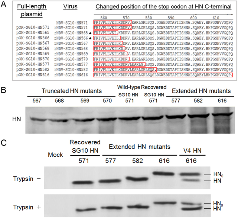
To investigate the role of the length of HN protein in NDV virulence and pathogenesis, the full-length plasmids encoding 10 different length variants of the HN protein were constructed to rescue recombinant viruses (Fig. 1A). Nine viable viruses were recovered with these 10 full-length cDNAs and the full-length cDNA encoding the 565aa HN protein did not recover a viable virus. The supernatants from transfected BSR T7/5 cells were passaged twice in embryonated specific-pathogen-free (SPF) chicken eggs. The allantoic fluids with positive HA titers were processed to isolate the viral RNAs, which were subjected to RT-PCR and sequence analyses. Nucleotide sequencing confirmed the presence of the mutations introduced into the HN genes. These results indicated that nine recombinant viruses were rescued, but rNDV-SG10-HN565 was not (Fig. 1A).
Figure 1. Recovery and identification of recombinant viruses.
(A) Alteration of the length of the HN C-terminus by site-directed mutagenesis. “·”, stop codon; and amino acids at the carboxy terminus of the HN protein are boxed. The two recombinant viruses that could not be successfully recovered are indicated with black triangles. (B) Western blot analysis for HN proteins of recombinant viruses using polyclonal antibody raised against the NDV SG10 strain. (C) The effect of protease treatment on the extended HN mutants. Western blot analysis of HN proteins in Vero cells infected with extended HN mutants processed in the presence and absence of trypsin. Mock infection served as negative control and the V4 strain was used as positive control.
To further determine the stability of the rescued viruses, eight rescued viruses excluding rNDV-SG10-HN566, were successively passaged 10 times through 9-day-old SPF eggs, and the mutated regions in the HN gene were sequenced from the allantoic fluid of each passage. However, a sequence revertant to the original amino acid residue was detected in rNDV-SG10-HN566 at the third passage. The sequencing results indicated that the eight rescued viruses were stable, except rNDV-SG10-HN566. Therefore, these eight rescued viruses, together with the wild-type virus (NDV-SG10-HN571), were used in subsequent experiments.
The various lengths of HN protein were analyzed by western blotting using a polyclonal anti-SG10 strain serum. This confirmed that the correct mutant HN proteins were present where expected (Fig. 1B). Furthermore, to analyse whether the extended HN proteins require the cleavage of amino acids from the C-terminus, western blot analysis of extended HN mutants processed in the presence or absence of trypsin was performed. The results showed that these extended HN proteins could not be proteolytically processed in the presence of trypsin (5 μg/ml) in Vero cells. However, the avirulent strain V4 with HN protein of 616aa, could be observed two band (HN0 and HN) in the absence or presence of trypsin (Fig. 1C). This demonstrated that protease treatment could not trim off the HN616 protein extension mutations of SG10 strain.
Pathogenicity of the recombinant viruses
To evaluate the influence of the length of the HN protein on NDV pathogenicity, the wild-type and rescued viruses were examined with the standard pathogenicity tests used for NDV: the mean death time (MDT) test in 9-day-old embryonated SPF chicken eggs and the intracerebral pathogenicity index (ICPI) test in 1-day-old SPF chicks in vivo. As summarized in Table 1, the MDT values for all eight rescued viruses ranged from 39.0 to 43.5 h, which were very similar to the wild-type NDV-SG10-HN571 value (42 h), and the results of the ICPI test were consistent with the results of the MDT. The ICPI values for all eight rescued viruses and NDV-SG10-HN571 were >1.90. Based on both the MDT and ICPI values, all these viruses were classified as velogenic strains. When the viruses were used to inoculate BSR T7/5 cells at a multiplicity of infection (MOI) of 0.01 PFU/cell. The cytopathogenic effects (CPEs) were observed in the cells infected with each virus at 48 h post-infection (hpi) (see Supplementary Fig. S1). These results indicate that the length of the HN protein has no obvious effect on NDV pathogenicity.
Table 1. Pathogenicity of the parental and recombinant viruses.
| Virus | MDT (h)a | ICPI scoreb |
|---|---|---|
| NDV-SG10-HN571 | 42.0 | 1.95 |
| rNDV-SG10-HN571 | 42.0 | 1.93 |
| rNDV-SG10-HN567 | 40.8 | 1.94 |
| rNDV-SG10-HN568 | 42.0 | 1.93 |
| rNDV-SG10-HN569 | 43.5 | 1.94 |
| rNDV-SG10-HN570 | 39.0 | 1.95 |
| rNDV-SG10-HN577 | 40.8 | 1.91 |
| rNDV-SG10-HN582 | 40.0 | 1.99 |
| rNDV-SG10-HN616 | 43.2 | 2.00 |
aMDT, the mean death times (hour) in embryonated chicken eggs. Criteria for classifying the virulence of NDVs: virulent viruses, <60 h; moderately virulent viruses, 60–90 h; avirulent viruses, >90 h.
bICPI, intracerebral pathogenicity index, determined by inoculating groups of 10 1-day-old SPF chicks with fresh infective allantoic fluid via the intracerebral route. The birds were observed for clinical symptoms and mortality daily for 8 days. At each observation, the birds were scored 0 if normal, 1 if sick, and 2 if dead. The ICPI is the mean score per bird per observation. Criteria for classifying the virulence of NDVs: virulent viruses, 1.5–2.0; moderately virulent viruses, 0.7–1.5; avirulent viruses, 0.0–0.7.
Growth properties of the mutant viruses
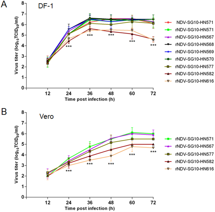
To investigate whether the length of the NDV HN protein affects viral replication in cell culture, the growth characteristics of the wild-type and recombinant viruses were evaluated from their multicycle growth kinetics in a chicken embryo fibroblast cell line (DF-1), and an African green monkey kidney cell line (Vero). The multistep growth curves for the recombinant HN mutant viruses in DF-1 cells (Fig. 2A) showed that the replication kinetics of all the HN-truncated mutant viruses (rNDV-SG10-HN567, rNDV-SG10-HN568, rNDV-SG10-HN569, and rNDV-SG10-HN570) were similar to those of wild-type NDV-SG10-HN571 and rNDV-SG10-HN571, whereas the two HN-extended mutant viruses (rNDV-SG10-HN582 and rNDV-SG10-HN616) showed delayed growth and significantly lower viral yields than the wild-type and parental recombinant viruses at 24 hpi (p < 0.001). rNDV-SG10-HN577 replicated to titers that were intermediate between the extremes of rNDV-SG10-HN570 and rNDV-SG10-HN616. To verify the growth characteristics of the recombinant viruses, Vero cells were infected with five representatives of the HN length mutant viruses (rNDV-SG10-HN571, rNDV-SG10-HN567, rNDV-SG10-HN577, rNDV-SG10-HN582 and rNDV-SG10-HN616). As shown in Fig. 2B, the trends in the viral growth curves in Vero cells were similar to those in DF-1 cells, although the growth rates of the five HN mutant viruses in Vero cells were lower than those in DF-1 cells. These results indicate that the HN protein truncation mutations negligibly influenced the replication efficiency of NDV in cells, but that the extension mutations substantially reduced the efficiency of NDV replication.
Figure 2. Growth characteristics of recombinant NDVs.
Multiple-cycle growth kinetics were used to assess the differences in the growth of these viruses. (A) DF-1 cells were infected with rNDV-SG10-HN571, rNDV-SG10-HN567, rNDV-SG10-HN568, rNDV-SG10-HN569, rNDV-SG10-HN570, rNDV-SG10-HN577, rNDV-SG10-HN582, or rNDV-SG10-HN616 at an MOI of 0.01 PFU/cell, and assayed as described in the Methods. (B) Vero cells were infected with rNDV-SG10-HN571, rNDV-SG10-HN567, rNDV-SG10-HN577, rNDV-SG10-HN582, or rNDV-SG10-HN616 at an MOI of 0.01 PFU/cell, and assayed as described in the Methods. Briefly, supernatant samples were collected at 12-h intervals for 72 h, and the viral titers were determined with a TCID50 limiting dilution assay, and calculated with the method of Reed and Muench. Means and standard deviations were shown from three independent experiments. Asterisks indicate the significance of the difference between the recombinant viral titer and the parental viral titer. P values were calculated with Tukey’s test (95% confidence levels). ***p < 0.001, extremely significant.
Hemagglutination (HA) titers of recombinant viruses
To evaluate the influence of the length of the HN protein on NDV HA titer, HA assay of the five representative HN length mutant viruses were performed. As shown in Table 2, at the virus titers of 7.0 and 6.0 log10 TCID50/ml, the rNDV-SG10-HN567 HA titer was slightly lower than parental virus rNDV-SG10-HN571, whereas the HA titers of HN-extended mutant viruses (rNDV-SG10-HN577, rNDV-SG10-HN582 and rNDV-SG10-HN616) were higher than the parental virus, especially the rNDV-SG10-HN582 HA titer existed significant differences (p < 0.05). The result suggests that the extension of HN protein leads to a moderate increase for the HA titer of NDV.
Table 2. Hemagglutination (HA) titers of the parental and recombinant viruses.
| Virus | HA titersa (log2) of different virus titersb (log10 TCID50/ml) | |
|---|---|---|
| 7.0 | 6.0 | |
| rNDV-SG10-HN571 | 5.17 ± 0.58 | 2.17 ± 0.58 |
| rNDV-SG10-HN567 | 4.67 ± 0.29 | 1.17 ± 0.76 |
| rNDV-SG10-HN577 | 5.33 ± 0.29 | 2.33 ± 0.29 |
| rNDV-SG10-HN582 | 7.17 ± 0.58* | 4.33 ± 0.29* |
| rNDV-SG10-HN616 | 6.17 ± 0.76 | 3.17 ± 0.58 |
aHA titers in microplate format were expressed in log2 of the mean ± standard deviation from three independent tests.
bVirus titers were expressed in log10 TCID50/ml on DF-1 cells. Asterisks at the same virus titer level indicate statistically significant differences (p < 0.05) of the HA titers compared with rNDV-SG10-HN571. P values were calculated with Tukey’s test (95% confidence levels).
Hemadsorption and neuraminidase activities of the recombinant viruses and expressed HN protein mutants
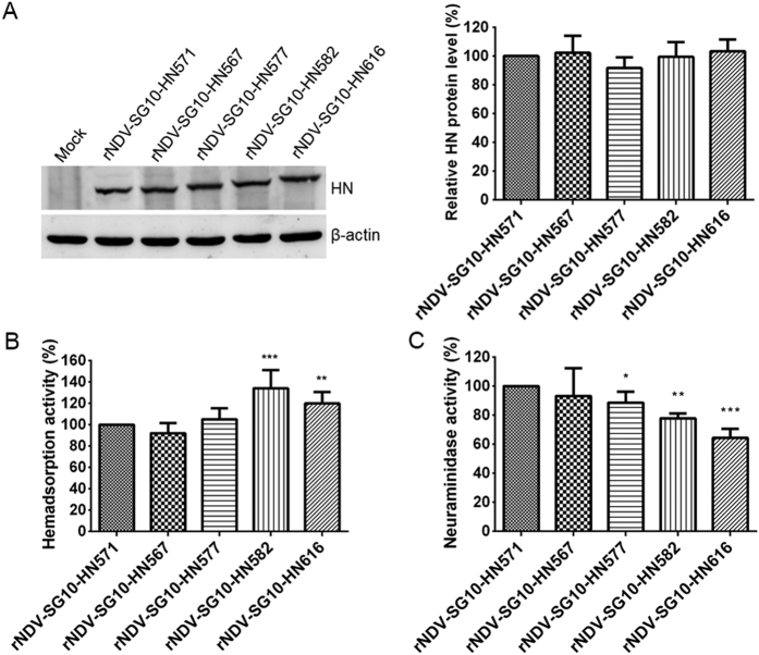
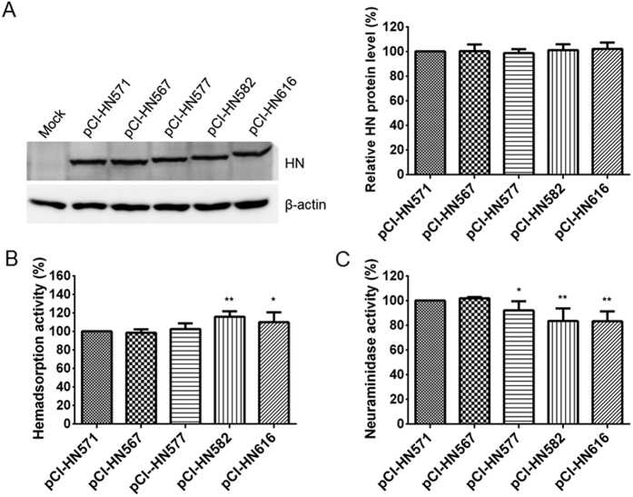
To determine whether the length of the NDV HN protein modulates the biological activities of NDV in cultured cells, the five representative HN length mutant viruses were assessed for their hemadsorption (HAd) and neuraminidase (NA) activities. Firstly, the HN protein expression of five representative HN length mutant viruses was measured in Vero cells. The HN expression of the various HN length mutants was similar to that of parental HN (Fig. 3A). Subsequently, the biological activities of each recombinant virus were analyzed and calculated as a percentage of that of rNDV-SG10-HN571, whose biological activities were deemed to be 100%. The HAd activity of rNDV-SG10-HN582 was 134% of that of rNDV-SG10-HN571, which was the greatest increase in all the mutants, followed by 120% for rNDV-SG10-HN616. The HAd activities of rNDV-SG10-HN567 and rNDV-SG10-HN577 did not differ significantly from those of rNDV-SG10-HN571 (Fig. 3B). All the extended HN length mutant viruses showed significantly reduced NA activity relative to that of the parental recombinant virus, with the following values (relative to rNDV-SG10-HN571, 100%): rNDV-SG10-HN577, 89%; rNDV-SG10-HN582, 78%; and rNDV-SG10-HN616, 64% (Fig. 3C). Furthermore, the HAd and NA activities of HN mutants at the protein level were performed using HN expression plasmids (pCI-HN571, pCI-HN567, pCI-HN577, pCI-HN582, pCI-HN616). The result was almost the same as the viral level. The HAd activities of HN582 and HN616 proteins were markedly increased than those of HN571 under the similar HN expression (Fig. 4A,B). The NA activities of all extended HN protein mutants were significantly decreased than those of HN571 (p < 0.05 or 0.01) (Fig. 4C). These results suggested that the extension of HN protein increased the hemadsorption capacity, but reduced the NA activity at viral and protein levels.
Figure 3. HAd and NA activities of parental and recombinant viruses.
(A) The amount of each HN protein was determined by western blot analysis at 24 h post-infection of Vero cells by using anti-SG10 serum. HAd (B) and NA activities (C) were examined in Vero cells infected with the virus at an MOI of 0.1 PFU/cell. HN protein expression level, HAd and NA activities of these viruses are expressed as percentages of the values for rNDV-SG10-HN571, which are deemed to be 100%. Each bar represents the mean and standard deviation of three independent experiments. Asterisks indicate the significance of the differences between the biological activity of a recombinant virus and that of the parental virus. P values were calculated with Tukey’s test (95% confidence levels). *p < 0.05, significant; **p < 0.01, very significant; ***p < 0.001, extremely significant.
Figure 4. HAd and NA activities of expressed HN protein mutants.
(A) The amount of each HN protein was determined by western blot analysis at 24 h post-transfection of Vero cells by using anti-SG10 serum. HAd (B) and NA activities (C) were examined in Vero cells transfected with the 0.5 μg each of pCI-HN mutant plasmids. HN protein expression level, HAd and NA activities of these HN mutants are expressed as percentages of the values for expressed HN571 protein (pCI-HN571), which are deemed to be 100%. Each bar represents the mean and standard deviation of three independent experiments. Asterisks indicate the significance of the differences between the biological activity of an HN protein mutant and that of the HN571 protein. P values were calculated with Tukey’s test (95% confidence levels). *p < 0.05, significant; **p < 0.01, very significant.
Syncytium formation induced by recombinant viruses and expressed HN protein mutants
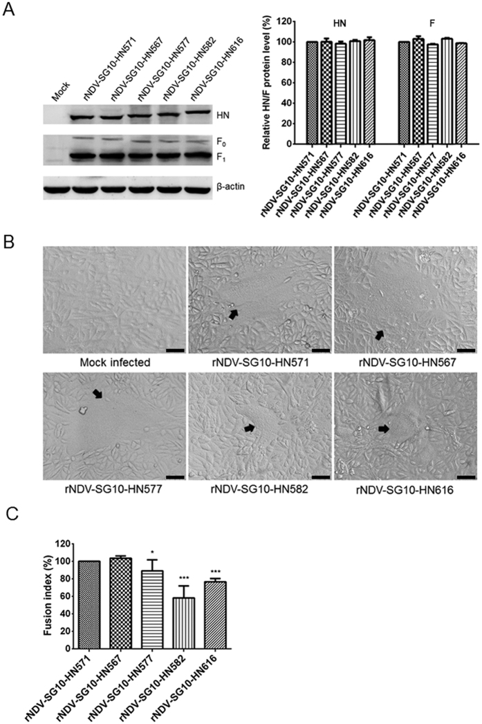
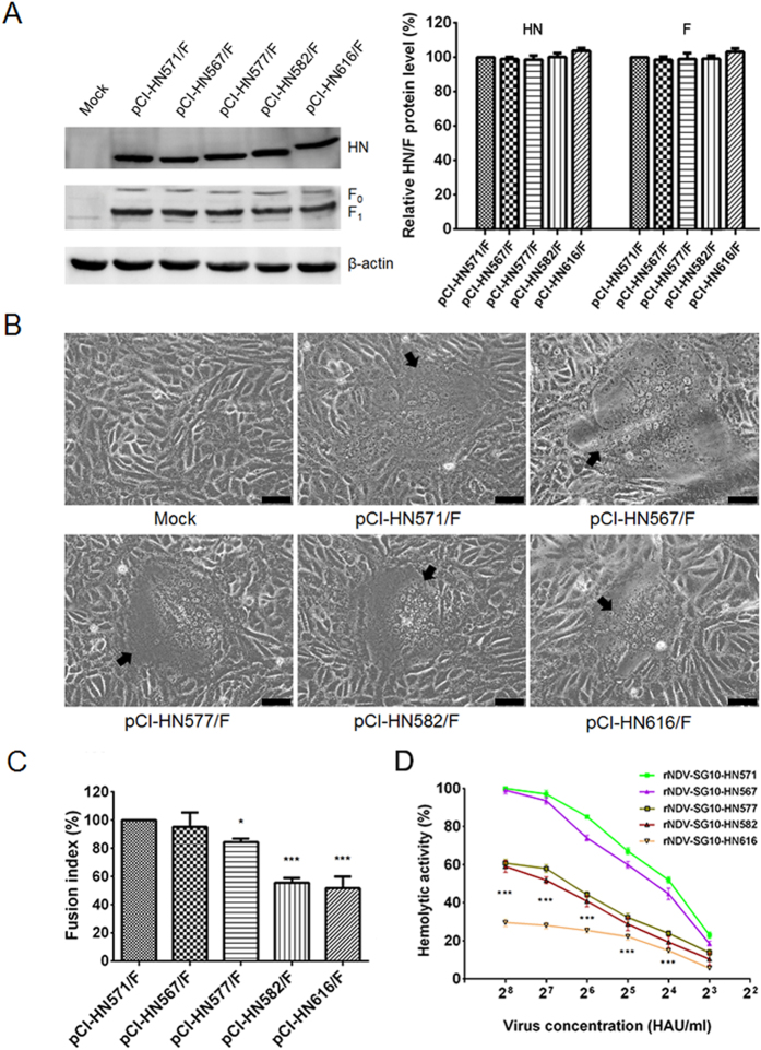
First, western blot analysis based on protein lysates, derived from Vero cells infected with recombinant viruses, indicated that the HN and F (F0 and F1) expressions of the various HN length mutants was similar to that of parental virus (Fig. 5A). Under these conditions, a fusion index assay was performed to measure the ability of the parental and recombinant viruses to induce syncytium formation in Vero cells. All the HN extension mutant viruses decreased the fusion ability in Vero cells, and the syncytia induced by rNDV-SG10-HN577, rNDV-SG10-HN582 and rNDV-SG10-HN616 were markedly smaller than those induced by the parental virus (Fig. 5B). Quantification of the fusogenic abilities of the mutants (fusion index compared with the parental level) showed that rNDV-SG10-HN577, rNDV-SG10-HN582, and rNDV-SG10-HN616 had significantly reduced fusogenic activity (89%, 58%, and 77%, respectively) (p < 0.05 or 0.001) (Fig. 5C). Subsequently, the fusogenic promotion activities of truncated or extended HN mutants at the protein level were performed using expression plasmids pCI-HN571, pCI-HN567, pCI-HN577, pCI-HN582, pCI-HN616 and pCI-F. The result was almost consistent with the viral level. The HN577, HN582 and HN616 showed significantly reduced fusogenic promotion activity (84%, 56%, and 52%, respectively) compared with HN571 under the similar HN and F expression (p < 0.05 or 0.001) (Fig. 6A–C). These results indicate that the extension of the HN protein seriously impairs the fusogenic ability at viral and protein levels, whereas truncation mutations of the HN protein have no statistically significant influence on the fusogenic capacity.
Figure 5. Syncytium formation of parental or recombinant viruses.
(A) The amount of each HN or F (F0 and F1) protein was determined by western blot analysis at 48 h post-infection of Vero cells by using anti-SG10 serum or anti-NDV F rabbit polyclonal antiserum. (B) At 48 h post-infection, Vero cell monolayers that had been infected with parental or recombinant NDV at an MOI of 0.1 PFU/cell in six-well plates were digitally photographed under an inverted microscope (IX73; Olympus) at ×100 magnification. Black arrows indicate syncytia. Bar = 50 μm. (C) The fusion index values of these viruses were calculated as the ratio of the total number of nuclei to the number of cells in which the nuclei were observed. All values are expressed relative to the value for rNDV-SG10-HN571 (100%). Each bar represents the mean and standard deviation of three independent experiments. Asterisks indicate the statistically significant differences. P values were calculated with Tukey’s test (95% confidence levels). *p < 0.05, significant; ***p < 0.001, extremely significant.
Figure 6. Syncytium formation in Vero cells co-transfected with expressed plasmids and hemolytic activities of parental or recombinant viruses.
(A) The amount of each HN or F (F0 and F1) protein was determined by western blot analysis at 48 h post-transfection of Vero cells by using anti-SG10 serum or anti-NDV F rabbit polyclonal antiserum. (B) At 48 h post-transfection, Vero cell monolayers that had been co-transfected with 1 μg each of pCI-F and pCI-HN mutant plasmids in six-well plates were digitally photographed under an inverted microscope (IX73; Olympus) at ×100 magnification. Black arrows indicate syncytia. Bar = 50 μm. (C) The fusion index values of these HN mutants were calculated as the ratio of the total number of nuclei to the number of cells in which the nuclei were observed. All values are expressed relative to the value for pCI-HN571/F (100%). (D) The hemolytic values for all the viruses were expressed as percentages of the values for rNDV-SG10-HN571 at 28 hemagglutination unit/ml, which were considered to be 100%. Each bar represents the mean and standard deviation of three independent experiments. Asterisks indicate the significance of the difference between the fusion index of a recombinant virus and that of the parental virus. P values were calculated with Tukey’s test (95% confidence levels). *p < 0.05, significant; ***p < 0.001, extremely significant.
Hemolytic activities of the recombinant viruses
To evaluate the influence of the length of the HN protein on NDV hemolytic activity, five representative HN length mutant viruses were tested at virus concentration ranged from 23–28 hemagglutination unit/ml (HAU/ml). As shown in Fig. 6D, the hemolytic activity of rNDV-SG10-HN567 was slightly lower than parental virus rNDV-SG10-HN571, whereas HN-extended mutant viruses (rNDV-SG10-HN577, rNDV-SG10-HN582 and rNDV-SG10-HN616) were significantly decreased than that of rNDV-SG10-HN571 (p < 0.001). The rNDV-SG10-HN616 hemolytic activity had the greatest reduction in all the mutants. These results indicate that the extension of the HN protein mutations seriously impairs the hemolytic activity, which is consistent with the fusogenic ability at viral and protein levels.
Discussion
The HN protein of NDV plays important roles in viral invasion and maturation, with three distinct activities: receptor binding, NA activity (receptor cleavage) and fusion promotion18,35,36,37,38. The HN protein has been shown to contribute greatly to NDV pathogenesis22,23,39. Previous studies have also suggested that even a single point variation in the different regions of the HN protein can markedly affect the biological activities of the protein or the virus40,41,42,43,44. In addition, for other paramyxovirus, measlesvirus attachment (H) protein stalk-length variation through deletion or insertion of heptad repeat (HR) elements at positions flanking this section including residues 111, 114, and 118 could alter fusion complexes function45. Vesicular stomatitisvirus (VSV) is a nonsegmented, negative-sense single-stranded RNA virus, as well as NDV. Changes in the transmembrane domain (TMD) length of VSV spike protein G could affect secretory transport dynamics and the capacity of the transport machinery to concentrate cargo46. However, the contribution of HN protein length diversity to NDV has not been thoroughly investigated. In this study, a series of recombinant NDVs containing truncated and extended HN proteins were generated with a reverse genetics technique to evaluate the contribution of the HN protein length to the pathogenicity, replication and biological characteristics of NDV in vivo and in vitro. The extension of the HN protein increased the HA titer, HAd activity and reduced the NA activity, fusion index, hemolytic activity and replication capacity of NDV, without any observable changes in viral pathogenicity.
The HN protein length diversity of NDV mainly arised from differences in the position of the stop codon of the HN ORF26,27,28. To explore the distribution of various HN protein length in NDVs of different clades and virulence, we analyzed all NDV HN sequences available in the National Center for Biotechnology Information (NCBI). As shown in Supplementary Table S1, there were 12 different HN protein length, with different length showing a clade and virulence preference. Most class І (76.1%) and class II genotype І (75.4%) viruses had HN protein of 616aa, 89.5% of class II genotype II viruses had HN protein of 577aa, and HN protein of class II genotype III (100%), IV (100%), V (100%), VI (97.9%), VII (92.3%), VIII (50.0%), IX (96.2%) contained 571aa. The virulence of 347 isolates is known, 84.8% of these virulent isolates have HN protein of 571aa, while most low-virulence strains contain HN proteins of 577aa (33.0%) and 616aa (56.7%). In the present study, a genotype VIId NDV strain (SG10) with a 571aa HN protein was used as the model virus to construct the recombinant NDVs encoding truncated or extended HN proteins47. The MDT and ICPI results indicated that mutations affecting the length of HN did not alter the virulence of the mutant viruses in embryonated chicken eggs or 1-day-old chickens. These results are consistent with a previous report of an avirulent NDV strain (Clone-30) with a 577aa HN protein29. This suggests that NDV pathogenicity is not influenced by the length of the HN protein, in either velogenic or lentogenic strains. The 570aa HN protein is the shortest length detected, as previously reported. A recombinant NDV expressing a 567aa HN protein (rNDV-SG10-HN567) was successfully recovered in this study. During the rescue of the recombinant virus, a recombinant NDV containing a 565aa HN protein (rNDV-SG10-HN565) was not recovered and a sequence revertant of rNDV-SG10-HN566 was recovered after passage in embryonated SPF chicken eggs. These results indicate that the length of the HN protein cannot be reduced indefinitely, and we suppose that this may relate to the evolution of NDV.
The Ulster, Queensland, V4 strains or some other avirulent strains with HN616, produce an HN protein precursor (HN0 protein) which requires proteolytic activation before the virons can attach to the receptors on host cells48,49. Schuy et al. reported that the HN0 protein of the Ulster strain was cleaved by trypsin in vitro and in vivo to generate the HN protein of approximately 571 amino acids50. In the present study, the extended HN protein of rNDV-SG10-HN616 was not cleaved by trypsin in vitro. These may well be closely related to the extended amino acid sequences and NDV virulence. The originally protein non-coding region was forced to be a coding region. Hence, the extended amino acid residues are likely to be unnatural. The extended 45 amino acid residues of rNDV-SG10-HN616 and avirulent strains with HN616 vary considerably with homology <45%. In addition, rNDV-SG10-HN616 is still a virulent strain. The results are in accordance with the viewpoint that only some avirulent NDV strains produce an HN0 protein precursor12,48,49.
The growth characteristics of the recombinant viruses containing a series of truncated or extended HN proteins were assessed based on their multicycle growth kinetics in DF-1 and Vero cells. The recombinant viruses expressing extended HN proteins (577aa, 582aa, and 616aa) showed reduced viral titers in vitro compared with those of the parental virus and the recombinant viruses containing truncated HN proteins. Interestingly, the reduced efficiency of viral replication in the cells did not reduce the virulence of NDV. These data seem to contradict the consistency between NDV replication and pathogenicity previously reported51,52, but probably attributable to differences in viral reproduction in vivo and in vitro. For example, Kim et al. reported that mutations at the fusion protein cleavage site of avian paramyxovirus conferred increased replication in vitro but did not increase its replication or pathogenicity in chickens and ducks53. Therefore, future studies should focus on the replication capacity and tissue tropism in vivo of recombinant viruses containing HN proteins of different lengths. What is more, these results seem to suggest that the replication ability of these viruses is not closely related to their pathogenicity. And the data are similar to the report that NDV M protein affected viral replication in vitro, but only had a minor effect on virulence54. Although the exact mechanism is not clear, it is likely to be related to the balance among the HAd, NA and hemolytic activities which ultimately determines the outcome of infection. We suppose that the decrease of NA and hemolytic activities mainly results in the impairment of replication ability, while the increase of HAd activity is likely to maintain the NDV virulence. Another plausible explanation is that when viral replication reaches a certain level, the higher virus titer does not have greater impact on the virulence. In this study, all the viruses maintain at high replication levels although the different virus titers exist. Thus, the replication of the two HN-extended mutant viruses (rNDV-SG10-HN582 and rNDV-SG10-HN616) was decreased almost 1.0 log10 compared with the parental strain at 24 hpi in DF-1 and Vero cells, but it is insufficient to alter the virulence.
Our in vitro studies of NDV support the hypothesis that the length of the HN protein affects some biological functions of the virus. Extension of the HN protein increased NDV HAd activity, reduced its NA activity and fusion index. These results emphasize the importance of the 571aa HN protein in the NA and fusogenic activities of NDV strain SG10. The impairment of the NA and fusogenic activities induced by the extension of the HN protein is consistent with the viral replication observed in cells. However, the HAd activity of HN appeared to be independent of its NA and fusogenic activities. Our results suggest that these altered activities could affect viral growth in vitro and the extension of HN allows viral attachment but prevents virion release during the life cycle of NDV.
Extension of the HN protein likely reduces the interaction between HN and F, leading to the lower fusion indices and hemolytic activities of the recombinant viruses expressing extended HN proteins. The exact mechanism is unclear, but it is likely to be related to the second receptor-binding site (site II), which is engaged by the C-terminal extension of the NDV HN protein, because it plays an active role in F activation10,19,55. Our data showed that the recombinant viruses containing HN extension mutations displayed substantially reduced viral titers in vitro. It is possible that, although these viruses had higher receptor affinity, their fusogenic and NA activities were diminished, reducing the efficiency of their replication in infected monolayers. Interestingly, the extension of the HN protein did not alter NDV virulence, as shown with MDT and ICPI tests, which seems to indicate that the fusogenic and NA activities of NDV are not related to its pathogenicity, or that these various assays have different sensitivities. These results also suggest that the three functions of HN are flexible and balanced at the virus level.
In summary, the contributions of HN protein length to the pathogenicity, replication and biological characteristics of NDV were investigated using recombinant viruses expressing truncated or extended HN proteins, which were rescued with a reverse genetics method. The extension of the HN protein affected the hemadsorption, neuraminidase and fusogenic activities of NDV. The balance among the three functions of HN determines the biological characteristics of NDV. We have also shown that the length of the HN protein cannot be reduced indefinitely and does not affect the virulence of NDV. These findings may also be applicable to other paramyxoviruses.
Materials and Methods
Viruses and cells
The recombinant viruses were generated in our laboratory from a full-length cDNA copy of the velogenic NDV strain rSG10, as described previously47. The NDV strain SG10 (an epidemic virulent strain in China of genotype VII) and V4 (an avirulent strain of genotype I) were used as the control. BSR T7/5 cells stably expressing the T7 phage RNA polymerase, a chicken embryo fibroblast cell line (DF-1), and an African green monkey kidney cell line (Vero) were all grown in Dulbecco’s modified eagle medium (DMEM, Gibco, Grand Island, NY, USA) with 10% fetal bovine serum (FBS, Gibco), and maintained in DMEM with 2% FBS at 37 °C under a 5% CO2 atmosphere.
Mutation of full-length NDV cDNA and virus rescue
The previously generated full-length cDNA clone, pOK-rSG10-HN57147, was used as the backbone upon which to construct a series of mutants containing truncated or extended HN genes, as illustrated in Fig. 1A. To change the NDV HN protein length, the position of the stop codon in the HN ORF was altered with site-directed mutagenesis. All the used primer sequences are listed in Supplementary Table S2. The resulting full-length plasmids encoding the truncated or extended HN proteins were used to recover the recombinant viruses with a reverse genetics procedure. Virus rescue was performed as described previously47.
Construction of HN and F protein expression plasmids
The HN and F genes of rNDV-SG10-HN571, rNDV-SG10-HN567, rNDV-SG10-HN577, rNDV-SG10-HN582 and rNDV-SG10-HN616 were amplified from full-length cDNA clones as described above by RT-PCR and were inserted into protein-expression plasmid pCI-neo (pCI) (Promega, Madison, USA) between the XhoI and MluI restriction sites. The plasmids obtained were named pCI-HN571, pCI-HN567, pCI-HN577, pCI-HN582, pCI-HN616 and pCI-F. Primers used to amplify the HN and F genes are listed in Supplementary Table S3.
Reverse transcription (RT)-PCR, sequence analysis
Viral RNA was extracted from 100 μl of allantoic fluid with the Simply P Total RNA Extraction kit (BioFlux, Hangzhou, China), according to the manufacturer’s instructions. The fragments that included the altered HN sequences were amplified with RT-PCR using specific primers T-F and T-R (see Supplementary Table S2). The PCR products were sequenced directly with specific primers by Tsingke (Beijing, China). The nucleotide sequences were assembled, aligned, and compared with the DNASTAR program (Madison, WI, USA).
Western blot analysis
Total cell protein lysates were extracted from infected or transfected Vero cells with ice-cold RIPA lysis buffer (50 mMTris [pH 7.4], 150 mM NaCl, 1% Triton X-100, 1% sodium deoxycholate). Cellular proteins were separated by 10% sodium dodecyl sulfate-polyacrylamide gel electrophoresis (SDS-PAGE) and transferred to a polyvinylidenedifluoride (PVDF) membrane (Amersham Biosciences, Germany). Each PVDF membrane was blocked with 5% (w/v) nonfat dry milk and 0.1% Tween 20 in Tris buffered saline (TBST) and subsequently incubated with a primary antibody overnight at 4 °C. Primary antibodies were SG10 polyclonal serum or anti-NDV F rabbit polyclonal antiserum (both diluted 1:100 and preserved in our laboratory), β-actin mouse monoclonal antibody (diluted 1:1000, Beyotime Biotechnology, Beijing, China). After being washed with TBST, the membranes were incubated for 1 h with corresponding horseradish peroxidase (HRP)-conjugated anti-chicken, anti-rabbit or anti-mouse antibody at a 1:10000 dilution (Bioss Biotechnology, Beijing, China). HRP presence was detected using a Western Lightning chemiluminescence kit (CWBIO, Beijing, China) in accordance with the manufacturer’s protocol. The load protein was normalized to β-actin. Protein bands were quantified by densitometry using ImageJ software (National Institute of Mental Health, Bethesda, MD, USA).
Viral pathogenicity
The pathogenicity of the wild-type and recombinant viruses was determined with standard pathogenicity tests for NDV: MDT in 9-day-old embryonated SPF chicken eggs and ICPI in 1-day-old SPF chicks2. Their pathogenicity of the mutant NDVs in BSR T7/5 cells was examined by evaluating CPEs they exerted.
Viral growth characteristics
The growth dynamics of nine NDVs, including the wild-type, parental, and recombinant viruses, were evaluated in DF-1 cells. The growth characteristics of five of these viruses were then confirmed in Vero cells. Cells in duplicate wells of six-well plates were infected with NDVs at an MOI of 0.01 PFU/cell, and then incubated with DMEM containing 2% FBS at 37 °C in 5% CO2. The culture supernatants were collected at 12 h intervals until 72 h after infection. The viral titers in the collected supernatants were quantified in DF-1 and Vero cells and expressed as median tissue culture infective doses (TCID50), using the endpoint method of Reed and Muench56.
Hemagglutination (HA) titers of recombinant viruses and Hemadsorption (HAd) assay
HA titres of recombinant viruses at the different virus titers level (6.0 and 7.0 log10 TCID50/ml) were obtained by using the microplate HA test. The HAd activity was assayed as the ability of the recombinant viruses to adsorb to chicken red blood cells (CRBCs), as described previously36, with modifications. Confluent monolayers of Vero cells in 24-well plates were inoculated with virus at an MOI of 0.1 PFU/cell. At 24 h post-infection (hpi), the medium was removed and the cells were washed with cold PBS and then incubated for 30 min at 4 °C with a 2% (vol/vol) suspension of CRBCs. After the unbound CRBCs were removed with two washes of ice-cold PBS, the CRBCs bound to the virus-infected cells were lysed with a red-blood-cell lysis solution (0.145 M NH4Cl, 17 mM Tris-HCl). The lysate was clarified by centrifugation and transferred to 96-well plates. Absorbance was measured at 549 nm with a Spectramax M5 ELISA reader (Molecular Devices, Sunnyvale, CA, USA). The HAd assay at the protein level was performed with monolayers of Vero cells transfected using 0.5 μg each of pCI-HN mutant plasmids (pCI-HN571, pCI-HN567, pCI-HN577, pCI-HN582, pCI-HN616) as above described.
NA assay
The NA activity was determined with a fluorimetric procedure, as described by Potier et al.37, with some modifications. Vero cell monolayers in 96-well plates were inoculated with NDVs at an MOI of 0.1 PFU/cell. At 24 hpi, the cells were washed with cold PBS and overlaid with 30 μl of substrate mix (one volume of 325 mM2-N-morpholinoethanesulfonic acid [MES; pH 6.4], two volumes of 0.5 mM 2′-(4-methylumbelliferyl)-α-D-N-acetylneuraminic acid [MUN; Sigma, St. Louis, MO, USA], and three volumes of 10 mM calcium chloride) per well, to give a final concentration of 100 μM MUN in the assay. Cells were incubated at 37 °C for 30 min with shaking, and the reaction was terminated by the addition of 0.014 M sodium hydroxide in 83% (vol/vol) ethanol. The fluorescence intensity was measured at an excitation wavelength of 360 nm and an emission wavelength of 450 nm with a Spectramax M5 ELISA reader (Molecular Devices). The NA assay at the protein level was performed with monolayers of Vero cells transfected using the same expression plasmids utilized in the HAd assay.
Fusion index assay
The fusogenic abilities of the parental and recombinant viruses were examined in Vero cells, as described previously22. Confluent (106 cells/ml) Vero cell monolayers in six-well plates were infected with NDVs at an MOI of 0.1 PFU/cell and maintained in DMEM with 2% FBS at 37 °C under 5% CO2. After the CPE was monitored for 48–72 h, the medium was removed and the cells were washed once with 0.02% EDTA, and then incubated with 1 ml of EDTA for 2 min at room temperature. The cells were washed with PBS and fixed with methanol for 20 min at room temperature, and then stained with hematoxylin-eosin. Fusion was quantitated with the fusion index: the ratio of the total number of nuclei to the number of cells in which these nuclei were observed (i.e., the mean number of nuclei per cell). The fusion index assay at the protein level was performed with monolayers of Vero cells co-transfected using 1 μg each of pCI-F and pCI-HN mutant plasmids. The HAd, NA and fusion index values for all the viruses or HN protein mutants were expressed as percentages of the values for the parental virus (rNDV-SG10-HN571) or the expressed HN571 protein (pCI-HN571), which were considered to be 100%.
Hemolytic titrations
The hemolytic activities of the parental and recombinant viruses were performed according to previously published methods57. In brief, the allantoic fluids of the rescued viruses were clarified by centrifugation (4 °C, 20 min, 500 g). Supernatant fluids of different NDVs were diluted to the same hemagglutination unit/ml (HAU/ml) based on the HA titer. The diluted virus sample 0.5 ml was mixed with 1 ml of 1% suspension of chicken erythrocytes. After incubated on ice for 20 min with occasional shaking, the erythrocytes were sedimented by centrifugation at 500 g for 3 min, washed with phosphate-buffered saline (PBS) and resuspended in the same buffer (0.5 ml). After incubation for 60 min at 37 °C, tubes were then centrifuged at 200 g for 5 min. Supernatant fluids were then transferred to 96-well plates and their hemoglobin contents were measured as absorbance at 549 nm with a Spectramax M5 ELISA reader (Molecular Devices). As for the positive control, 0.03 M NH4OH was added to the assay and for the negative control, the PBS was used. The hemolytic values for all the viruses were expressed as percentages of the values for rNDV-SG10-HN571 at 28 HAU/ml, which were considered to be 100%.
Statistical analysis
All statistical significance of differences was performed using Prism 5.0 program (GraphPad Software Inc., San Diego, CA, USA). Statistical differences between repeats were determined using the analysis of variance (ANOVA) method followed by Tukey’s test. Statistical significance was set at p < 0.05 (*), p < 0.01 (**) and p < 0.001 (***), respectively.
Additional Information
How to cite this article: Jin, J. et al. Contribution of HN protein length diversity to Newcastle disease virus virulence, replication and biological activities. Sci. Rep. 6, 36890; doi: 10.1038/srep36890 (2016).
Publisher’s note: Springer Nature remains neutral with regard to jurisdictional claims in published maps and institutional affiliations.
Supplementary Material
Acknowledgments
This study was supported by the Beijing Agriculture Innovation Consortium of Poultry Research System (BAIC04-2016). The funders had no role in study design, data collection and interpretation, or the decision to submit the work for publication.
Footnotes
Author Contributions J.J. and G.Z. contributed to the design of experiments. J.J., J.Z., Y.R. and G.Z. contributed to the analysis and interpretation of data. J.J. and Q.Z. wrote the paper and created the figures. All authors discussed the results. J.Z. and G.Z. supervised the work. G.Z. administered the project.
References
- Alexander D. J. Newcastle disease. Br. Poult. Sci. 42, 5–22 (2001). [DOI] [PubMed] [Google Scholar]
- Alexander D. J. & Swayne D. E. Newcastle disease virus and other avian paramyxoviruses. A laboratory manual for the isolation and identification of avian pathogens 4, 156–163 (1998). [Google Scholar]
- Mayo M. A summary of taxonomic changes recently approved by ICTV. Arch. Virol. 147, 1655–1656 (2002). [DOI] [PubMed] [Google Scholar]
- Miller P. J. & Koch G. Newcastle disease, other avian paramyxoviruses, and avian metapneumovirus infections. Diseases of poultry 13th ed. 89–138 (2013). [Google Scholar]
- Knipe D., Lamb R. & Parks G. Paramyxoviridae: the viruses and their replication. Fields virology 5th ed. 1449–1496 (2007). [Google Scholar]
- Steward M., Vipond I. B., Millar N. S. & Emmerson P. T. RNA editing in Newcastle disease virus. J. Gen. Virol. 74, 2539–2547 (1993). [DOI] [PubMed] [Google Scholar]
- Connaris H. et al. Probing the Sialic Acid Binding Site of the Hemagglutinin-Neuraminidase of Newcastle Disease Virus: Identification of Key Amino Acids Involved in Cell Binding, Catalysis, and Fusion. J. Virol. 76, 1816–1824 (2002). [DOI] [PMC free article] [PubMed] [Google Scholar]
- Connolly S. A., Leser G. P., Jardetzky T. S. & Lamb R. A. Bimolecular complementation of paramyxovirus fusion and hemagglutinin-neuraminidase proteins enhances fusion: implications for the mechanism of fusion triggering. J. Virol. 83, 10857–10868 (2009). [DOI] [PMC free article] [PubMed] [Google Scholar]
- Scheid A. & Choppin P. W. Isolation and purification of the envelope proteins of Newcastle disease virus. J. Virol. 11, 263–271 (1973). [DOI] [PMC free article] [PubMed] [Google Scholar]
- Porotto M. et al. The second receptor binding site of the globular head of the Newcastle disease virus hemagglutinin-neuraminidase activates the stalk of multiple paramyxovirus receptor binding proteins to trigger fusion. J. Virol. 86, 5730–5741 (2012). [DOI] [PMC free article] [PubMed] [Google Scholar]
- Yuan P. et al. Structure of the Newcastle disease virus hemagglutinin-neuraminidase (HN) ectodomain reveals a four-helix bundle stalk. Proc. Nat. l Acad. Sci. USA 108, 14920–14925 (2011). [DOI] [PMC free article] [PubMed] [Google Scholar]
- Yuan P., Paterson R. G., Leser G. P., Lamb R. A. & Jardetzky T. S. Structure of the ulster strain newcastle disease virus hemagglutinin-neuraminidase reveals auto-inhibitory interactions associated with low virulence. PLoS Pathog. 8, e1002855 (2012). [DOI] [PMC free article] [PubMed] [Google Scholar]
- McGinnes L., Sergel T. & Morrison T. Mutations in the transmembrane domain of the HN protein of Newcastle disease virus affect the structure and activity of the protein. Virology 196, 101–110 (1993). [DOI] [PubMed] [Google Scholar]
- Stone-Hulslander J. & Morrison T. G. Mutational analysis of heptad repeats in the membrane-proximal region of Newcastle disease virus HN protein. J. Virol. 73, 3630–3637 (1999). [DOI] [PMC free article] [PubMed] [Google Scholar]
- Crennell S., Takimoto T., Portner A. & Taylor G. Crystal structure of the multifunctional paramyxovirus hemagglutinin-neuraminidase. Nat. Struct. Biol. 7, 1068–1074 (2000). [DOI] [PubMed] [Google Scholar]
- Iorio R. M. et al. Structural and functional relationship between the receptor recognition and neuraminidase activities of the Newcastle disease virus hemagglutinin-neuraminidase protein: receptor recognition is dependent on neuraminidase activity. J. Virol. 75, 1918–1927 (2001). [DOI] [PMC free article] [PubMed] [Google Scholar]
- Melanson V. R. & Iorio R. M. Addition of N-glycans in the stalk of the Newcastle disease virus HN protein blocks its interaction with the F protein and prevents fusion. J. Virol. 80, 623–633 (2006). [DOI] [PMC free article] [PubMed] [Google Scholar]
- Melanson V. R. & Iorio R. M. Amino acid substitutions in the F-specific domain in the stalk of the newcastle disease virus HN protein modulate fusion and interfere with its interaction with the F protein. J. Virol. 78, 13053–13061 (2004). [DOI] [PMC free article] [PubMed] [Google Scholar]
- Zaitsev V. et al. Second sialic acid binding site in Newcastle disease virus hemagglutinin-neuraminidase: implications for fusion. J. Virol. 78, 3733–3741 (2004). [DOI] [PMC free article] [PubMed] [Google Scholar]
- Bousse T. L. et al. Biological significance of the second receptor binding site of Newcastle disease virus hemagglutinin-neuraminidase protein. J. Virol. 78, 13351–13355 (2004). [DOI] [PMC free article] [PubMed] [Google Scholar]
- Porotto M. et al. Inhibition of parainfluenza virus type 3 and Newcastle disease virus hemagglutinin-neuraminidase receptor binding: effect of receptor avidity and steric hindrance at the inhibitor binding sites. J. Virol. 78, 13911–13919 (2004). [DOI] [PMC free article] [PubMed] [Google Scholar]
- Kim S. H., Subbiah M., Samuel A. S. & Collins P. L. & Samal S. K. Roles of the fusion and hemagglutinin-neuraminidase proteins in replication, tropism, and pathogenicity of avian paramyxoviruses. J. Virol. 85, 8582–8596 (2011). [DOI] [PMC free article] [PubMed] [Google Scholar]
- Huang Z. et al. The Hemagglutinin-Neuraminidase Protein of Newcastle Disease Virus Determines Tropism and Virulence. J. Virol. 78, 4176–4184 (2004). [DOI] [PMC free article] [PubMed] [Google Scholar]
- Dortmans J. C., Koch G., Rottier P. J. & Peeters B. P. Virulence of Newcastle disease virus: what is known so far? Vet. Res. 42, 122 (2011). [DOI] [PMC free article] [PubMed] [Google Scholar]
- de Leeuw O. S., Koch G., Hartog L., Ravenshorst N. & Peeters B. P. Virulence of Newcastle disease virus is determined by the cleavage site of the fusion protein and by both the stem region and globular head of the haemagglutinin-neuraminidase protein. J. Gen. Virol. 86, 1759–1769 (2005). [DOI] [PubMed] [Google Scholar]
- Gould A. R. et al. Newcastle disease virus fusion and haemagglutinin-neuraminidase gene motifs as markers for viral lineage. Avian Pathol. 32, 361–373 (2003). [DOI] [PubMed] [Google Scholar]
- Murulitharan K., Yusoff K., Omar A. R. & Molouki A. Characterization of Malaysian velogenic NDV strain AF2240-I genomic sequence: a comparative study. Virus Genes 46, 431–440 (2013). [DOI] [PubMed] [Google Scholar]
- Zhang Y. Y., Shao M. Y., Yu X. H., Zhao J. & Zhang G. Z. Molecular characterization of chicken-derived genotype VIId Newcastle disease virus isolates in China during 2005–2012 reveals a new length in hemagglutinin-neuraminidase. Infect. Genet. Evol. 21, 359–366 (2014). [DOI] [PubMed] [Google Scholar]
- Romer-Oberdorfer A., Werner O., Veits J., Mebatsion T. & Mettenleiter T. C. Contribution of the length of the HN protein and the sequence of the F protein cleavage site to Newcastle disease virus pathogenicity. J. Gen. Virol. 84, 3121–3129 (2003). [DOI] [PubMed] [Google Scholar]
- Herczeg J. et al. Two novel genetic groups (VIIb and VIII) responsible for recent Newcastle disease outbreaks in Southern Africa, one (VIIb) of which reached Southern Europe. Arch. Virol. 144, 2087–2099 (1999). [DOI] [PubMed] [Google Scholar]
- Liu X. F., Wan H. Q., Ni X. X., Wu Y. T. & Liu W. B. Pathotypical and genotypical characterization of strains of Newcastle disease virus isolated from outbreaks in chicken and goose flocks in some regions of China during 1985–2001. Arch. Virol. 148, 1387–1403 (2003). [DOI] [PubMed] [Google Scholar]
- Miller P. J., Decanini E. L. & Afonso C. L. Newcastle disease: evolution of genotypes and the related diagnostic challenges. Infect. Genet. Evol. 10, 26–35 (2010). [DOI] [PubMed] [Google Scholar]
- Miller P. J., Kim L. M., Ip H. S. & Afonso C. L. Evolutionary dynamics of Newcastle disease virus. Virology 391, 64–72 (2009). [DOI] [PubMed] [Google Scholar]
- Perozo F., Marcano R. & Afonso C. L. Biological and phylogenetic characterization of a genotype VII Newcastle disease virus from Venezuela: efficacy of field vaccination. J. Clin. Microbiol. 50, 1204–1208 (2012). [DOI] [PMC free article] [PubMed] [Google Scholar]
- Mirza A. M., Deng R. & Iorio R. M. Site-directed mutagenesis of a conserved hexapeptide in the paramyxovirus hemagglutinin-neuraminidase glycoprotein: effects on antigenic structure and function. J. Virol. 68, 5093–5099 (1994). [DOI] [PMC free article] [PubMed] [Google Scholar]
- Li J., Quinlan E., Mirza A. & Iorio R. M. Mutated Form of the Newcastle Disease Virus Hemagglutinin-Neuraminidase Interacts with the Homologous Fusion Protein despite Deficiencies in both Receptor Recognition and Fusion Promotion. J. Virol. 78, 5299–5310 (2004). [DOI] [PMC free article] [PubMed] [Google Scholar]
- Potier M., Mameli L., Belisle M., Dallaire L. & Melancon S. Fluorometric assay of neuraminidase with a sodium (4-methylumbelliferyl-α-d-N-acetylneuraminate) substrate. Anal. Biochem. 94, 287–296 (1979). [DOI] [PubMed] [Google Scholar]
- Mirza A. M., Sheehan J. P., Hardy L. W., Glickman R. L. & Iorio R. M. Structure and function of a membrane anchor-less form of the hemagglutinin-neuraminidase glycoprotein of Newcastle disease virus. J. Biol. Chem. 268, 21425–21431 (1993). [PubMed] [Google Scholar]
- Morrison T. G. The three faces of paramyxovirus attachment proteins. Trends Microbiol. 9, 103–105 (2001). [DOI] [PubMed] [Google Scholar]
- Mirza A. M. & Iorio R. M. A mutation in the stalk of the newcastle disease virus hemagglutinin-neuraminidase (HN) protein prevents triggering of the F protein despite allowing efficient HN-F complex formation. J. Virol. 87, 8813–8815 (2013). [DOI] [PMC free article] [PubMed] [Google Scholar]
- Estevez C., King D. J., Luo M. & Yu Q. A single amino acid substitution in the haemagglutinin-neuraminidase protein of Newcastle disease virus results in increased fusion promotion and decreased neuraminidase activities without changes in virus pathotype. J. Gen. Virol. 92, 544–551 (2011). [DOI] [PubMed] [Google Scholar]
- McGinnes L. W. & Morrison T. G. The role of individual oligosaccharide chains in the activities of the HN glycoprotein of Newcastle disease virus. Virology. 212, 398–410 (1995). [DOI] [PubMed] [Google Scholar]
- Liu B. et al. Two single amino acid substitutions in the intervening region of Newcastle disease virus HN protein attenuate viral replication and pathogenicity. Sci. Rep. 5, 13038 (2015). [DOI] [PMC free article] [PubMed] [Google Scholar]
- Kim S. H., Xiao S., Paldurai A., Collins P. L. & Samal S. K. Role of C596 in the C-terminal extension of the haemagglutinin-neuraminidase protein in replication and pathogenicity of a highly virulent Indonesian strain of Newcastle disease virus. J. Gen. Virol. 95, 331–336 (2014). [DOI] [PMC free article] [PubMed] [Google Scholar] [Retracted]
- Paal T. et al. Probing the spatial organization of measles virus fusion complexes. J. Virol. 83, 10480–10493 (2009). [DOI] [PMC free article] [PubMed] [Google Scholar]
- Dukhovny A., Yaffe Y., Shepshelovitch J. & Hirschberg K. The length of cargo-protein transmembrane segments drives secretory transport by facilitating cargo concentration in export domains. J. Cell Sci. 122, 1759–1767 (2009). [DOI] [PubMed] [Google Scholar]
- Liu M. M. et al. Generation by reverse genetics of an effective attenuated Newcastle disease virus vaccine based on a prevalent highly virulent Chinese strain. Biotechnol. Lett. 37, 1287–1296 (2015). [DOI] [PubMed] [Google Scholar]
- Nagai Y., Klenk H. D. & Rott R. Proteolytic cleavage of the viral glycoproteins and its significance for the virulence of Newcastle disease virus. Virology 72, 494–508 (1976). [DOI] [PubMed] [Google Scholar]
- Hodder A. N., Selleck P. W., White J. R. & Gorman J. J. Analysis of pathotype-specific structural features and cleavage activation of Newcastle disease virus membrane glycoproteins using antipeptide antibodies. J. Gen. Virol. 74, 1081–1091 (1993). [DOI] [PubMed] [Google Scholar]
- Schuy W., Garten W., Linder D. & Klenk H. D. The carboxyterminus of the hemagglutinin-neuraminidase of Newcastle disease virus is exposed at the surface of the viral envelope. Virus Res. 1, 415–426 (1984). [DOI] [PubMed] [Google Scholar]
- Dortmans J. C., Rottier P. J., Koch G. & Peeters B. P. Passaging of a Newcastle disease virus pigeon variant in chickens results in selection of viruses with mutations in the polymerase complex enhancing virus replication and virulence. J. Gen. Virol. 92, 336–345 (2011). [DOI] [PubMed] [Google Scholar]
- Khattar S. K., Yan Y., Panda A., Collins P. L. & Samal S. K. A Y526Q mutation in the Newcastle disease virus HN protein reduces its functional activities and attenuates virus replication and pathogenicity. J. Virol. 83, 7779–7782 (2009). [DOI] [PMC free article] [PubMed] [Google Scholar]
- Kim S. H., Xiao S., Shive H., Collins P. L. & Samal S. K. Mutations in the fusion protein cleavage site of avian paramyxovirus serotype 4 confer increased replication and syncytium formation in vitro but not increased replication and pathogenicity in chickens and ducks. PLoS One 8, e50598 (2013). [DOI] [PMC free article] [PubMed] [Google Scholar] [Retracted]
- Dortmans J. C., Rottier P. J., Koch G. & Peeters B. P. The viral replication complex is associated with the virulence of Newcastle disease virus. J. Virol. 84, 10113–10120 (2010). [DOI] [PMC free article] [PubMed] [Google Scholar]
- Mahon P. J., Mirza A. M. & Iorio R. M. Role of the two sialic acid binding sites on the newcastle disease virus HN protein in triggering the interaction with the F protein required for the promotion of fusion. J. Virol. 85, 12079–12082 (2011). [DOI] [PMC free article] [PubMed] [Google Scholar]
- Reed L. J. & Muench H. A simple method of estimating fifty per cent endpoints. Am. J. Epidemiol. 27, 493–497 (1938). [Google Scholar]
- Bratt M. A. & Clavell L. A. Hemolytic interaction of Newcastle disease virus and chicken erythrocytes. I. Quantitative comparison procedure. Appl. Microbiol. 23, 454–460 (1972). [DOI] [PMC free article] [PubMed] [Google Scholar]
Associated Data
This section collects any data citations, data availability statements, or supplementary materials included in this article.


