Abstract
Enshrined in this review are the biogenic fabrication and applications of coated and uncoated iron and iron oxide nanoparticles. Depending on their magnetic properties, they have been used in the treatment of cancer, drug delivery system, MRI, and catalysis and removal of pesticides from potable water. The polymer-coated iron and iron oxide nanoparticles are made biocompatible, and their slow release makes them more effective and lasting. Their cytotoxicity against microbes under aerobic/anaerobic conditions has also been discussed. The magnetic moment of superparamagnetic iron oxide nanoparticles changes with their interaction with biomolecules as a consequence of which their size decreases. Their biological efficacy has been found to be dependent on the shape, size, and concentration of these nanoparticles.
Keywords: Iron, Iron oxide nanoparticles, Biosynthesis, Drug delivery system, Cytotoxicity
Review
Introduction
Many physical and chemical methods have been developed for the fabrication of nanoparticles. However, the chemicals used in these procedures leave toxic residues and pollute the environment. Therefore, biogenic synthesis of nanoparticles using fungi, bacteria, actinomycetes, algae, and higher plants have emerged as potential nanofactories which are cost effective and environment friendly [1–8]. The metal and metal oxide nanoparticles are widely used in agriculture, drug delivery, cosmetics, photonic crystals, analysis, food, coatings, paints, catalysis, and material science [1, 9–12].
At present, the application of iron oxide nanoparticles in medical and other sectors appears to be increasing fast [1, 13–17]. Antibacterial activity of Argemone mexicana treated with iron oxide nanoparticles against Proteus mirabilis and Escherichia coli has been reported [18]. Iron nanoparticles fabricated using five different plants (Lawsonia inermis, Gardenia jasminoides, Azadirachta indica, Camellia sinensis leaf extract, and Cinnamon zeylanicum bark extract) were found to be toxic to many bacterial strains [19].
Table 1 summarizes the physical parameters of iron and iron oxide nanoparticles. Biogenic fabrication of the iron and iron oxide nanoparticles using extracts of different parts of a variety of plants such as Euphorbia milli, Tridax procumbense, Tinospora cordifolia, Datura innoxia, Calotropis procera, and Cymbopogon citratus have also been reported [44]. Magnetic iron oxide nanoparticles have great potential as a drug carrier and MRI agent and in tissue repair and in the treatment of tumor [45]. They are also used as pigments in paints and ceramics and as catalysts for the manufacture of ammonia by Haber’s process and oxidation of alcohols to aldehydes [46–48] and other chemicals. The toxicity of iron oxide nanoparticles can be employed in inhibiting the growth of bacteria, fungi, and other pathogens [49].
Table 1.
Size and morphology of iron and iron oxide nanoparticles fabricated from plant system
| Reference | Plant | Morphology | Size (nm) |
|---|---|---|---|
| [20] | Green tea | Spherical | 70–80 |
| [21] | Green tea | Spherical | 5–15 |
| [22] | Tea powder | Differs according to the quantity of tea extract | – |
| [23] | Green tea | Irregular clusters | 40–60 |
| [24] | Green tea, oolong tea, and black tea | Irregular spherical | 20–40 |
| [25] | Oolong tea | Spherical | 40–50 |
| [26] | Sorghum bran | Spherical | 40–50 |
| [27] | Eucalyptus | Spherical | 50–80 |
| [28] | Eucalyptus | Cubic | 40–60 |
| [29] | Eucalyptus | Spherical | 20–80 |
| [30] | Pomegranate | – | 100–200 |
| [31] | Plantain peel | Spherical | Less than 50 |
| [32] | Banana peel | – | 10–25 |
| [33] | Tangerine peel | Spherical | 50 |
| [34] | Dodonaea viscosa | Spherical | 50–60 |
| [35] | Tridax procumbens | Irregular spheres | 80–100 |
| [36] | Grape seed proanthocyanidin (GSP) | – | Around 30 |
| [37] | Pomegranate, mulberry, and cherry | – | 10–30 |
| [38] | Vine leaves, black tea leaves, and grape marc | – | 15–45 |
| [39] | Terminalia chebula | Chain-like | Less than 80 |
| [40] | Eucalyptus tereticornis | Spherical | 40–60 |
| [40] | Melaleuca nesophila | Spherical | 40–60 |
| [40] | Rosmarinus officinalis | Aggregates like grapes | – |
| [19] | Lawsonia inermis | Distorted hexagonal-like appearance | 21 |
| [19] | Gardenia jasminoides | Shattered rock-like | 32 |
| [41] | Amaranthus dubius | Spherical | 43 to 220 |
| [42] | Kappaphycus alvarezii | Spherical | 14.7 |
| [43] | Padina pavonica | – | 10–19.5 |
| [43] | Sargassum acinarium | – | 21.6–27.4 |
Fe2(SO4)3, FeSO4·H2O, and FeSO4·7H2O are used as herbicides, micronutrient for crops, an electrolyte in dry batteries, a supplement in animal feed, and as a galvanizer. These salts are also used in the purification of water, sewage treatment, and in textiles [50]. Iron nanoparticles, in the absence of air, act as better antimicrobial agent than in the presence of oxygen. It is because rusting of iron occurs in the presence of oxygen and water.
Although conflicting reports with reference to toxicity of iron oxide nanoparticles have been received, even then, they are useful in trace amounts [51–56]. However, the experimental evidences gathered thus far indicate that superparamagnetic iron oxide nanoparticles coated with R–COOH or R–NH2 are less toxic than bare nanoparticles [57, 58]. For maximum performance, nanoparticles must be of uniform size and there should not be much variation in temperature because the magnetic moment varies with temperature due to the alignment of spins of free electrons [59].
The application of magnetic nanoparticles is not restricted to only material science but has expanded its tentacles in almost all areas of science such as agriculture, biomedical, and engineering [60–62]. The shape and size of these nanoparticles can be controlled if pH, temperature, and concentration of all components, in the presence of a surfactant, are monitored [63–66]. Their properties vary with their dimensions [67, 68]. The nanoparticles should be of different sizes for different uses. For instance, in case of biomedical application, the iron and iron oxide nanoparticles should exhibit superparamagnetic behavior at ambient temperature [69–71] but for their use as therapeutic agent and in diagnosis, they should be uniformly smooth and stable at physiological pH [72]. Fe2O3 and Fe3O4 are commonly employed for biomedical application. The degree of structural variation depends on the process of synthesis of the nanoparticles. The superparamagnetism of nanoparticles exists even in the absence of external magnetic field. Iron and iron oxide nanoparticles are used in the removal of organic substances in aqueous medium [73–75].
Although iron and iron oxide nanoparticles are extensively used in MRI, immunoassay, drug delivery system, catalysis, and magnetic material in biology and medicine, their application in today’s life is more significant [76]. The surfaces of nanoparticles used in drug delivery are generally functionalized with drugs, protein, and genetic materials [77, 78]. Since these nanoparticles have increased surface area, they reduce the quantity of the drug to minimum and also reduce the adverse effect of the drug on normal cells [79–82].
Besides their technological applications, iron and magnetic iron oxide nanoparticles are of great fundamental scientific interest. In recent years, emphasis has been given to target drug delivery by superparamagnetic iron oxide nanoparticles [83, 84] and in vivo application such as detoxification and hyperthermia. In order to reduce the toxicity of these nanoparticles, they are generally coated with nontoxic and biocompatible materials. These nanoparticles can bind with drugs, enzymes, and antibodies and subsequently directed to a specific organ or tissue through an external magnetic field [85]. Magnetization of nanoparticle is therefore essential. It is of prime importance that the nanoparticles should be selected from among the transition metal ions which are highly magnetic in nature. Nonfunctionalized iron oxide nanoparticles have been used for labeling leucocytes, lymphocytes, etc. [86–88]. Cellular uptake of iron oxide nanoparticles can be increased by coating them with dendrimers [89]. Magnetic nanoparticle conjugate is made of iron oxide nanoparticles, covalently bind with methotrexate, and can act both as a contrast agent in MRI and drug carrier.
An attempt has been made to review the biogenic fabrication of magnetic iron and iron oxide nanoparticles and their application in drug delivery and cancer therapy and as a sensor for the detection of pesticides.
Biogenic Fabrication of Magnetic Nanoparticles
Fabrication of iron oxide nanoparticles at room temperature, using tea (C. sinensis) polyphenols has been reported [21]. These nanoparticles showed highest rate of bromothymol blue degradation in comparison to Fe-ethylenediaminetetraacetic acid (Fe-EDTA) and Fe-ethylenediamine-disuccinic acid (Fe-EDDS). In another study, Muthukumar and Matheswaran [90] obtained iron oxide nanoparticles using Amaranthus spinosus leaf aqueous extracts. These nanoparticles were spherical with rhombohedral phase structure, smaller in size with large surface, and less aggregation than those produced with sodium borohydride. In this, the photocatalytic and antioxidant activities of the leaf extract as well as the sodium borohydride-mediated iron oxide nanoparticles were also studied. A. spinosus leaf extract-mediated iron oxide nanoparticles exhibited better photocatalytic and antioxidant activities than those produced by sodium borohydride. Iron nanoparticles synthesized using green tea extracts have been shown to act as Fenton-like catalysts for the degradation of cationic dyes such as methylene blue and anionic dyes like methyl orange [23]. It has been found that iron nanoparticles synthesized from green tea extract removed almost 100 % of methylene blue and methyl orange at an initial dye concentration of 10 and 100 mg L−1. However, when iron nanoparticles were synthesized using the conventional borohydride reduction method, the efficiency was somewhat less for methylene blue (96.3 % for 10 mg L−1 and 86.6 % for 100 mg L−1) and significantly less in the case of methyl orange (61.6 % for 10 mg L−1 and 47.1 % for 100 mg L−1). Fe3O4 nanoparticles were synthesized by hydrothermal method using aloe vera plant extract [91]. Authors have reported that with increase in the reaction temperature and time resulted in magnetite nanoparticles with increased crystallinity and saturated magnetization. Herrera-Becerra et al. [92] have synthesized iron oxide nanoparticles by exposing pretreated and milled powder of Medicago sativa to the salt solution of ferrous ammonium sulfate. For this fabrication, 48 h was given. At pH 10, smaller particles with greater proportion of Fe2O3 were produced whereas larger nanoparticles were produced at lower pH (pH 5).
Lunge et al. [93] have synthesized magnetic iron oxide nanoparticles of 2–25 nm with cuboid/pyramid structure using tea waste template. They exhibited high adsorption capacity for arsenic. It showed very low cost (Rs. 136 per kg). These nanoparticles may be reused up to 5 cycles and regenerated using NaOH. The estimated cost of As(III) removal from water was estimated to be negligible. Leaf extracts of 26 plants were used for the production of nanoscale zero-valent iron particles [37]. The optimum temperature (80 °C) was noted; however, for the extraction time and leaf mass, solvent volume ratio was varied according to the leaf type. Thakur and Karak [32] used banana peel ash extract to synthesize iron oxide nanoparticles; and aqueous extract of Colocasia esculenta leaves was used to reduce graphene oxide. Iron oxide formation was validated by XRD (peaks at 30.15, 36.2, 43.32, 53.89, and 29) and FTIR (stretching Fe–O) (Fig. 1a, b). In this study, the nanohybrids exhibited a good reusability with insignificant decrease in efficiency even after the third cycle [32].
Fig. 1.
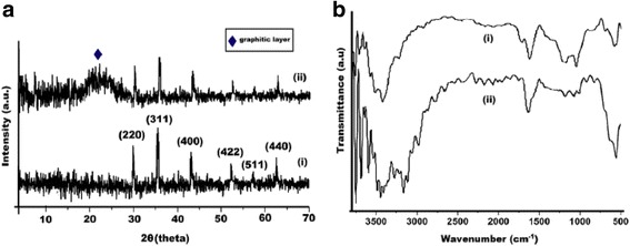
a XRD patterns of (i) iron oxide nanoparticles and (ii) iron oxide/reduced graphene oxide nanohybrid and b FTIR spectra of (i) reduced graphene oxide nanohybrid [94] and (ii) iron oxide/reduced graphene oxide nanohybrid [32]
Au–Fe3O4 composite with magnetic core was primarily produced by co-precipitation of Fe2+ and Fe3+. Further, Eucalyptus camaldulensis was used for the reduction of Au+3 on the surface of magnetite nanoparticles and for the functionalization of the Au–Fe3O4 nanocomposite particles [95]. UV–vis spectra showed a redshift due to the surface plasmon resonance of Au. The highest absorbance was observed for gold nanoparticles at 530 nm (solid line curve) whereas Au– composite nanoparticles showed a peak at 608 nm (dotted line) which agreed with previous reports (Fig. 2) [96–99]. It has also been observed by many other researchers [96–98, 100].
Fig. 2.
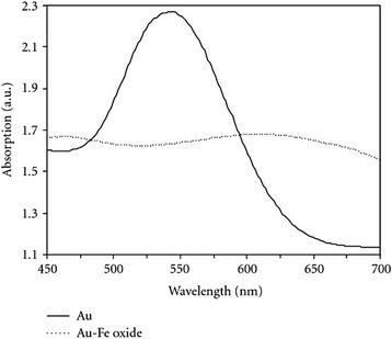
Absorbance spectra of gold nanoparticles (solid line) and magnetite-gold composite nanoparticles (dotted line) [95]
Senthil and Ramesh [35] synthesized iron oxide nanoparticles by the reduction of ferric chloride solution using Tridax procumbens leaf extract containing carbohydrates with an aldehyde group as a reducing agent. Further, these nanoparticles exhibited antibacterial activity against Pseudomonas aeruginosa.
Recently, Ehrampoush et al. [33] have reported that the size of iron oxide nanoparticles synthesized from tangerine peel is dependent on its concentration. The size of nanoparticles decreases with increasing concentration of tangerine peel extract from 2 to 6 %. In addition, they have reported that these nanoparticles can be a good adsorbent for the removal of cadmium from wastewater.
Biomedical Application of Magnetic Nanoparticles
Therapeutic Applications
Hyperthermia is a simple technique used to destroy tumor cells by raising their temperature to 41–45 °C. Since the living cells can undergo repair, they are rejuvenated while damage of the tumor cells is an irreversible process [101–103]. This magnetic hyperthermia is more effective when the iron oxide nanoparticles (IONP) are small and uniform. Their surface is generally made biocompatible by coating them with organic polymers or bioactive molecules for their slow release.
Cellular Labeling and Cell Separation
In vivo cell separation can be done by cell labeled with ferro paramagnetic substances [104] as the labeled cells can be detected by MRI [105]. They can be labeled by one of the two techniques: (a) attaching magnetic particles to the cell surface [106] or (b) internalizing biocompatible magnetic particles by fluid phase endocytosis [107]. The most appropriate technique for cell labeling is to modify the nanoparticle surface with a suitable ligand such as transferrin, lactoferrin, albumin, and insulin which are generally biocompatible. Such receptors have been shown [108] to internalize without disturbing the nanoparticles. Gupta and Gupta [66] have demonstrated that supramagnetic nanoparticles derivatized with proteins like lactoferrin, transferrin, and ceruloplasmin have strong affinity for receptors on the human fibroblasts surface, which inhibit the phagocytosis. These nanoparticles of less than 20 nm size have high magnetization value. Their influence on dermal fibroblast has been assessed in terms of adhesion viability and morphology by SEM and TEM images.
The interaction of the protein-coated nanoparticle is size dependent since different particles respond differently. Although there is a significantly visible difference in the interaction between fibroblast cell and coated/uncoated supramagnetic nanoparticle, no attempt has been made to realize the magnetic moment value of iron oxide nanoparticles, which changes as a consequence of its binding with proteins and other biomolecules. Iron in the trivalent state in Fe2O3 is in high spin state with five unpaired electrons in its d orbital but the moment it is coated with proteins, it goes to low spin state with a consequent change in the repulsion and magnetic moment value from 5.91 BM to 1.73 BM corresponding to one unpaired electron. It is due to the complex formation of Fe3+ with protein which being a strong ligand forces the electrons to be paired up. As a result, the Fe3+ is reduced in size but surface area increases due to complexation with the protein. The magnetic moment and reduction in size of Fe2O3 nanoparticle seems to be the key factor in the process of internalization and phagocytosis. It has been suggested that tissue repair can be done either by welding or soldering when polymer-coated nanoparticles are placed between two tissue surfaces. Temperature greater than 50 °C is produced by denaturation of tissue and also by absorption of light by coated nanoparticle [109].
Tissue Repair
The above method suggested for tissue repair is not convincing as the same temperature is produced to destroy the tumor cell which is supposed to join the two damaged normal cells [66]. The hypothesis first shows denaturation and then connecting the cells through other proteins. It appears as if the denaturation has twofold purposes; some proteins are disintegrated from the cell at 50 °C and some proteins remain unaffected which subsequently join the cells together. When gold/silica-coated Fe2O3 nanoparticles are coated on the tissues, they may prevent further damage but it seems unlikely that two unit cells are joined together. However, the self-repairing of the damaged tissue is a natural process that does not require raising the temperature of the tissues.
Treatment of Cancer
Wu et al. [109] have shown that gold-coated iron nanoparticles suppress cancer cell growth in oral and colorectal cancer cells in vivo and in vitro [110]. Although the healthy cells are equally exposed to iron nanoparticle, they are not much affected and the replication of cancer cells is inhibited. The cytotoxicity is due to the magnetic properties of the elemental iron nanoparticle; the oxidation of which is delayed by coating them with gold. As the oxidation of iron nanoparticles begins, the cytotoxicity decreases towards cancer cells. In fact, the gold coating slowly dissolves to release the iron nanoparticle. The reactive oxygen species (ROS) is generated which triggers the process of cytotoxicity. It has also been observed that the addition of ROS scavenger does not protect the cancer cells from the nanoparticles with an iron core and gold shell (Fe@Au)-induced cytotoxicity.
Decrease in mitochondrial membrane potential in cancerous cells occurs when treated with Fe@Au, although it is not clearly known as to how it interferes with the normal function of the mitochondria. Since the mitochondria are redox sensitive, they are targeted by Fe@Au. Iron is slowly oxidized, due to which, perhaps the mitochondrial membrane potential decreases. The cytotoxicity of Fe@Au towards cancer cells is an irreversible process, while the healthy cells are also affected but they recover within 24 h. The oxidation of iron nanoparticles and generation of ROS are simultaneous processes (Fig. 3).
Fig. 3.
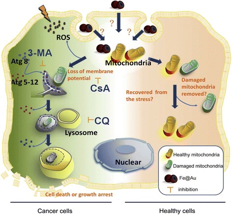
Fe@Au induced a cancer cell-specific cytotoxicity through the mitochondria-mediated autophagy [109]
Fe@Au caused a shock to the mitochondria within 4 h, but the cancer cells could not recover from the damage caused by them. Further, it caused a sequential autophagy and inhibited the cancer cell growth.
Drug Delivery
Mahmoudi et al. [58] have studied the application of supraparamagnetic iron oxide nanoparticle (SPION) in drug delivery. The drugs are bound on SPION surface or encapsulated in magnetic liposomes and microspheres. SPIONs can deliver peptides, DNA, chemotherapeutics, and radioactive and hyperthermic drugs. They are designed such that the drug or ligand is bound to its surface and guided with an external magnetic field to the desired site. The nanoparticles enter the target cells and deliver the drug there (Fig. 4).
Fig. 4.
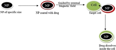
Drug delivery through nanoparticles on the target cells
When the drug is dissolved inside the target cell, the SPIONs are exposed to other cells. This process can reduce the quantity of drug, absorption time, and interaction of drug with nontarget cells. However, it is essential that iron nanoparticles should be magnetic and smaller than the target cells so that they can easily diffuse into them. Since large quantity of SPIONs cause agglomeration, their high concentration may be avoided. From in vivo experiments, it has been shown that in most cases, the drug is immediately delivered due to the bursting of the nanobubbles carrying the drug, as a consequence of which insufficient quantity of the drug reaches the target cells.
Mahmoudi et al. [111] synthesized cross-linked polyethylene glycol co-fumarate-coated iron oxide nanoparticles and loaded them with tamoxifen to see if it can reduce the burst time. Interestingly, it was found to reduce the burst time by 21 %. In a similar experiment, Guo et al. [112] loaded monodispersed SPION having a mesoporous structure, with doxorubicin, and observed that it had very high drug loading capacity and slow release. The drug release can be controlled by permeability, temperature, sensitivity, pH, surface functionality, and biodegradability of nanoparticles [113].
Colloidal magnetic nanoparticles are used in drug delivery at a desired target without interacting with other living cells. In the case of breast cancer (BT 20 cells), polyethylene glycol (PEG)-coated nanoparticles ranging between 10 and 100 nm were found to penetrate into the cells [114]. It is believed that since PEG is appreciably soluble in both polar and nonpolar solvents, it releases the magnetic nanoparticles at the tumor cells. Gupta and Gupta [66] have shown that the efficacy of magnetic microspheres in the targeted delivery of the incorporated drug is mainly due to magnetic effects. However, it is true for magnetic nanoparticle only, but innocuous and biocompatible nanoparticles of other metals such as gold and silver of smaller size [115] are known to be more effective than the larger particles of the same metal as drug carrier [116]. This technique is cost effective and reduces the quantum of drug to be transported to the site of use. Also, it protects the normal healthy cells from adverse effects of the drug.
Since the magnetic iron oxide nanoparticles are an excellent drug carrier, they are used in chemotherapy. However, iron oxide nanoparticles do not influence the human fibroblasts cells (IMR-90). It means that they are selective towards cancer cells. Khan et al. [117] demonstrated that when both the cancer cells (A549) and normal cells are exposed to iron oxide nanoparticles in a concentration range of 10–100 μg/ml for 24–48 h, necrosis of cancer cells occurs leading to their death. Loss of mitochondrial membrane potential and depletion of ATP suggest that necrosis is the major cause of cytotoxicity of cancer cells rather than apoptosis. ROS generation has been evidenced in A549 cells which are concentration and time dependent.
A variety of bimetallic nanoparticles of the type MFe2O4 (where M = divalent Mg, Fe, Co, Ni, Cu, and Zn) containing two metal ions has been reported for biomedical applications. Their magnetic properties are dependent on the number of unpaired electrons in the d orbital of transition metal ions. Multifunctional magnetic nanoparticles (MNPs) can be prepared by coating them with gold, silica, zinc oxide, polymer, liposome, etc. They can be further functionalized to make MNPs stable and multifunctional [118]. Xu and Sun [119] have attempted to deliver cisplatin to solid tumor through Fe3O4 HMNPs.
They have produced small pores in the polycrystalline nanoparticles by heating in oleic acid which allows the drug to be diffused easily in these pores. It has been shown schematically that the drug is delivered via ligand exchange. However, it is known that cisplatin is labile in aqueous medium and can react with water as shown below followed by exchange with surfactant.
Magnetic targeting of diseased cells by SPION has been done in some cases [120]. In order to increase the target yield, SPIONs are generally coated with polymers and functionalized by attaching carboxyl groups, biotin, avidin, carbodiimide, or any other biomolecule [121–123]. When the drug is carried at the target cell (tumor cell), it has to be released either by external force or by changes in pH, osmotic pressure, or temperature [124]. The drug is then picked up by tumor cells and penetrates via diffusion [125]. For such application, it is essential for the SPIONs to be stable at neutral pH and physiological conditions. The stability of the colloidal solution is dependent on the dimension of the nanoparticle and their aggregation may be prevented by coating them with an appropriate substance [126]. Larger particles (larger than 10 nm) cannot penetrate the endothelium [127] under normal conditions, but they can easily penetrate the tumor cells and inflamed cells [128]. When the coated nanoparticles enter the tumor cells, the coating is dissolved in the biological fluid and they are exposed to other cellular components [111, 129].
However, if the concentration is increased, aggregation of nanoparticles may occur leading to greater magnetic interaction. It is also believed that agglomeration of nanoparticles in the capillaries may block their passage [130]. Since the biological pH and the isoelectric point of SPION at pH 7 are the same [131, 132], they influence the colloidal stability of the SPIONs [133, 134].
It is essential that the drug-carrying spherical particles must always be smaller than the RBC and the blood capillaries where the drug is injected. The nanoparticles after drug delivery are exposed to normal cells, and therefore, it is essential that they should be nontoxic to them.
Effect of Internalization of Nanoparticles
Calero and others [135] have studied the effect of internalization of magnetic iron oxide nanoparticle on HeLa cells in vitro. They also assessed the damage of normal healthy cells and production of ROS. The internalization was found to be dependent on the type of coating of MNPs and their concentration. It was, however, noticed that besides the increasing concentration of MNPs (0.05, 0.1, and 0.5 mg ml−1), the uptake of APS-coated iron oxide nanoparticles by cells was higher than those coated with AF or dimercaptosuccinic acid (DMSA) [136]. The charge and surface of MNPs are important since positively charged particle surface is attracted towards negatively charged surface by default. Calero et al. [135] have observed that APS-coated positively charged nanoparticles are capable of penetrating easily into the HeLa cell than the DMSA- and AD-coated MNPs. It can be understood that positively charged MNPs are smaller than the negatively charged species, and therefore, being smaller in size, they can easily diffuse into the cells which has also been demonstrated by Kenzaoui et al. [137] in a separate experiment. The entrance of MNPs follows endocytosis [138, 139]. Although substantial number of MNPs accumulates in the cytoplasm and does not reach the nucleus, genotoxic damage of iron oxide MNPs due to the production of ROS occurs, irrespective of the cell type, coating, or their concentration. Such nanosized magnetic materials may therefore be used in medical diagnosis, especially in the identification of cancer cells, MRI, and carrier for drug delivery.
Tumor Treatment
It was observed from a study of cancer cells exposed to iron oxide nanoparticles that the cell death occurs by necrosis rather than apoptosis. Interestingly, it was also found that when normal human fibroblasts cells were exposed to iron oxide nanoparticles, insignificant cell death occurred. It demonstrates that these nanoparticles can be safely used in the treatment of tumors without damaging the healthy cells.
The ROS generation in this system was examined by the probe when cancer cells A549 were treated with iron oxide nanoparticles. The maximum ROS was generated after 24 h at a rate of 100 μg/ml iron oxide nanoparticles which subsequently induced autophagy [117].
Antibacterial Activity
Effect of iron nanoparticles on the deactivation of Escherichia coli has been studied by Lee et al. [140] under aerobic and anaerobic conditions. It was observed that in the absence of oxygen, the inactivation of E. coli was at maximum when exposed to 9 mg L−1 of iron nanoparticles for 10 min.
In air-saturated solution containing as high as 90 mg L−1, iron nanoparticles in E. coli solution exposed for 90 min had negligible inactivation of the bacteria. This is particularly due to the presence of oxygen which oxidizes the Fe0→Fe2+ and also the absence of hydroxyl radical. The iron nanoparticles are oxidized to FeO and Fe2O3 which form a film on the surface of the nanoparticles preventing the lower layer from further corrosion [141].
However, when a chelating agent such as PO4 3− ion is added in an air saturated system, the biocidal activity of iron nanoparticle is reduced because Fe(III) forms an insoluble metal chelate with PO4 3− ion. On the contrary, when oxalate ion, C2O4 2−, is added, the bactericidal activity is enhanced because it forms a soluble complex with the iron ion. This is also evidenced and monitored from a change in color from black to yellow.
Since Fe(II) is highly susceptible to oxidation by air, it does not stay stable unless stabilized by an acid.
Superparamagnetic iron oxide nanoparticles are frequently used as magnetic drug targeting, MRI, tissue repair, etc. [142–146]. They are useful in drug delivery. Since they can be guided by external electric field to the desired target, they can stay there when the magnetic field is cut off. Fe3O4 can be synthesized by a variety of procedures. Their size and shape may be controlled by monitoring the pH, temperature, and concentration of the reacting components. Coating with a suitable substance can prevent their agglomeration. SPIONs (800 nm) were introduced to the fibroblast cells (Fig. 5) to examine the changes in their morphology [147].
Fig. 5.
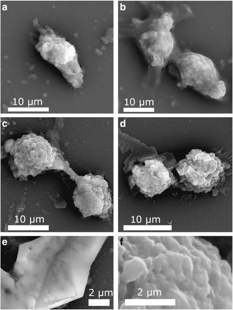
SEM results for a control L929 cells and the cells interacted with b nanobeads, c nanoworms, and d nanospheres. Panels e and f illustrate the higher-magnification image of the surface of control L929 and the one interacted with nanospheres, respectively [156]
It was found that the toxicity increases with the shape of SPIONs such as nanobeads, nanowires, and nanospheres. The deformation of the cell increases with the concentration of SPIONs. The TEM images of fibroblast cells exposed to SPION showed that smaller nanoparticles penetrate into the cell (Fig. 6) while larger ones have not been traced into them. The size is, therefore, of prime importance in such cases, although the coating also influences the size of the nanoparticles.
Fig. 6.
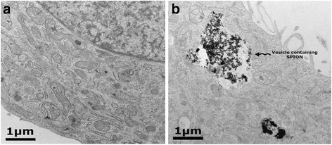
TEM images of L929 cells for a control and b cells exposed to SPION with vesicle-containing SPION nanospheres [156]
Although efforts are being made to produce SPION coated with organic molecules for use in human system, only dextran-coated SPIONs have so far been approved by Food and Drug Administration (FDA) [135].
Mahmoudi et al. [49] have evaluated the toxicity of bare SPION and those coated with –COOH and –NH2 against human cell lines HCM (heart), BE-2C (brain), and 293T (kidney). The toxicity of bare SPION was found to be higher than those coated with organic molecules due to their greater affinity to absorb proteins, vitamins, amino acids, and ions causing change in pH of the medicine [148–150]. Since the human cell contains proteins, vitamins, and amino acids, they have affinity to bind with SPION whereas the SPIONs already coated with these groups do not have vacant space for chelation with them. The toxicity of the coated SPIONs is, therefore, less than those of uncoated ones. The low toxicity of coated SPION is useful in the detection of cancer cells because they do not damage the normal cells. One type of SPION is toxic to certain types of cells while the other types produce insignificant effect. For instance, Mahmoudi and others [49] have reported that negative SPIONs did not produce significant changes on the cytoskeleton of heart cells compared to high toxic effect on the cytoskeleton of both kidney and brain cells.
Environmental Application of Magnetic Nanoparticles
Removal of Dyes
The iron nanoparticles produced from green tea leaf extract have been shown to contain iron oxide and oxohydroxide [23]. They have been used as a Fenton-like catalyst for the removal of organic dyes such as methylene blue and methyl orange in aqueous medium. It has been shown that they are highly effective in the removal of cationic and anionic dyes over a wide range of concentrations (10–200 mg L−1). Also, the iron nanoparticles synthesized from green tea leaf extract are more effective than those produced from borohydride reduction. Shahwan et al. [23] have reported that the iron nanoparticles were washed with ethyl alcohol to remove NaCl from the sample. Since NaCl is an ionic salt, it is soluble only in water and cannot be removed by washing with ethyl alcohol. It was also noted that the pH of methylene blue and that of methyl orange containing iron nanoparticles was around 8.30. However, the pH started declining immediately after the addition of H2O2 until it became constant at 3.11 after about 6 h when the dye was completely removed.
Pesticide Detection Sensor
Because of their application in agriculture, pesticides have become one of the major environmental pollutants. Iron oxide nanoparticle (Fe3O4) being chemically and biologically neutral have been coated with catalysts, enzymes, or even antibodies to be used as biosensors [151]. In a recently published paper, Chauhan et al. [152] have modified Fe3O4 nanoparticle using poly(indole-5-carboxylicacid) by preparing nanobiocomposite for its use as a sensor for the determination of pesticides such as malathion and chlorpyrifos in a wide range of concentrations (0.1–70 nm).
Iron nanoparticles in the elemental state have also been used in the purification of ground water. It has been tested against reductive dehalogenation of organochlorine pesticides and insecticides [153–155] and several other toxic substances such as chromium [156] and arsenic [157]. Since agglomeration of iron nanoparticle occurs, it was modified using surfactants. This not only reduces the organochlorine pesticides but also prevents corrosion. Mukherjee et al. [158] have shown that after accepting H+, the hepatochlor pH increases which is true but the term reduction used by them is incorrect as the acceptance of proton, by definition, is oxidation.
Pesticides used in agriculture are sometime harmful to other animals and plants. Their reduction to innocuous chemicals by iron nanoparticle is a simple strategy to make them useful. Polyhalogenated and nitroaromatic compounds are generally reduced by metal nanoparticles, metal sulfides, quinine, and vitamin B12. Metal nanoparticles can also be used for the reduction of nonhalogenated pesticides and azo dyes. The findings made by Keum and Li [159] suggest remediation, but it is too expensive to be used in the field. Also, it leaves toxic residues in the environment.
Conclusions
Iron and iron oxide magnetic nanoparticles can be fabricated by plant extracts or microbes such as fungi and microalgae. They can be coated with water soluble polymers for greater solubility. For instance, polyvinyl alcohol-coated nanoparticles are prevented from aggregation; therefore, they can easily diffuse through the semi-permeable membrane in a living system. Their shape and size may be controlled by maintaining the temperature, pH, and concentration of the reacting components. Their cytotoxicity varies with shape such as spherical, beads, and rods. The hydrodynamic size and the paramagnetic/diamagnetic nature of iron oxide nanoparticles make them specific for specific application. For example, nanoparticles with larger hydrodynamic size have lower cytotoxicity. Superparamagnetic iron oxide nanoparticles have great potential for use in instruments and medical devices, as drug carrier, and in the treatment of many diseases. Microbes and plant extracts containing alkaloids, flavonoids, saponins, ketones, aldehydes and phenols, or reducing acids like citric acid and ascorbic acids can be used for nanoparticle synthesis. Iron oxide nanoparticles can be used as an inexpensive material for the removal of dyes from textile industries and tanneries and in the treatment of contamination in wastewater and purification of ground water. Since the properties of iron oxide nanoparticles solely depend on the number of unpaired electrons and particle size in the presence and absence of magnetic field, it can be altered by coating them with polymers and by applying external magnetic field. These nanoparticles can be selectively used for the separation of magnetic materials from a huge deposit of nonmagnetic substances.
Acknowledgements
The authors are thankful to the publishers for the permission to adopt the figures in this review.
Authors’ Contributions
KSS, AR, and AH gathered the research data. KSS, AH, AR, and T analyzed these data and wrote this review paper. All authors read and approved the final manuscript.
Competing Interests
The authors declare that they have no competing of interests.
Contributor Information
Khwaja Salahuddin Siddiqi, Email: ks_siddiqi@yahoo.co.in.
Aziz ur Rahman, Email: rahman.mdi@gmail.com.
Tajuddin, Email: drtajuddinamu@gmail.com.
Azamal Husen, Email: adroot92@yahoo.co.in.
References
- 1.Husen A, Siddiqi KS. Phytosynthesis of nanoparticles: concept, controversy and application. Nano Res Lett. 2014;9:229. doi: 10.1186/1556-276X-9-229. [DOI] [PMC free article] [PubMed] [Google Scholar]
- 2.Husen A, Siddiqi KS. Plants and microbes assisted selenium nanoparticles: characterization and application. J Nanobiotechnol. 2014;12:28. doi: 10.1186/s12951-014-0028-6. [DOI] [PMC free article] [PubMed] [Google Scholar]
- 3.Mohanraj S, Kodhaiyolii S, Rengasamy M, Pugalenthi V. Green synthesized iron oxide nanoparticles effect on fermentative hydrogen production by Clostridium acetobutylicum. Appl Microbiol Biotechnol. 2014;173:318–331. doi: 10.1007/s12010-014-0843-0. [DOI] [PubMed] [Google Scholar]
- 4.Herlekar M, Barve S, Kumar R. Plant-mediated green synthesis of iron nanoparticles. J Nanopar. 2014;2014:140614. doi: 10.1155/2014/140614. [DOI] [Google Scholar]
- 5.Makarov VV, Makarova SS, Love AJ, Sinitsyna OV, Dudnik AO, Yaminsky IV, Taliansky ME, Kalinina NO. Biosynthesis of stable iron oxide nanoparticles in aqueous extracts of Hordeum vulgare and Rumex acetosa plants. Langmuir. 2014;30:5982–5988. doi: 10.1021/la5011924. [DOI] [PubMed] [Google Scholar]
- 6.Siddiqi KS, Husen A (2016) Green synthesis, characterization and uses of palladium/platinum nanoparticles. Nano Res Lett 11:482 [DOI] [PMC free article] [PubMed]
- 7.Siddiqi KS, Husen A. Fabrication of metal nanoparticles from fungi and metal salts: scope and application. Nano Res Lett. 2016;11:98. doi: 10.1186/s11671-016-1311-2. [DOI] [PMC free article] [PubMed] [Google Scholar]
- 8.Siddiqi KS, Husen A. Fabrication of metal and metal oxide nanoparticles by algae and their toxic effects. Nano Res Lett. 2016;11:363. doi: 10.1186/s11671-016-1580-9. [DOI] [PMC free article] [PubMed] [Google Scholar]
- 9.Rai M, Ingle A. Role of nanotechnology in agriculture with special reference to management of insect pests. Appl Microbiol Biotechnol. 2012;94:287–293. doi: 10.1007/s00253-012-3969-4. [DOI] [PubMed] [Google Scholar]
- 10.Husen A, Siddiqi KS. Carbon and fullerene nanomaterials in plant system. J Nanobiotechnol. 2014;12:16. doi: 10.1186/1477-3155-12-16. [DOI] [PMC free article] [PubMed] [Google Scholar]
- 11.Siddiqi KS, Husen A. Engineered gold nanoparticles and plant adaptation potential. Nano Res Lett. 2016;11:400. doi: 10.1186/s11671-016-1607-2. [DOI] [PMC free article] [PubMed] [Google Scholar]
- 12.Mohammadinejad R, Karimi S, Iravani S, Varma RS. Plant-derived nanostructures: types and applications. Green Chem. 2016;18:20. doi: 10.1039/C5GC01403D. [DOI] [Google Scholar]
- 13.Huber DL. Synthesis, properties, and applications of iron nanoparticles. Small. 2005;1:482–501. doi: 10.1002/smll.200500006. [DOI] [PubMed] [Google Scholar]
- 14.Laurent S, Bridot JL, Elst LV, Muller RN. Magnetic iron oxide nanoparticles for biomedical applications. Future Med Chem. 2010;2:427–49. doi: 10.4155/fmc.09.164. [DOI] [PubMed] [Google Scholar]
- 15.Mahmoudi M, Sant S, Wang B, Laurent S, Sen T. Superparamagnetic iron oxide nanoparticles (SPIONs): development, surface modification and applications in chemotherapy. Adv Drug Delivery Rev. 2011;63:24–46. doi: 10.1016/j.addr.2010.05.006. [DOI] [PubMed] [Google Scholar]
- 16.Hola K, Markova Z, Zoppellaro G, Tucek J, Zboril R. Tailored functionalization of iron oxide nanoparticles for MRI, drug delivery, magnetic separation and immobilization of biosubstances. Biotechnol Adv. 2015;33:1162–1176. doi: 10.1016/j.biotechadv.2015.02.003. [DOI] [PubMed] [Google Scholar]
- 17.Martínez-Cabanas M, López-García M, Barriada JL, Herrero R, Sastre de Vicente ME. Green synthesis of iron oxide nanoparticles. Development of magnetic hybrid materials for efficient As(V) removal. Chem Eng J. 2016;301:83–91. doi: 10.1016/j.cej.2016.04.149. [DOI] [Google Scholar]
- 18.Arokiyaraj S, Saravanan M, Udaya Prakash NK. Enhanced antibacterial activity of iron oxide magnetic nanoparticles treated with Argemone mexicana L. leaf extract: an in vitro study. Mat Res Bull. 2013;48:3323–3327. doi: 10.1016/j.materresbull.2013.05.059. [DOI] [Google Scholar]
- 19.Naseem T, Farrukh MA. Antibacterial activity of green synthesis of iron nanoparticles using Lawsonia inermis and Gardenia jasminoides leaves extract. J Chem. 2015;2015:912342. doi: 10.1155/2015/912342. [DOI] [Google Scholar]
- 20.Huang L, Luo F, Chen Z, Megharaj M, Naidu R. Green synthesized conditions impacting on the reactivity of Fe NPs for the degradation of malachite green. Spect Acta Part A: Mol Biomol Spect. 2015;137:154–159. doi: 10.1016/j.saa.2014.08.116. [DOI] [PubMed] [Google Scholar]
- 21.Hoag GE, Collins JB, Holcomb JL, Hoag JR, Nadagouda MN, Varma RS. Degradation of bromothymol blue by ‘greener’ nano-scale zero-valent iron synthesized using tea polyphenols. J Mater Chem. 2009;19:8671–8677. doi: 10.1039/b909148c. [DOI] [Google Scholar]
- 22.Nadagouda MN, Castle AB, Murdock RC, Hussain SM, Varma RS. In vitro biocompatibility of nanoscale zerovalent iron particles (NZVI) synthesized using tea polyphenols. Green Chem. 2010;12:114–122. doi: 10.1039/B921203P. [DOI] [Google Scholar]
- 23.Shahwan T, Abu Sirriah S, Nairat M, Boyac E, Eroğlu AE, Scott TB, Hallam KR. Green synthesis of iron nanoparticles and their application as a Fenton-like catalyst for the degradation of aqueous cationic and anionic dyes. Chem Eng J. 2011;172:258–266. doi: 10.1016/j.cej.2011.05.103. [DOI] [Google Scholar]
- 24.Kuang Y, Wang Q, Chen Z, Megharaj M, Naidu R. Heterogeneous Fenton-like oxidation of monochlorobenzene using green synthesis of iron nanoparticles. J Coll Inter Sci. 2013;410:67–73. doi: 10.1016/j.jcis.2013.08.020. [DOI] [PubMed] [Google Scholar]
- 25.Huang L, Weng X, Chen Z, Megharaj M, Naidu R. Synthesis of iron-based nanoparticles using oolong tea extract for the degradation of malachite green. Spect Acta Part A: Mol Biomol Spect. 2014;117:801–804. doi: 10.1016/j.saa.2013.09.054. [DOI] [PubMed] [Google Scholar]
- 26.Njagi EC, Huang H, Stafford L, Genuino H, Galindo HM, Collins JB, Hoag GE, Suib SL. Biosynthesis of iron and silver nanoparticles at room temperature using aqueous sorghum bran extracts. Langmuir. 2011;27:264–271. doi: 10.1021/la103190n. [DOI] [PubMed] [Google Scholar]
- 27.Madhavi V, Prasad TNVKV, Reddy AVB, Ravindra Reddy B, Madhavi G. Application of phytogenic zerovalent iron nanoparticles in the adsorption of hexavalent chromium. Spect Acta Part A: Mol Biomol Spect. 2013;116:17–25. doi: 10.1016/j.saa.2013.06.045. [DOI] [PubMed] [Google Scholar]
- 28.Wang Z. Iron complex nanoparticles synthesized by eucalyptus leaves. ACS Sus Chem Eng. 2013;1:1551–1554. doi: 10.1021/sc400174a. [DOI] [Google Scholar]
- 29.Wang T, Jin X, Chen Z, Megharaj M, Naidu R. Green synthesis of Fe nanoparticles using eucalyptus leaf extracts for treatment of eutrophic wastewater. Sci Total Environ. 2014;466–467:210–213. doi: 10.1016/j.scitotenv.2013.07.022. [DOI] [PubMed] [Google Scholar]
- 30.Rao A, Bankar A, Kumar AR, Gosavi S, Zinjarde S. Removal of hexavalent chromium ions by Yarrowia lipolytica cells modified with phyto-inspired Fe0/Fe3O4 nanoparticles. J Conta Hydrol. 2013;146:63–73. doi: 10.1016/j.jconhyd.2012.12.008. [DOI] [PubMed] [Google Scholar]
- 31.Venkateswarlu S, Rao YS, Balaji T, Prathima B, Jyothi NVV. Biogenic synthesis of Fe3O4 magnetic nanoparticles using plantain peel extract. Mat Lett. 2013;100:241–244. doi: 10.1016/j.matlet.2013.03.018. [DOI] [Google Scholar]
- 32.Thakur S, Karak N. One-step approach to prepare magnetic iron oxide/reduced graphene oxide nanohybrid for efficient organic and inorganic pollutants removal. Mat Chem Phys. 2014;144:425–432. doi: 10.1016/j.matchemphys.2014.01.015. [DOI] [Google Scholar]
- 33.Ehrampoush MH, Miria M, Salmani MH, Mahvi AH. Cadmium removal from aqueous solution by green synthesis iron oxide nanoparticles with tangerine peel extract. J Environ Health Sci Eng. 2015;13:84. doi: 10.1186/s40201-015-0237-4. [DOI] [PMC free article] [PubMed] [Google Scholar]
- 34.Kiruba Daniel SCG, Vinothini G, Subramanian N, Nehru K, Sivakumar M. Biosynthesis of Cu, ZVI, and Ag nanoparticles using Dodonaea viscosa extract for antibacterial activity against human pathogens. J Nanopar Res. 2013;15:1319. doi: 10.1007/s11051-012-1319-1. [DOI] [Google Scholar]
- 35.Senthil M, Ramesh C. Biogenic synthesis of Fe3O4 nanoparticles using Tridax procumbens leaf extract and its antibacterial activity on Pseudomonas aeruginosa. Dig J Nanomat Biostruc. 2012;7:1655–1661. [Google Scholar]
- 36.Narayanan S, Sathy BN, Mony U, Koyakutty M, Nair SV, Menon D. Biocompatible magnetite/gold nanohybrid contrast agents via green chemistry for MRI and CT bioimaging. ACS App Mat Inter. 2012;4:251–260. doi: 10.1021/am201311c. [DOI] [PubMed] [Google Scholar]
- 37.Machado S, Pinto SL, Grosso JP, Nouws HPA, Albergaria JT, Delerue-Matos C. Green production of zero-valent iron nanoparticles using tree leaf extracts. Sci Total Environ. 2013;445–446:1–8. doi: 10.1016/j.scitotenv.2012.12.033. [DOI] [PubMed] [Google Scholar]
- 38.Machado S, Stawiński W, Slonina P, Pinto AR, Grosso JP, Nouws HPA, Albergaria JT, Delerue-Matos C. Application of green zero-valent iron nanoparticles to the remediation of soils contaminated with ibuprofen. Sci Total Environ. 2013;461–462:323–329. doi: 10.1016/j.scitotenv.2013.05.016. [DOI] [PubMed] [Google Scholar]
- 39.Kumar KM, Mandal BK, Kumar KS, Reddy PS, Sreedhar B. Biobased green method to synthesise palladium and iron nanoparticles using Terminalia chebula aqueous extract. Spect Acta Part A: Mol Biomol Spect. 2013;102:128–133. doi: 10.1016/j.saa.2012.10.015. [DOI] [PubMed] [Google Scholar]
- 40.Wang Z, Fang C, Megharaj M. Characterization of iron-polyphenol nanoparticles synthesized by three plant extracts and their fenton oxidation of azo dye. ACS Sust Chem Eng. 2014;2:1022–1025. doi: 10.1021/sc500021n. [DOI] [Google Scholar]
- 41.Harshiny M, Iswarya CN, Matheswaran M. Biogenic synthesis of iron nanoparticles using Amaranthus dubius leaf extract as a reducing agent. Powder Technol. 2015;286:744–749. doi: 10.1016/j.powtec.2015.09.021. [DOI] [Google Scholar]
- 42.Yew YP, Shameli K, Miyake M, Kuwano N, Khairudin NBBA, Mohamad SEB, Lee KX. Green synthesis of magnetite (Fe3O4) nanoparticles using seaweed (Kappaphycus alvarezii) extract. Nanoscale Res Lett. 2016;11:276. doi: 10.1186/s11671-016-1498-2. [DOI] [PMC free article] [PubMed] [Google Scholar]
- 43.El-Kassas HY, Aly-Eldeen MA, Gharib SM. Green synthesis of iron oxide (Fe3O4) nanoparticles using two selected brown seaweeds: characterization and application for lead bioremediation. Acta Oceanologica Sin. 2016;35:89–98. doi: 10.1007/s13131-016-0880-3. [DOI] [Google Scholar]
- 44.Shah S, Dasgupta S, Chakraborty M, Vadakkekara R, Hajoori M. Green synthesis of iron nanoparticles using plant extracts. Int J Biol Pharm Res. 2014;5:549–552. [Google Scholar]
- 45.Indira TK, Laksmi PK. Magnetic nanoparticles—a review. Int J Pharm Sci Nanotechnol. 2010;3:1035–1042. [Google Scholar]
- 46.Park SJ, Kim S, Lee S, Khim ZG, Char K, Hyeon T. Synthesis and magnetic studies of uniform iron nanorods and nanospheres. J Am Chem Soc. 2000;122:8581–8582. doi: 10.1021/ja001628c. [DOI] [Google Scholar]
- 47.Dumestre F, Chaudret B, Amiens C, Fromen MC, Casanove MJ, Renaud P, Zurcher P. Shape control of thermodynamically stable cobalt nanorods through organometallic chemistry. Angew Chem Int Ed. 2002;41:4286–4289. doi: 10.1002/1521-3773(20021115)41:22<4286::AID-ANIE4286>3.0.CO;2-M. [DOI] [PubMed] [Google Scholar]
- 48.Dumestre F, Chaudret B, Amiens C, Renaud P, Fejes P. Superlattices of iron nanocubes synthesized from Fe[N(SiMe3)(2)](2) Science. 2004;303:821–823. doi: 10.1126/science.1092641. [DOI] [PubMed] [Google Scholar]
- 49.Mahmoudi M, Laurent S, Shokrgozar MA, Hosseinkhani M. Toxicity evaluations of superparamagnetic iron oxide nanoparticles: cell “vision” versus physicochemical properties of nanoparticles. ACS Nano. 2011;5:7263–7726. doi: 10.1021/nn2021088. [DOI] [PubMed] [Google Scholar]
- 50.United States Environmental Protection Agency . Office of prevention, pesticides and toxic substances. 1993. pp. 1–4. [Google Scholar]
- 51.Karlsson HL, Cronholm P, Gustafsson J, Moller L. Copper oxide nanoparticles are highly toxic: a comparison between metal oxide nanoparticles and carbon nanotubes. Chem Res Toxicol. 2008;21:1726–1732. doi: 10.1021/tx800064j. [DOI] [PubMed] [Google Scholar]
- 52.Gupta AK, Berry C, Gupta M, Curtis A. Receptor mediated targeting of magnetic nanoparticles using insulin as a surface ligand to prevent endocytosis. IEEE Trans Nanobiosci. 2003;2:256–261. doi: 10.1109/TNB.2003.820279. [DOI] [PubMed] [Google Scholar]
- 53.Gupta AK, Wells S. Surface-modified superparamagnetic nanoparticles for drug delivery: preparation, characterization, and cytotoxicity studies. IEEE Trans Nanobiosci. 2004;3:66–73. doi: 10.1109/TNB.2003.820277. [DOI] [PubMed] [Google Scholar]
- 54.Hussain SM, Hess KL, Gearhart JM, Geiss KT, Schlager JJ. In vitro toxicity of nanoparticles in BRL 3A rat liver cells. Toxicol in Vitro. 2005;19:975–983. doi: 10.1016/j.tiv.2005.06.034. [DOI] [PubMed] [Google Scholar]
- 55.Arbab AS, Bashaw LA, Miller BR, Jordan EK, Lewis BK, Kalish H, Frank JA. Characterization of biophysical and metabolic properties of cells labeled with superparamagnetic iron oxide nanoparticles and transfection agent for cellular MR imaging. Radiology. 2003;229:838–846. doi: 10.1148/radiol.2293021215. [DOI] [PubMed] [Google Scholar]
- 56.Arbab AS, Yocum GT, Wilson LB, Parwana A, Jordan EK, Kalish H, Frank JA. Comparison of transfection agents in forming complexes with ferumoxides, cell labeling efficiency, and cellular viability. Mol Imaging. 2004;3:24–32. doi: 10.1162/153535004773861697. [DOI] [PubMed] [Google Scholar]
- 57.Mahmoudi M, Milani AS, Stroeve P. Synthesis, surface architecture and biological response of superparamagnetic iron oxide nanoparticles for application in drug delivery: a review. Int J Biomed Nanosci Nanotechnol. 2010;1:164–201. doi: 10.1504/IJBNN.2010.034651. [DOI] [Google Scholar]
- 58.Mahmoudi M, Azadmanesh K, Shokrgozar MA, Journeay WS, Laurent S. Effect of nanoparticles on the cell life cycle. Chem Rev. 2011;111:3407–3432. doi: 10.1021/cr1003166. [DOI] [PubMed] [Google Scholar]
- 59.Issa B, Obaidat IM, Albiss BA, Haik Y. Magnetic nanoparticles: surface effects and properties related to biomedicine applications. Int J Mol Sci. 2013;14:21266–21305. doi: 10.3390/ijms141121266. [DOI] [PMC free article] [PubMed] [Google Scholar]
- 60.Tartaj P, Morales MDD, Veintemillas-Verdaguer S, Gonzalez-Carreno T, Serna CJ. The preparation of magnetic nanoparticles for applications in biomedicine. J Phys D Appl Phys. 2003;36:R182–R197. doi: 10.1088/0022-3727/36/13/202. [DOI] [Google Scholar]
- 61.Faraji M, Yamini Y, Rezaee M. Magnetic nanoparticles: synthesis, stabilization, functionalization, characterization and applications. J Iran Chem Soc. 2010;7:1–37. doi: 10.1007/BF03245856. [DOI] [Google Scholar]
- 62.Niemeyer CM. Nanoparticles, proteins, and nucleic acids: biotechnology meets materials science. Angew Chem Int Ed. 2001;40:4128–4158. doi: 10.1002/1521-3773(20011119)40:22<4128::AID-ANIE4128>3.0.CO;2-S. [DOI] [PubMed] [Google Scholar]
- 63.Cornell RM, Schertmann U. Iron oxides in the laboratory: preparation and characterization. Weinheim: VCH; 1991. [Google Scholar]
- 64.Chen S, Feng J, Guo X, Hong J, Ding W. One-step wet chemistry for preparation of magnetite nanorods. Mat Lett. 2005;59:985–988. doi: 10.1016/j.matlet.2004.11.043. [DOI] [Google Scholar]
- 65.Bae DS, Han KS, Cho SB, Choi SH. Synthesis of ultrafine Fe3O4 powder by glycothermal process. Mat Lett. 1998;37:255–258. doi: 10.1016/S0167-577X(98)00101-3. [DOI] [Google Scholar]
- 66.Gupta AK, Gupta M. Synthesis and surface engineering of iron oxide nanoparticles for biomedical applications. Biomaterials. 2005;26:3995–4021. doi: 10.1016/j.biomaterials.2004.10.012. [DOI] [PubMed] [Google Scholar]
- 67.Kwon G, Naito M, Yokoyama M, Okano T, Sakurai Y, Kataoka K. Block copolymer micelles for drug delivery: loading and release of doxorubicin. J Control Release. 1997;48:195–201. doi: 10.1016/S0168-3659(97)00039-4. [DOI] [Google Scholar]
- 68.Kwon GS, Suwa S, Yokoyama M, Okano T, Sakurai Y, Kataoka K. Physical entrapment of adriamycin in AB block copolymer micelles. Pharm Res. 1995;12:192–195. doi: 10.1023/A:1016266523505. [DOI] [PubMed] [Google Scholar]
- 69.Akbarzadeh A, Samiei M, Davaran S. Magnetic nanoparticles: preparation, physical properties, and applications in biomedicine. Nanoscale Res Lett. 2012;7:144. doi: 10.1186/1556-276X-7-144. [DOI] [PMC free article] [PubMed] [Google Scholar]
- 70.Morcos SK. Nephrogenic systemic fibrosis following the administration of extracellular gadolinium based contrast agents: is the stability of the contrast agent molecule an important factor in the pathogenesis of this condition? Br J Radiol. 2007;80:73–76. doi: 10.1259/bjr/17111243. [DOI] [PubMed] [Google Scholar]
- 71.Ersoy H, Rybicki FJ. Biochemical safety profiles of gadolinium-based extracellular contrast agents and nephrogenic systemic fibrosis. J Magn Reson Imaging. 2007;26:1190–1197. doi: 10.1002/jmri.21135. [DOI] [PMC free article] [PubMed] [Google Scholar]
- 72.Denizot B, Tanguy G, Hindre F, Rump E, Lejeune JJ, Jallet P. The preparation of magnetite nanoparicles for biomedical. J Colloid Interface Sci. 1999;209:66. doi: 10.1006/jcis.1998.5850. [DOI] [PubMed] [Google Scholar]
- 73.Shin S, Yoon H, Jang J. Polymer-encapsulated iron oxide nanoparticles as highly efficient Fenton catalysts. Catal Commun. 2008;10:178–182. doi: 10.1016/j.catcom.2008.08.027. [DOI] [Google Scholar]
- 74.Xu L, Wang J. A heterogeneous Fenton-like system with nanoparticulate zerovalent iron for removal of 4-chloro-3-methyl phenol. J Hazard Mater. 2011;186:256–264. doi: 10.1016/j.jhazmat.2010.10.116. [DOI] [PubMed] [Google Scholar]
- 75.Zelmanov G, Semiat R. Iron(3) oxide-based nanoparticles as catalysts in advanced organic aqueous oxidation. Water Res. 2008;42:492–498. doi: 10.1016/j.watres.2007.07.045. [DOI] [PubMed] [Google Scholar]
- 76.Teja AS, Pei-Yoong K. Synthesis, properties, and applications of magnetic iron oxide nanoparticles. Prog Cry Grow Charac Mat. 2009;55:22–45. doi: 10.1016/j.pcrysgrow.2008.08.003. [DOI] [Google Scholar]
- 77.Neuberger T, Schopf B, Hofmann H, Hofmann M, Rechenberg B. Superparamagnetic nanoparticles for biomedical applications: possibilities and limitations of a new drug delivery system. J Magnet Mag Mat. 2005;293:483–496. doi: 10.1016/j.jmmm.2005.01.064. [DOI] [Google Scholar]
- 78.Kohler N, Sun C, Wang J, Zhang M. Methotrexate-modified superparamagnetic nanoparticles and their intracellular uptake into human cancer cells. Langmuir. 2005;21:8858–8864. doi: 10.1021/la0503451. [DOI] [PubMed] [Google Scholar]
- 79.Tartaj P, Morales MD, Veintemillas-Verdaguer S, Gonzalez-Carreno T, Serna CJ. The preparation of magnetic nanoparticles for applications in biomedicine. J Phys D: App Phys. 2003;36:182–197. doi: 10.1088/0022-3727/36/13/202. [DOI] [Google Scholar]
- 80.Jurgons R, Seliger C, Hilpert A, Trahms L, Oden-bach S, Alexiou C. Drug loaded magnetic nanoparticles for cancer therapy. J Phys: Cond Mat. 2006;18:S2893–S2902. [Google Scholar]
- 81.Ai H, Flask C, Weinberg B, Shuai XT, Pagel MD, Farrell D, Duerk J, Gao J. Magnetite-loaded polymeric micelles as ultrasensitive magnetic-resonance probes. Adv Mat. 2005;17:1949–1952. doi: 10.1002/adma.200401904. [DOI] [Google Scholar]
- 82.Gonzales M, Krishnan KM. Synthesis of magnetoliposomes with monodisperse iron oxide nanocrystal cores for hyperthermia. J Magnet Mag Mat. 2005;293:265–270. doi: 10.1016/j.jmmm.2005.02.020. [DOI] [Google Scholar]
- 83.Jain TK, Morales MA, Sahoo SK, Leslie-Pelecky DL, Labhasetwar V. Iron oxide nanoparticles for sustained delivery of anticancer agents. Mol Pharm. 2005;2:194–205. doi: 10.1021/mp0500014. [DOI] [PubMed] [Google Scholar]
- 84.Chourpa I, Douziech-Eyrolles L, Ngaboni-Okassa L, Fouquenet JF, Cohen-Jonathan S, Souce M, Marchais H, Dubois P. Molecular composition of iron oxide nanoparticles, precursors for magnetic drug targeting, as characterized by confocal Raman microspectroscopy. Analyst. 2005;130:1395–1405. doi: 10.1039/b419004a. [DOI] [PubMed] [Google Scholar]
- 85.Chastellain M, Petri A, Gupta A, Rao KV, Hofmann H. Superparamagnetic silica-iron oxide nanocomposites for application in hyperthermia. Adv Eng Mat. 2004;6:235–241. doi: 10.1002/adem.200300574. [DOI] [Google Scholar]
- 86.Kalish H, Arbab AS, Miller BR, Lewis BK, Zywicke HA, Bulte´ JWM, Bryan LH, Frank JA. Combination of transfection agents and magnetic resonance contrast agents for cellular imaging: relationship between relaxivities, electrostatic forces, and chemical composition. Magn Reson Med. 2003;50:275–282. doi: 10.1002/mrm.10556. [DOI] [PubMed] [Google Scholar]
- 87.Moore A, Marecos E, Bogdanov A, Weissleder R. Tumoral distribution of long-circulating dextran-coated iron oxide nanoparticles in a rodent model. Radiology. 2000;214:568–574. doi: 10.1148/radiology.214.2.r00fe19568. [DOI] [PubMed] [Google Scholar]
- 88.Sipe JC, Filippi M, Martino G, Furlan R, Rocca MA, Rovaris M, Bergami A, Zyroff J, Scotti G, Comi G. Method for intracellular magnetic labeling of human mononuclear cells using approved iron contrast agents. Magn Reson Imaging. 1999;17:1521–1523. doi: 10.1016/S0730-725X(99)00085-5. [DOI] [PubMed] [Google Scholar]
- 89.Strable E, Bulte JWM, Moskowitz B, Vivekanandan K, Allen M, Douglas T. Designed fabrication of multi functional magnetic gold nano shells and their application to magnetic resonance imaging and photothermal therapy. Chem Mater. 2001;13:2201–2209. doi: 10.1021/cm010125i. [DOI] [Google Scholar]
- 90.Muthukumar H, Matheswaran M. Amaranthus spinosus leaf extract mediated FeO nanoparticles: physicochemical traits, photocatalytic and antioxidant activity. ACS Sust Chem Eng. 2015;3:3149–3156. doi: 10.1021/acssuschemeng.5b00722. [DOI] [Google Scholar]
- 91.Phumying S, Labuayai S, Thomas C, Amornkitbamrung V, Swatsitang E, Maensiri S. Aloe vera plant-extracted solution hydrothermal synthesis and magnetic properties of magnetite (Fe3O4) nanoparticles. App Phys A. 2013;111:1187–1193. doi: 10.1007/s00339-012-7340-5. [DOI] [Google Scholar]
- 92.Herrera-Becerra R, Zorrilla C, Ascencio JA. Production of iron oxide nanoparticles by a biosynthesis method: an environmentally friendly route. J Phys Chem. 2007;111:16147–16153. [Google Scholar]
- 93.Lunge S, Singh S, Sinha A. Magnetic iron oxide (Fe3O4) nanoparticles from tea waste for arsenic removal. J Magnetism Mag Mat. 2014;356:21–31. doi: 10.1016/j.jmmm.2013.12.008. [DOI] [Google Scholar]
- 94.Thakur S, Karak N. Green reduction of graphene oxide by aqueous phytoextracts. Carbon. 2012;50:5331–5339. doi: 10.1016/j.carbon.2012.07.023. [DOI] [Google Scholar]
- 95.Haratifar EAD, Shahverdi HR, Shakibaie M, Moghaddam KM, Amini M, Montazeri H, Shahverdi AR. Semi-biosynthesis of magnetite-gold composite nanoparticles using an ethanol extract of Eucalyptus camaldulensis and study of the surface chemistry. J Nanomat. 2009;2009:962021. doi: 10.1155/2009/962021. [DOI] [Google Scholar]
- 96.Kinoshita T, Seino S, Mizukoshi Y, Otome Y, Nakagawa T, Okitsu K, Yamamoto TA. Magnetic separation of amino acids by gold/iron-oxide composite nanoparticles synthesized by gamma-ray irradiation. J Magnetism Mag Mat. 2005;293:106–110. doi: 10.1016/j.jmmm.2005.01.050. [DOI] [Google Scholar]
- 97.Yu H, Chen M, Rice PM, Wang SX, White RL, Sun S. Dumbbell-like bifunctional Au-Fe3O4 nanoparticles. Nano Lett. 2005;5:379–382. doi: 10.1021/nl047955q. [DOI] [PubMed] [Google Scholar]
- 98.Lim J, Tilton RD, Eggeman A, Majetich SA. Design and synthesis of plasmonic magnetic nanoparticles. J Magnetism Mag Mat. 2007;311:78–83. doi: 10.1016/j.jmmm.2006.10.1169. [DOI] [Google Scholar]
- 99.Zhang XF, Dong XL, Huang H, Lv B, Zhu XG, Lei JP, Ma S, Liu W, Zhang ZD. Synthesis, structure and magnetic properties of SiO2-coated Fe nanocapsules. Mat Sci Eng A. 2007;454–455:211–215. doi: 10.1016/j.msea.2006.11.010. [DOI] [Google Scholar]
- 100.Kinoshita T, Seino S, Okitsu K, Nakayama T, Nakagawa T, Yamamoto TA. Magnetic evaluation of nanostructure of gold-iron composite particles synthesized by a reverse micelle method. J Alloy Comp. 2003;359:46–50. doi: 10.1016/S0925-8388(03)00198-1. [DOI] [Google Scholar]
- 101.Kim J, Park S, Lee JE, Jin SM, Lee JH, Lee IS, Yang I, Kim JS, Kim SK, Cho MH, Hyeon T. Designed fabrication of multi functional magnetic gold nano shells and their application to magnetic resonance imaging and photothermal therapy. Angew Chem Int Ed. 2006;45:7754–7758. doi: 10.1002/anie.200602471. [DOI] [PubMed] [Google Scholar]
- 102.Green M. Organometallic based strategies for metal nanocrystal synthesis. Chem Commun. 2005;24:3002–3011. doi: 10.1039/b501835h. [DOI] [PubMed] [Google Scholar]
- 103.Olsvik O, PopovicT SE, Cudjoe KS, Hornes E, Ugelstad J, Uhlen M. Magnetic separation techniques in diagnostic microbiology. Clin Microbiol Rev. 1994;7:43–54. doi: 10.1128/CMR.7.1.43. [DOI] [PMC free article] [PubMed] [Google Scholar]
- 104.Yeh TC, Zhang W, Ldstad ST, Ho C. Intracellular labeling of T-cells with superparamagnetic contrast agents. Magn Reson Med. 1993;30:617–625. doi: 10.1002/mrm.1910300513. [DOI] [PubMed] [Google Scholar]
- 105.Handgretinger R, Lang P, Schumm M, Taylor G, Neu S, Koscielnak E, Niethammer D, Klingebiel T. Isolation and transplantation of autologous peripheral CD34+ progenitor cells highly purified by magnetic-activated cell sorting. Bone Marrow Trans. 1998;21:987–993. doi: 10.1038/sj.bmt.1701228. [DOI] [PubMed] [Google Scholar]
- 106.Schoepf U, Marecos E, Jain R, Weissleder R. Intracellular magneticlabelling of lymphocytes for in vivo trafficking studies. Biotechniques. 1998;24:642–651. doi: 10.2144/98244rr01. [DOI] [PubMed] [Google Scholar]
- 107.Weissleder R, Cheng HC, Bogdanova A, Bogdanov A. Magnetically labelled cells can be detected by MR imaging. J Magn Reson Imaging. 1997;7:258–263. doi: 10.1002/jmri.1880070140. [DOI] [PubMed] [Google Scholar]
- 108.Lobel B, Eyal O, Kariv N, Katzir A. Temperature controlled CO2 laser welding of soft tissues: urinary bladder welding in different animal models (rats, rabbits, and cats) Lasers Surg Med. 2000;26:4–12. doi: 10.1002/(SICI)1096-9101(2000)26:1<4::AID-LSM3>3.0.CO;2-J. [DOI] [PubMed] [Google Scholar]
- 109.Wu YN, Li-Xing Y, Xuan-Yu S, I-Chen L, Joanna MB, Ratinac KR, Dong-Hwang C, Pall T, Dar-Bin S, Filip B. The selective growth inhibition of oral cancer by iron core-gold shell nanoparticles through mitochondria-mediated autophagy. Biomaterials. 2011;32:4565–4573. doi: 10.1016/j.biomaterials.2011.03.006. [DOI] [PubMed] [Google Scholar]
- 110.Wu YN, Chen DH, Shi XY, Lian CC, Wang TY, Yeh CS, Ratinac KR, Thordarson P, Braet F, Shieh DB. Cancer-cell-specific cytotoxicity of non-oxidized iron elements in iron core-gold shell nanoparticles. Nanomedicine: NBM. 2011;7:420–427. doi: 10.1016/j.nano.2011.01.002. [DOI] [PubMed] [Google Scholar]
- 111.Mahmoudi M, Simchi A, Imani M, Hafeli UO. Superparamagnetic iron oxide nanoparticles with rigid cross-linked polyethylene glycol fumarate coating for application in imaging and drug delivery. J Phys Chem C. 2009;113:8124–8131. doi: 10.1021/jp900798r. [DOI] [Google Scholar]
- 112.Guo S, Li D, Zhang L, Li J, Wang E. Monodisperse mesoporous superparamagnetic single-crystal magnetite nanoparticles for drug delivery. Biomaterials. 2009;30:1881–1889. doi: 10.1016/j.biomaterials.2008.12.042. [DOI] [PubMed] [Google Scholar]
- 113.Butoescu N, Seemayer CA, Palmer G, Guerne PA, Gabay C, Doelker E, Jordan O. Magnetically retainable microparticles for drug delivery to the joint: efficacy studies in an antigen-induced arthritis model in mice. Arthritis Res Therapy. 2009;11:R72. doi: 10.1186/ar2701. [DOI] [PMC free article] [PubMed] [Google Scholar]
- 114.Zhang Y, Kohler N, Zhang M. Surface modification of superparamagnetic magnetite nanoparticles and their intracellular uptake. Biomaterials. 2002;23:1553–1561. doi: 10.1016/S0142-9612(01)00267-8. [DOI] [PubMed] [Google Scholar]
- 115.Chouly C, Pouliquen D, Lucet I, Jeune P, Pellet JJ. Development of superparamagnetic nanoparticles for MRI: effect of particles size, charge and surface nature on biodistribution. J Microencapsul. 1996;13:245–255. doi: 10.3109/02652049609026013. [DOI] [PubMed] [Google Scholar]
- 116.Chatterjee J, Haik Y, Chen CJ. Size dependent magnetic properties of iron oxide nanoparticles. J Magnetism Mag Mat. 2003;257:113–118. doi: 10.1016/S0304-8853(02)01066-1. [DOI] [Google Scholar]
- 117.Khan MI, Mohammad A, Patil G, Naqvi SAH, Chauhan LKS, Ahmad I. Induction of ROS, mitochondrial damage and autophagy in lung epithelial cancer cells by iron oxide nanoparticles. Biomaterials. 2012;33:1477–1488. doi: 10.1016/j.biomaterials.2011.10.080. [DOI] [PubMed] [Google Scholar]
- 118.Wang L, Park HY, Lim SII, Schadt MJ, Mott D, Luo J, Wang X, Zhong CJ. Core@shell nanomaterials: gold-coated magnetic oxide nanoparticles. J Mat Chem. 2008;18:2629–2635. doi: 10.1039/b719096d. [DOI] [Google Scholar]
- 119.Xu C, Sun S. New forms of superparamagnetic nanoparticles for biomedical applications. Adv Drug Del Rev. 2013;65:732–743. doi: 10.1016/j.addr.2012.10.008. [DOI] [PubMed] [Google Scholar]
- 120.Langer R. Drug delivery and targeting. Nature. 1998;392(6679):5–10. [PubMed] [Google Scholar]
- 121.Koneracka M, Kopcansky P, Antalik M, Timko M, Ramchand CN, Lobo D, Mehta RV, Upadhyay RV. Immobilization of proteins and enzymes to fine magnetic particles. J Magnetism Mag Mat. 1999;201:427–430. doi: 10.1016/S0304-8853(99)00005-0. [DOI] [Google Scholar]
- 122.Mehta RV, Upadhyay RV, CharlesSW RCN. Direct binding of protein to magnetic particles. Biotechnol Tech. 1997;11:493–496. doi: 10.1023/A:1018457915380. [DOI] [Google Scholar]
- 123.Koneracka M, Kopcansky P, Timko M, Ramchand CN, Sequeira A, Trevan M. Direct binding procedure of proteins and enzymes to fine magnetic particles. J Mol Cat B, Enz. 2002;18:13–18. doi: 10.1016/S1381-1177(02)00016-4. [DOI] [Google Scholar]
- 124.Alexiou C, Arnold W, Klein RJ, Parak FG, Hulin P, Bergemann C, Erhardt W, Wagenpfeil S, Lubbe AS. Locoregional cancer treatment with magnetic drug targeting. Can Res. 2000;60:6641–6648. [PubMed] [Google Scholar]
- 125.Widder KJ, Senyei AE, Scarpelli DG. Magnetic microspheres—model system for site specific drug delivery in vivo. Proc Soci Experi Biol Med. 1978;158:141–146. doi: 10.3181/00379727-158-40158. [DOI] [PubMed] [Google Scholar]
- 126.Tartaj P, Serna CJ. Synthesis of monodisperse superparamagnetic Fe/silica nanospherical composites. J Am Chem Soci. 2003;125:15754–15755. doi: 10.1021/ja0380594. [DOI] [PubMed] [Google Scholar]
- 127.Barratt G, Courraze G, Couvreur P. Polymeric biomaterials. In: Dumitriu S, editor. Headquarters. New York: Marcel Dekker, Inc.; 2002. [Google Scholar]
- 128.Moghimi SM, Hunter AC, Murray JC. Long-circulating and target-specific nanoparticles: theory to practice. Pharmaco Rev. 2001;53:283–318. [PubMed] [Google Scholar]
- 129.Gupta AK, Naregalkar RR, Vaidya VD, Gupta M. Recent advances on surface engineering of magnetic iron oxide nanoparticles and their biomedical applications. Nanomedicine. 2007;2:23–39. doi: 10.2217/17435889.2.1.23. [DOI] [PubMed] [Google Scholar]
- 130.Hong R, Li JH, Wang J, Li HZ. Comparison of schemes for preparing magnetic Fe3O4 nanoparticles. China Partic. 2007;5:186–191. doi: 10.1016/j.cpart.2007.01.011. [DOI] [Google Scholar]
- 131.Bacri JC, Perzynski R, Salin D, Cabuil V, Massart R. Ionic ferrofluids: a crossing of chemistry and physics. J Magnetism Mag Mat. 1990;85:27–32. doi: 10.1016/0304-8853(90)90010-N. [DOI] [Google Scholar]
- 132.Douziech-Eyrolles L, Marchais H, Herve K, Munnier E, Souce M, Linassier C, Dubois P, Chourpa I. Nanovectors for anticancer agents based on superparamagnetic iron oxide nanoparticles. Int J Nanomed. 2007;2:541–550. [PMC free article] [PubMed] [Google Scholar]
- 133.Akerman ME, Chan WCW, Laakkonen P, Bhatia SN, Ruoslahti E. Nanocrystal targeting in vivo. Proc Nat Aca Sci USA. 2002;99:12617–12621. doi: 10.1073/pnas.152463399. [DOI] [PMC free article] [PubMed] [Google Scholar]
- 134.LaVan DA, McGuire T, Langer R. Small-scale systems for in vivo drug delivery. Nat Biotechnol. 2003;21:1184–1191. doi: 10.1038/nbt876. [DOI] [PubMed] [Google Scholar]
- 135.Calero M, Gutiérrez L, Salas G, Ya L, Lázaro A, Acedo P, Morales M, Miranda R, Villanueva A. Efficient and safe internalization of magnetic iron oxide nanoparticles: two fundamental requirements for biomedical applications. Nanomedicine: Nanotechnol, Biol Med. 2014;10:733–743. doi: 10.1016/j.nano.2013.11.010. [DOI] [PubMed] [Google Scholar]
- 136.Villanueva A, Cañete M, Roca AG, Calero M, Veintemillas-Verdaguer S, Serna CJ, Morales Mdel P, Miranda R. The influence of surface functionalization on the enhanced internalization of magnetic nanoparticles in cancer cells. Nanotechnology. 2009;20:115103. doi: 10.1088/0957-4484/20/11/115103. [DOI] [PubMed] [Google Scholar]
- 137.Kenzaoui BH. Evaluation of uptake and transport of cationic and anionic ultra small iron oxide nanoparticles by human colon cells. Int J Nanomedicine. 2012;7:1275–1286. doi: 10.2217/nnm.12.99. [DOI] [PMC free article] [PubMed] [Google Scholar]
- 138.Hoet PHM, Brüske-Hohlfeld I, Salata OV. Nanoparticles—known and unknown health risks. J Nanobiotechnol. 2004;2:12. doi: 10.1186/1477-3155-2-12. [DOI] [PMC free article] [PubMed] [Google Scholar]
- 139.Iversen TG, Skotland T, Sandvig K. Endocytosis and intracellular transport of nanoparticles: present knowledge and need for future studies. Nano Today. 2011;6:176–185. doi: 10.1016/j.nantod.2011.02.003. [DOI] [Google Scholar]
- 140.Lee C, Kim JY, Lee WI, Nelson KL, Yoon J, Sedlak DL. Bactericidal effect of zero-valent iron nanoparticles on Escherichia coli. Environ Sci Technol. 2008;42:4927–4933. doi: 10.1021/es800408u. [DOI] [PMC free article] [PubMed] [Google Scholar]
- 141.Davenport AJ, Oblonsky LJ, Ryan MP, Toney MF. The structure of the passive film that forms on iron in aqueous environments. J Electrochem Soc. 2000;147:2162–2173. doi: 10.1149/1.1393502. [DOI] [Google Scholar]
- 142.Häfeli U, Schutt W, Teller J, Zborowski M. Scientific and clinical applications of magnetic microspheres. New York: Plenum Press; 1997. [Google Scholar]
- 143.Lian S, Wang E, Kang Z, Bai Y, Gao L, Jiang M, Hu C, Xu L. Synthesis of magnetite nanorods and porous hematite nanorods. Solid State Commun. 2004;129:485–490. doi: 10.1016/j.ssc.2003.11.043. [DOI] [Google Scholar]
- 144.Zaitsev VS, Filimonov DS, Presnyakov IA, Gambino RJ, Chu B. Physical and chemical properties of magnetite and magnetite-polymer nanoparticles and their colloidal dispersions. J Coll Inter Sci. 1999;212:49–57. doi: 10.1006/jcis.1998.5993. [DOI] [PubMed] [Google Scholar]
- 145.Kang YS, Risbud S, Rabolt JF, Stroeve P. Synthesis and characterization of nanometer-size Fe3O4 and g-Fe2O3 particles. Chem Mater. 1996;8(9):2209–2211. doi: 10.1021/cm960157j. [DOI] [Google Scholar]
- 146.Wang YXJ, Xuan S, Port M, Idee JM (2013) Recent advances in superparamagnetic iron oxidenanoparticles for cellular imaging and targeted therapy research. Curr Pharm Des 19:6575–6593 [DOI] [PMC free article] [PubMed]
- 147.Mahmoudi M, Simchi A, Milani AS, Stroeve P. Cell toxicity of superparamagnetic iron oxide nanoparticles. J Coll Inter Sci. 2009;336:510–518. doi: 10.1016/j.jcis.2009.04.046. [DOI] [PubMed] [Google Scholar]
- 148.Mahmoudi M, Simchi A, Imani M. Cytotoxicity of uncoated and polyvinyl alcohol coated superparamagnetic iron oxide nanoparticles. J Phys Chem C. 2009;113:9573–9580. doi: 10.1021/jp9001516. [DOI] [Google Scholar]
- 149.Mahmoudi M, Simchi A, Imani M, Shokrgozar MA, Milani AS, Hafeli UO, Stroeve P. A new approach for the in vitro identification of the cytotoxicity of superparamagnetic iron oxide nanoparticles. Colloid Surf B. 2010;75:300–309. doi: 10.1016/j.colsurfb.2009.08.044. [DOI] [PubMed] [Google Scholar]
- 150.Mahmoudi M, Simchi A, Imani M, Milani AS, Stroeve P. An in vitro study of bare and poly(ethylene glycol)-cofumarate-coated superparamagnetic iron oxide nanoparticles: a new toxicity identification procedure. Nanotechnology. 2009;20:225104. doi: 10.1088/0957-4484/20/22/225104. [DOI] [PubMed] [Google Scholar]
- 151.Ali A, AlSalhi MS, Atif M, Ansari AA, Israr MQ, Sadaf JR, Ahmed E, Nur O, Willander M. Potentiometric urea biosensor utilizing nano biocomposite of chitosan-iron oxide magnetic nanoparticles. J Phys. 2013;414:1–11. [Google Scholar]
- 152.Chauhan N, Narang J, Jain U. Amperometric acetylcholinesterase biosensor for pesticides monitoring utilising iron oxide nanoparticles and poly(indole-5-carboxylic acid) J Exper Nanosci. 2016;11:111–122. doi: 10.1080/17458080.2015.1030712. [DOI] [Google Scholar]
- 153.Wang CB, Zhang WX. Synthesizing nanoscale iron particles for rapid and complete dechlorination of TCE and PCBs. Environ Sci Technol. 1997;31:2154–2156. doi: 10.1021/es970039c. [DOI] [Google Scholar]
- 154.Lien HL, Zhang WX. Nanoscale iron particles for complete reduction of chlorinated ethenes. Coll Surf A: Physicochem Eng Asp. 2001;191:97–105. doi: 10.1016/S0927-7757(01)00767-1. [DOI] [Google Scholar]
- 155.Zhang WX. Nanoscale iron particles for environmental remediation: an overview. J Nanopart Res. 2003;5:323–332. doi: 10.1023/A:1025520116015. [DOI] [Google Scholar]
- 156.Hu J, Lo IM, Chen G. Removal of Cr(VI) by magnetite nanoparticle. Water Sci Technol. 2004;50:139–146. [PubMed] [Google Scholar]
- 157.Kanel SR, Greneche JM, Choi H. Arsenic (V) removal from groundwater using nano scale zero-valent iron as a colloidal reactive barrier material. Environ Sci Technol. 2006;40:2045–2050. doi: 10.1021/es0520924. [DOI] [PubMed] [Google Scholar]
- 158.Mukherjee R, Sinha A, LamaY KV. Utilization of zero valent iron (ZVI) particles produced from steel industry waste for in-situ remediation of ground water contaminated with organo-chlorine pesticide heptachlor. Int J Environ Res. 2015;9:19–26. [Google Scholar]
- 159.Keum YS, Li QX. Reduction of nitroaromatic pesticides with zero-valent iron. Chemosphere. 2004;54:255–263. doi: 10.1016/j.chemosphere.2003.08.003. [DOI] [PubMed] [Google Scholar]


