Abstract
Camera assistant plays a very important role in uniportal video-assisted thoracoscopic surgery (VATS), who acts as the eye of the surgeon, providing the VATS team with a stable and clear operating view. Thus, a good assistant should cooperate with surgeon and manipulate the camera expertly, to ensure eye-hand coordination. We have performed more than 100 uniportal VATS in the Department Of Thoracic Surgery in Ruijin Hospital. Based on our experiences, we summarized the method of holding camera, known as “ipsilateral, high, single-hand, sideways”, which largely improves the comfort and fluency of surgery.
Keywords: Camera assistant, uniportal video-assisted thoracoscopic surgery (uniportal VATS), Ruijin rule
Video-assisted thoracoscopic surgery (VATS), also known as minimally invasive thoracic surgery, has experienced great development since it was introduced to China in the 1990s (1). As VATS indications have been extended to various kinds of general thoracic surgeries, numerous patients benefit from it. Nowadays VATS lobectomy has become the standard operation for peripheral lung cancer (2-4), however, it is still developing all the time. So many thoracic surgeons are enthusiastic about the advances of minimally invasive surgery, and the number of ports have been progressively reduced, developing from 3- to 2-port, and now the latest uniportal VATS (3,5,6). Camera assistants who don’t operate on the patients directly, as the eye of the VATS team, should provide the surgeons with a stable image and enable them to control their own view direction. All the manipulation activities should be performed together by surgeons and camera assistants to ensure eye-hand coordination, which is an intricate surgical procedure and requires more skills for camera assistants. Camera assistants should be well acquainted with not only the working principles and the adjustment of endoscopic instruments, but also the surgical procedures and the intention of surgeons. Uniportal VATS requires more for camera assistant compared with the 3- or 2-port VATS. Camera assistant should expertly adjust the camera lens, focal length, angle and clarity, simultaneously avoid the collision of instruments and limbs to provide enough room for operating (7). So far, more than 100 uniportal VATS have been performed in the Department Of Thoracic Surgery in Ruijin Hospital, we summarized the method of holding camera, known as “ipsilateral, high, single-hand, sideways”, which largely improves the comfort and fluency of surgery. Here we will introduce our experience in the following content for your reference.
Fixation of the camera lens
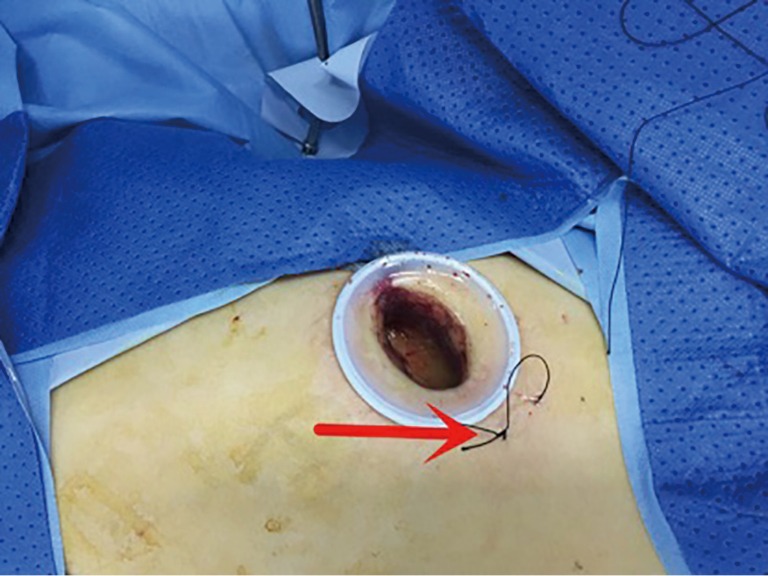
In uniportal VATS, the camera lens is entered into the patient’s body from the incision, so the endoscope should be placed above the incision as high as possible to provide enough room for surgeons. In the early stage, the camera assistant controls the camera completely. It is a big challenge for the camera assistant to ensure the quality of image and keep the camera straight simultaneously. The camera assistant has to use both hands to ensure stability of the camera. Fatigue caused by holding the camera for a long time reduces the concentration of the camera assistant, which increases the uncertainty of the surgery. In the later stage, we fix the shank of the camera on the margin of the incision with 7# suture to give the camera a fulcrum (Figure 1), and then the camera assistant can use singe hand to hold the camera, which decreases the level of fatigue and helps to stay in focus. Single-hand camera holding can also provide an important prerequisite for the sideways position.
Figure 1.

The fixation of the camera lens. 7# suture was used to make a fulcrum for camera shank, as the arrow pointed.
Position of the camera assistant
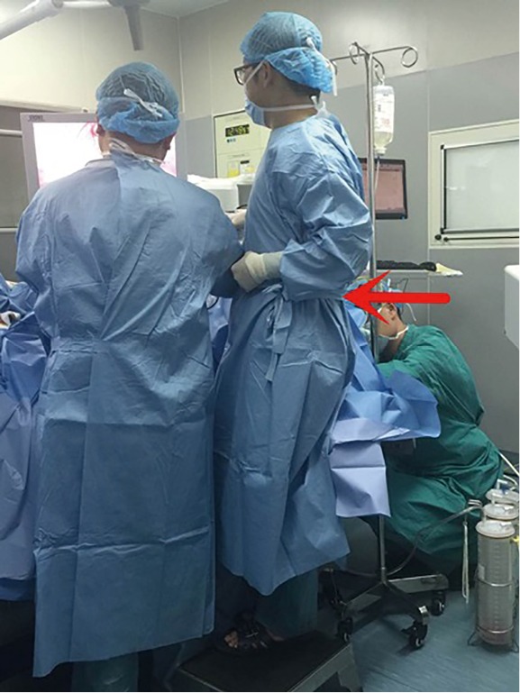
In the early stage, we found that contralateral camera holding requires the assistant to keep stooping and staring at the screen for a long time, which causes great uncomfortableness. However, we also found that the ipsilateral camera holding takes up too much operating space and causes more collision of instruments and limbs, which reduces comfort of the surgeon and fluency of the whole surgery. In the later stage, we improve the way of ipsilateral camera holding. When we operate on the upper and middle part of the thoracic cavity, the camera assistant stands in the caudal region of the patient. When we operate on the lower part of the thoracic cavity, the camera assistant stands in the rostral region of the patient on proper foot-stool to keep the upper limbs 15–20 cm higher than the surgeon (Figure 2). During the whole procedure, the camera assistant should use “ipsilateral and single-hand” position as often as possible. And then, it will give the surgeon more operating space, reduce the collision of instruments and limbs and increases the degree of comfort and concentration. The fluency and accuracy of the surgery can also be guaranteed.
Figure 2.

Position of the camera assistant. The camera assistant (as the arrow pointed) stands ipsilaterally on proper foot-stool to keep the upper limbs 15–20 cm higher than the surgeon.
Adjustment of the focal length
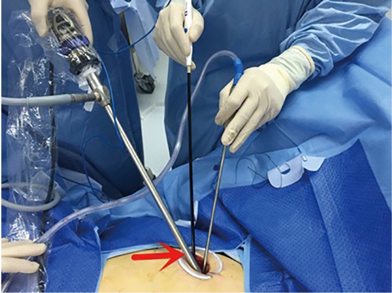
In VATS, we keep the camera lens at a macroscopic distance in the following conditions: thoracoscopic exploration, pulmonary lobe moving, instrument access, gauze and specimen withdraw, vessel or trachea cross and tissue incision by cut stapler, determination of the lobe or segment plane and observation of pulmonary ventilation in lung inflation, etc. But we keep the camera lens at a microscopic distance in these conditions: observation of small tumor, dealing with pleural adhesion, dissociating fine tissues such as vessels and lymph nodes, hemostasis and suture, etc. To adjust the camera lens in a proper distance, the camera assistant should be well acquainted with the surgical procedures, understand the intention and habits of the surgeons, stay in focus and be adaptable in any condition. In uniportal VATS, the endoscope is often fixed among incision protector and silk suture, which increases the resistance of movement (Figure 3). We can use paraffin to lubricate the endoscope to reduce the resistance, which improves control of the camera lens.
Figure 3.

Position of camera lens and surgical instruments for uniportal video-assisted thoracoscopic surgery (VATS).
Adjustment of the camera angle
The 30-degree camera lens has a unique advantage in VATS and an excellent camera assistant should take full use of it. Following are four perspectives of observation commonly used:
From anterior to posterior: it’s often used in the dissection of anterior mediastinum and the front of pulmonary hilum, such as the anterior hilum and the group 2 or 4 lymph node dissection, etc.;
From posterior to anterior: it’s often used in the dissection of posterior mediastinum and the back of pulmonary hilum, such as the posterior hilum and the group 7, 8 or 9 lymph node dissection;
From top to bottom: it’s often used in the dissection of the top of the hilum, such as free of the first branch of pulmonary artery and the group 5 or 6 lymph node dissection;
From bottom to top: it’s often used in hemostasis of thoracic wall and release of pleural adhesions.
At the same time, the adjustment of the camera angle also should be flexible. Sometimes we need to change the observation perspective even if we observe the same place. For example, when cutting the target tissues with cut stapler, we need to adjust the camera angle to assist the anvil to cross vessels or bronchi at an appropriate angle, which can avoid injuring the normal tissues and ensure the accuracy of surgery. In the adjustment of camera in uniportal VATS, apart from the principles above, the endoscope should be kept away from the surgeon to reduce the disturbance.
Cooperation of camera assistants and surgeons
A successful VATS is dependent on the tacit cooperation of the surgeon and the camera assistant, especially in emergency conditions. The adjustment of the camera should be natural and steady to avoid big movements when spinning or getting in and out. Also, the camera can’t be withdrawn without the surgeon’s permission. When facing the condition of angiorrhexis or blood splattering, camera assistant should keep calm and clean the camera lens swiftly, which can win more time to stop bleeding. Moreover, camera assistant needs to be more patient and concentrated when facing a long-time surgery with severe pleural adhesions, difficult separation of lymph nodes and vessels, complex tracheal or vascular reconstruction. In addition, an excellent camera assistant can expose the potential or undetected problems, helping to improve the safety of surgery.
Acknowledgements
None.
Informed Consent: Written informed consent was obtained from the patient for publication of this manuscript and any accompanying images.
Footnotes
Conflicts of Interest: The authors have no conflicts of interest to declare.
References
- 1.Hartwig MG, D'Amico TA. Thoracoscopic lobectomy: the gold standard for early-stage lung cancer? Ann Thorac Surg 2010;89:S2098-101. 10.1016/j.athoracsur.2010.02.102 [DOI] [PubMed] [Google Scholar]
- 2.Yan TD, Black D, Bannon PG, et al. Systematic review and meta-analysis of randomized and nonrandomized trials on safety and efficacy of video-assisted thoracic surgery lobectomy for early-stage non-small-cell lung cancer. J Clin Oncol 2009;27:2553-62. 10.1200/JCO.2008.18.2733 [DOI] [PubMed] [Google Scholar]
- 3.Rocco G, Martucci N, La Manna C, et al. Ten-year experience on 644 patients undergoing single-port (uniportal) video-assisted thoracoscopic surgery. Ann Thorac Surg 2013;96:434-8. 10.1016/j.athoracsur.2013.04.044 [DOI] [PubMed] [Google Scholar]
- 4.Kuritzky AM, Ryder BA, Ng T. Long-term survival outcomes of Video-assisted Thoracic Surgery (VATS) lobectomy after transitioning from open lobectomy. Ann Surg Oncol 2013;20:2734-40. 10.1245/s10434-013-2929-2 [DOI] [PubMed] [Google Scholar]
- 5.Anile M, Diso D, Mantovani S, et al. Uniportal video assisted thoracoscopic lobectomy: going directly from open surgery to a single port approach. J Thorac Dis 2014;6:S641-3. [DOI] [PMC free article] [PubMed] [Google Scholar]
- 6.Liu CY, Lin CS, Shih CH, et al. Single-port video-assisted thoracoscopic surgery for lung cancer. J Thorac Dis 2014;6:14-21. [DOI] [PMC free article] [PubMed] [Google Scholar]
- 7.Bertolaccini L, Rocco G, Viti A, et al. Geometrical characteristics of uniportal VATS. J Thorac Dis 2013;5 Suppl 3:S214-6. [DOI] [PMC free article] [PubMed] [Google Scholar]


