Smartphones have been used in anterior and posterior segment photography1, 2 and adaptors are available to enable the recording of surgical videos. The two-dimensional (2D) images can be of excellent quality but are lacking when compared to the stereoscopic three-dimensional (3D) view through the slit lamp or microscope eyepieces.
A number of companies (eg Google/Samsung/Zeiss) are using smartphones and head-mounted Virtual Reality (VR) headsets (eg Google Cardboard/Samsung Gear VR/Zeiss VR One) for the purpose of immersive 3D video gaming or movie viewing. These are effectively head-mounted stereoscopes into which a smartphone is placed to display the image. Using two smartphones and a VR headset we have developed a way of viewing slit lamp and surgical videos in 3D.
We connected two iPhone 4Ss to the slit lamp or operating microscope using 3D-printed eyepiece adaptors (Figure 1). Video of signs such as anterior chamber cells in anterior uveitis, and of cataract and strabismus surgery were recorded from left and right eyepieces. The two videos were synchronized and displayed side-by-side (left video on the left and the right video on the right) in a single video using video-editing software.
Figure 1.
Two phones attached to the slit lamp or microscope eyepieces record images from two perspectives to create a stereoscopic effect.
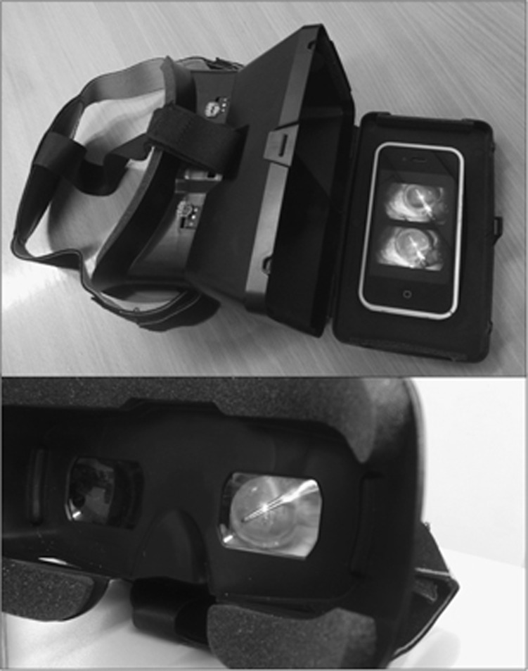
This side-by-side video is loaded onto the iPhone. It is then viewed in stereoscopic 3D using an inexpensive VR headset into which the phone is placed (Figure 2, Supplementary video).
Figure 2.
iPhone loaded into the VR headset for viewing of video with stereoscopic 3D effect.
Recently, novel methods have been used to record video of eye surgery, including Google Glass3 and two GoPro cameras to create a stereoscopic video.4 These methods are limited by the wide field of view, meaning the object of interest is quite small. This limits the capability of such methods to create a useful surgical educational video. The image quality with our technique is excellent with a good stereoscopic effect. Ophthalmic trainees can view surgery in the same way as when looking through the microscope assistant eyepieces. This allows a much more immersive and instructional view of surgery than is available with 2D surgical educational videos. In future, surgical trainees worldwide could view an online library of 3D instructional videos using a VR headset as if they were in theatre with the surgeon.
The authors declare no conflict of interest.
Footnotes
Supplementary Information accompanies this paper on Eye website (http://www.nature.com/eye)
Supplementary Material
References
- Barsam A, Bhogal M, Morris S, Little B. Anterior segment slit lamp photography using the iPhone. J Cataract Refract Surg 2010; 36(7): 1240–1241. [DOI] [PubMed] [Google Scholar]
- Haddock LJ, Kim DY, Mukai S. Simple, inexpensive technique for high-quality smartphone fundus photography in human and animal eyes. J Ophthalmol 2013; 2013: 518479. [DOI] [PMC free article] [PubMed] [Google Scholar]
- Rahimy E, Garg SJ. Google glass for recording Scleral Buckling Surgery. JAMA Ophthalmol 2015; 133(6): 710–711. [DOI] [PubMed] [Google Scholar]
- Birnbaum FA, Wang A, Brady CJ. Stereoscopic surgical recording using GoPro cameras: a low-cost means for capturing external eye surgery. JAMA Ophthalmol 2015; 133(12): 1483–1484. [DOI] [PMC free article] [PubMed] [Google Scholar]
Associated Data
This section collects any data citations, data availability statements, or supplementary materials included in this article.


